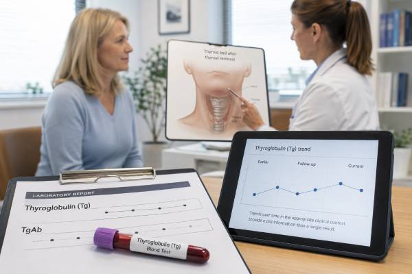
A thyroglobulin blood test measures a protein made only by normal thyroid follicular cells and most differentiated thyroid cancer cells. Its main role is monitoring papillary and follicular thyroid cancer after treatment, especially after total thyroidectomy with or without radioactive iodine. In that setting, a very low or undetectable thyroglobulin level is reassuring, while a rising trend can signal remaining or recurrent thyroid tissue. The number cannot be interpreted by itself. Thyroglobulin antibodies (TgAb) may make standard immunoassays falsely low, and thyroid-stimulating hormone (TSH) strongly affects how much thyroglobulin is produced. Surgery type, radioactive iodine, time since treatment, assay sensitivity, and imaging findings all change the meaning. When part of the thyroid remains after lobectomy, measurable thyroglobulin is expected and is less specific for cancer. Thyroglobulin is not a screening test for thyroid cancer in someone with an intact gland because benign thyroid tissue, goiter, nodules, thyroiditis, and Graves disease can all raise it.
- Thyroglobulin is primarily a follow-up marker for papillary and follicular thyroid cancer, not a general cancer screening test.
- After total thyroidectomy, an undetectable or very low Tg with negative TgAb is usually reassuring when imaging is also clear.
- A rising Tg trend is more important than one isolated value and should be compared using the same assay when possible.
- TgAb must be measured with every Tg because antibodies can cause falsely low or otherwise unreliable results.
- TSH stimulation can raise Tg, so suppressed and stimulated values are not directly comparable.
- Medullary thyroid cancer is monitored with calcitonin and CEA rather than thyroglobulin.
Table of Contents
- What the Thyroglobulin Test Measures
- When Thyroglobulin Testing Is Used
- High, Low, and Undetectable Results After Treatment
- TSH, Suppressed Tg, and Stimulated Tg
- Tg Antibodies and Assay Interference
- Preparation, Timing, and Follow-Up Testing
- Test Limits, Imaging, and Next Steps
What the Thyroglobulin Test Measures
Thyroglobulin is a large protein produced inside thyroid follicles. The gland uses it as a framework for making T4 and T3. Small amounts normally enter the bloodstream, so almost anyone with functioning thyroid tissue can have measurable Tg.
Papillary thyroid carcinoma and follicular thyroid carcinoma arise from follicular cells and usually retain the ability to make thyroglobulin. After most or all normal thyroid tissue has been removed, that tissue specificity turns Tg into a useful tumor marker. The less normal tissue remains, the more a new or increasing value points toward persistent thyroid cells that need evaluation.
What Tg does not measure
Thyroglobulin is not a thyroid function test. It does not show whether the circulating T4 and T3 supply is adequate. TSH and free T4 perform that role. A high Tg does not necessarily mean the thyroid is overactive, and a low Tg does not mean hypothyroidism.
It is also not a reliable diagnostic test for an untreated thyroid nodule. Benign thyroid tissue releases Tg, and large goiters can produce high values. Blood Tg cannot distinguish a benign nodule from papillary or follicular cancer. Ultrasound and, when indicated, fine-needle aspiration provide the relevant preoperative assessment.
Which cancers produce Tg
Tg is useful for most differentiated thyroid cancers:
- Papillary thyroid cancer
- Follicular thyroid cancer
- Oncocytic thyroid carcinoma, although production can vary
- Some poorly differentiated cancers that retain follicular-cell features
Anaplastic cancers may lose Tg production. Medullary thyroid cancer arises from C cells and is followed with calcitonin testing and carcinoembryonic antigen. Lymphoma and metastatic tumors in the thyroid are also not monitored with Tg.
Units and reference intervals
Tg is commonly reported in ng/mL or µg/L; these units are numerically equivalent. A general laboratory reference interval for someone with an intact thyroid is not the main target after cancer treatment. Post-treatment interpretation uses the surgery performed, TSH level, TgAb status, assay’s functional sensitivity, prior trend, and response-to-therapy category.
The lower limit of quantification matters. One assay may reliably measure down to 0.1 ng/mL, while another reports anything below 0.2 ng/mL as undetectable. “Undetectable” means below that method’s reporting capability, not proof that no thyroid cell exists anywhere.
When Thyroglobulin Testing Is Used
The clinical value changes dramatically before and after thyroid treatment.
After total thyroidectomy
Total thyroidectomy removes nearly all visible thyroid tissue but microscopic normal remnants often remain. A postoperative Tg provides early information about residual tissue and possible disease. Timing matters because Tg can be temporarily elevated after surgical manipulation and then decline as protein clears.
A very low Tg several weeks after surgery is favorable. A clearly elevated value may reflect a larger normal remnant, persistent cancer in the neck, or metastatic disease. It does not determine location, so ultrasound or other imaging may be needed.
After radioactive iodine
Radioactive iodine can ablate remaining normal thyroid tissue and treat iodine-avid cancer. Once both the gland and remnant are minimized, Tg becomes more specific. Follow-up commonly pairs Tg/TgAb with neck ultrasound and clinical risk assessment.
The 2025 American Thyroid Association guideline uses response-to-therapy assessment rather than one universal threshold for every patient. The significance of a detectable value depends on whether the person is low, intermediate, or high risk; whether radioiodine was given; and whether structural disease is present.
After lobectomy
A lobectomy leaves one thyroid lobe. That normal tissue continues to make Tg, often in amounts that vary with TSH, gland size, nodules, thyroiditis, and iodine status. A measurable or even increasing Tg is therefore less specific for recurrence than after total thyroidectomy.
Surveillance after lobectomy emphasizes neck ultrasound, examination, pathology risk, and TSH. Tg can contribute when a substantial and consistent trend develops, but no single low cutoff reliably separates normal remaining lobe from microscopic cancer. The clinician should not apply total-thyroidectomy targets to a lobectomy patient.
Fine-needle aspiration washout
When an abnormal neck lymph node is biopsied, the needle can be rinsed and the washout fluid tested for thyroglobulin. High Tg in a lymph-node aspirate supports metastatic differentiated thyroid cancer, especially when cytology is nondiagnostic or cystic. Blood contamination, an intact thyroid, assay method, and anti-Tg antibodies can complicate interpretation.
This is different from serum Tg and uses its own laboratory cutoff. The 2023 European Thyroid Association thyroid nodule guideline supports adding Tg washout when differentiated thyroid cancer metastasis is suspected.
Non-cancer uses
Tg may help distinguish endogenous thyroid hormone release from taking thyroid hormone. In destructive thyroiditis or Graves disease, Tg is usually present or elevated because thyroid tissue is releasing protein. In factitious thyrotoxicosis caused by taking exogenous hormone, Tg may be low. Antibodies can invalidate this distinction, and it is a specialist use rather than a routine test.
Tg can also support rare evaluations of congenital absence of thyroid tissue or defects in thyroglobulin production. These uses rely on a complete endocrine assessment.
High, Low, and Undetectable Results After Treatment
A Tg result is best treated as a time series. The same number can mean something different at the first postoperative check than five years into stable follow-up.
Undetectable or very low Tg
After total thyroidectomy and radioactive iodine, an undetectable basal Tg with negative TgAb and a normal neck ultrasound is strongly reassuring. It often fits an excellent response to therapy, especially when the assay is highly sensitive and TSH is in the intended range.
An undetectable value does not eliminate all recurrence risk. Very small tumors may make too little Tg to detect, poorly differentiated cells may lose Tg production, TgAb may hide the signal, or disease may be located in a site that is better detected by imaging. Follow-up intensity is adjusted to the original cancer risk and ongoing response.
After surgery without radioactive iodine, small normal remnants may leave a low measurable Tg. Stable or falling values are generally more reassuring than a single absolute threshold.
Detectable but stable Tg
A stable low-level Tg can reflect benign thyroid remnants. The clinician considers:
- Whether total thyroidectomy or lobectomy was performed
- Whether radioactive iodine was given
- Current TSH
- Time since surgery or ablation
- TgAb trend
- Ultrasound findings
- The assay’s measurement uncertainty
Small differences near the lower limit may be analytical noise. For example, a change from 0.10 to 0.14 ng/mL is not necessarily biologic progression. Repeating with the same assay after an appropriate interval is often more useful than immediate extensive imaging.
Rising Tg
A persistent increase raises concern for growing thyroid tissue or recurrent cancer. The rate of rise, doubling time, TSH stability, and magnitude all matter. A gradual rise across several same-assay measurements is more convincing than one jump after switching laboratories.
The next step may include high-resolution neck ultrasound, cross-sectional imaging, radioactive iodine imaging, or PET/CT depending on risk, Tg level, pathology, and iodine avidity. A rising Tg with no visible disease is called a biochemical incomplete response in some settings. It does not automatically mean immediate treatment; some values remain stable or decline, while others lead to structural findings later.
High Tg soon after surgery
Early postoperative Tg can be high from normal remnant tissue, surgical injury, or persistent disease. It is interpreted with the pathology report, extent of surgery, lymph-node involvement, imaging, and TSH. A high value may influence radioactive iodine planning, but no universal number replaces individualized staging.
High Tg with an intact thyroid
With the gland still present, Tg can rise from:
- Multinodular goiter
- Graves disease
- Hashimoto or subacute thyroiditis
- Iodine deficiency
- Thyroid stimulation from elevated TSH
- Recent biopsy, surgery, or radiofrequency ablation
- Benign adenomas and differentiated cancer
Because the causes overlap, serum Tg should not be used to decide whether a nodule is malignant.
Unexpectedly low Tg
Low Tg after extensive treatment may be desirable. Unexpectedly low Tg despite known structural disease suggests TgAb interference, a tumor that produces little Tg, a hook effect at extremely high concentrations, or an assay issue. The laboratory can perform dilution studies or use another method.
TSH, Suppressed Tg, and Stimulated Tg
TSH stimulates both normal and cancerous follicular thyroid cells to produce thyroglobulin. A Tg result cannot be compared fairly without knowing the TSH environment.
Basal or suppressed Tg
Basal Tg is measured while the person continues levothyroxine. In thyroid cancer follow-up, levothyroxine may keep TSH in a low or target range. A highly sensitive assay can detect very small amounts of Tg without interrupting treatment.
This approach is convenient and avoids hypothyroid symptoms. Many low-risk patients with an undetectable basal Tg, negative TgAb, and normal ultrasound do not need repeated stimulated testing.
Stimulated Tg
Stimulated Tg is measured after TSH rises. Stimulation can occur in two ways:
- Thyroid hormone withdrawal: Levothyroxine is stopped long enough for endogenous TSH to rise. This can cause fatigue, cognitive slowing, constipation, cold intolerance, and other hypothyroid symptoms.
- Recombinant human TSH: Thyrotropin alfa injections raise TSH while the patient stays on thyroid hormone.
Stimulated Tg is usually higher than basal Tg because residual thyroid cells receive a stronger signal. It may reveal disease that a less-sensitive basal assay misses. Its role is now more selective because modern assays have improved and risk-adapted follow-up avoids unnecessary testing.
Do not compare unlike conditions
A basal Tg of 0.2 ng/mL and a stimulated Tg of 1.5 ng/mL are not evidence of a sevenfold disease increase. The values were collected under different TSH stimulation. Trends should compare basal with basal or stimulated with stimulated under similar protocols.
Likewise, a change in TSH from 0.05 to 3.0 mIU/L can raise Tg even without a meaningful increase in tissue. The report should include a same-day TSH whenever Tg is used for surveillance.
TSH suppression and risk
Levothyroxine doses may suppress TSH after differentiated thyroid cancer, but the target depends on recurrence risk and response. Strong suppression can increase atrial fibrillation and bone loss. Current care balances cancer control with cardiovascular and skeletal harm rather than driving TSH as low as possible for everyone. The thyroid hormone monitoring panel helps track the replacement and suppression dose.
Tg Antibodies and Assay Interference
TgAb is the most important limitation of thyroglobulin testing. It should be measured every time Tg is measured, preferably in the same laboratory.
How TgAb affects immunometric Tg
Most routine Tg tests are immunometric assays. TgAb can bind thyroglobulin and prevent assay antibodies from recognizing it, often producing a falsely low result. A reported “undetectable” Tg is therefore not reassuring when TgAb is positive or newly rising.
The separate thyroglobulin antibody test has its own assay variability. A negative result on one platform does not guarantee absence of interference on every platform.
Following the TgAb trend
When Tg is unreliable, TgAb can serve as an indirect marker. Falling antibody concentrations after total thyroidectomy and treatment are generally reassuring. Persistent or rising TgAb may prompt closer imaging, especially if the rise is consistent and not explained by an assay change.
TgAb trends are qualitative, not exact tumor measurements. The concentration reflects immune activity as well as antigen exposure. Comparing values from different assays is especially problematic because calibration and sensitivity differ.
Radioimmunoassay and mass spectrometry
Tg radioimmunoassays may be less prone to falsely low results from TgAb but can have other biases and lower sensitivity. Liquid chromatography–tandem mass spectrometry measures Tg peptides and is designed to resist antibody interference. However, some patients with structural disease and TgAb still have low or undetectable mass-spectrometry Tg, so the method does not replace imaging or clinical judgment.
Heterophile antibodies and hook effect
Heterophile antibodies can create false high or false low results. In a hook effect, an extremely high Tg overwhelms the assay and paradoxically appears low. Laboratories can detect this by testing diluted samples. These issues are considered when Tg is incompatible with known disease burden.
Method changes
Different Tg assays can differ by twofold or more at the same true concentration. Use the same laboratory and method for serial follow-up when possible. If a method changes, establish a new baseline and avoid interpreting the first cross-platform difference as recurrence.
Preparation, Timing, and Follow-Up Testing
Fasting is usually unnecessary. Preparation depends on whether the test is basal or TSH-stimulated.
For a routine basal Tg
Continue levothyroxine unless instructed otherwise. Take it consistently and record whether the blood was drawn before or after the daily dose. Levothyroxine timing has less immediate effect on Tg than on free T4, but consistency helps interpret TSH.
Bring information about:
- Surgery type and date
- Radioactive iodine dose and date
- Original pathology and risk category
- Current levothyroxine dose and TSH target
- Prior Tg, TgAb, TSH, and imaging results
- Pregnancy status
- Biotin and other supplements
Biotin can interfere with some immunoassays. Many laboratories recommend stopping non-prescription biotin for at least 48 hours; high-dose prescribed biotin requires individualized guidance.
For stimulated testing
Follow the endocrine or nuclear-medicine protocol exactly. Recombinant TSH involves scheduled injections and timed blood sampling. Withdrawal protocols include medication changes and may use a low-iodine diet when radioactive iodine imaging or treatment is planned.
Do not stop levothyroxine independently. Severe hypothyroidism can impair driving, work, mood, heart function, and safety.
Timing after treatment
Tg is often checked several weeks after surgery and during subsequent response assessment. Follow-up may occur every 6 to 12 months at first, with longer or shorter intervals based on risk and trends. NICE recommends risk-adapted follow-up ranging from several years to lifelong surveillance when biochemical or structural disease persists.
What should be ordered together
A useful surveillance set commonly includes:
- Thyroglobulin
- TgAb
- TSH
- Sometimes free T4
- Neck ultrasound according to risk and prior findings
Ordering Tg without TgAb creates an avoidable blind spot. Ordering Tg without TSH removes essential stimulation context.
Test Limits, Imaging, and Next Steps
Thyroglobulin is highly useful after appropriate treatment, but it is not a stand-alone scan.
Tg-positive, imaging-negative disease
A rising Tg can precede visible disease. The clinician may repeat neck ultrasound, obtain contrast CT or MRI, use radioactive iodine imaging, or consider FDG PET/CT when Tg rises but iodine imaging is negative. The choice depends on tumor histology, Tg level and doubling time, prior radioiodine response, and likely disease location.
Immediate empiric treatment is not always necessary. Low stable Tg may be observed, while faster or higher rises justify more intensive investigation.
Imaging-positive, Tg-negative disease
Structural recurrence can occur with undetectable Tg, particularly with TgAb, small-volume lymph-node disease, or tumors that have lost differentiation. Suspicious ultrasound findings require evaluation regardless of a reassuring blood test. Fine-needle aspiration cytology and Tg washout can confirm nodal metastasis.
Lobectomy follow-up
After lobectomy, ultrasound and pathology-based risk carry more weight because normal tissue produces Tg. A slowly changing level may reflect TSH or lobe growth. A substantial, consistent rise plus an abnormal ultrasound is more concerning than an isolated value.
When to contact the care team
Contact the treating clinician when:
- Tg becomes newly detectable after being undetectable
- The value rises on two or more same-assay tests
- TgAb appears or rises consistently
- A neck lump, persistent hoarseness, swallowing difficulty, or unexplained cough develops
- TSH is far outside the intended cancer target
- A test was performed under a different stimulation condition or laboratory method
Most small changes are not emergencies. They need organized comparison, not panic. A useful report interpretation states the current Tg, TgAb, TSH, assay, surgery type, radioiodine history, prior trend, and imaging result together.
Thyroglobulin works best as a personalized marker. After enough normal tissue has been removed, the patient becomes their own reference: a stable low baseline is reassuring, and a reproducible upward departure directs the next evaluation.
References
– 2025 American Thyroid Association Management Guidelines for Adult Patients with Differentiated Thyroid Cancer 2025 (Guideline) – Thyroid cancer: assessment and management 2022 (Guideline) – Diagnostic, Theranostic and Prognostic Value of Thyroglobulin in Differentiated Thyroid Cancer 2024 (Review) – Revisiting Thyroglobulin Measurement: Current Methods and Emerging Alternative Approaches 2025 (Review) – Clinical use of thyroglobulin: not only thyroid cancer 2024 (Review) – 2023 European Thyroid Association Clinical Practice Guidelines for thyroid nodule management 2023 (Guideline)
Disclaimer
This article is educational and cannot determine whether an individual has persistent or recurrent thyroid cancer. Thyroglobulin interpretation depends on surgery, TSH, TgAb, assay method, pathology, and imaging. Review every trend with the clinician managing the thyroid cancer follow-up.