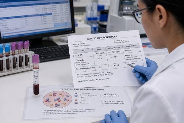
The free thyroxine index, usually shortened to FTI or FT4I, is a calculated estimate of biologically available thyroxine. It combines a total T4 result with a thyroid hormone binding estimate, most often the T3 uptake test. The calculation was developed to correct total T4 for changes in thyroxine-binding globulin and other transport proteins. A high FTI usually supports excess thyroid hormone, while a low FTI can support thyroid hormone deficiency, but the result must be read with TSH and the clinical setting. Direct free T4 assays have replaced FTI in many laboratories, yet the index can still help when binding proteins are abnormal, direct free T4 appears inconsistent, or a historical panel uses total T4 and T3 uptake. Pregnancy, severe illness, inherited binding-protein disorders, medicines, and assay limitations can still affect the calculation. FTI is an estimate rather than a direct measurement, so a flagged result should be confirmed when it conflicts with symptoms or other thyroid tests.
- FTI is calculated from total T4 and a binding-protein estimate; it is not a separately measured hormone.
- A high FTI can support hyperthyroidism, while a low FTI can support hypothyroidism when the TSH pattern agrees.
- The index helps correct total T4 when thyroxine-binding globulin is high or low, but the correction is imperfect.
- Modern laboratories usually prefer direct free T4, although FTI remains useful in selected binding-protein and pregnancy-related situations.
- The normal interval depends on the laboratory’s formula and component assays, so values from different laboratories should not be compared directly.
Table of Contents
- How the Free Thyroxine Index Works
- Why an FTI Test Is Ordered
- Total T4, T3 Uptake, and the Calculation
- Normal Range and Common Result Patterns
- Causes of a High FTI
- Causes of a Low FTI
- Limitations, Pregnancy, and Interference
- Next Steps After an Abnormal FTI
How the Free Thyroxine Index Works
Thyroxine, or T4, circulates in two forms. More than 99.9% is attached to transport proteins, mainly thyroxine-binding globulin, or TBG. A tiny fraction remains free and can enter cells or be converted to the more active hormone T3. Total T4 measures both forms, so it rises when binding proteins increase and falls when they decrease even if the thyroid gland is working normally.
The free thyroxine index was designed to separate a true change in thyroid hormone activity from a change caused mainly by binding proteins. It combines total T4 with a measurement that moves in the opposite direction when binding capacity changes. Traditionally, that second measurement is T3 resin uptake, now usually called T3 uptake.
When TBG is high, more labeled T3 binds to unoccupied sites in the patient’s serum and less is taken up by the test resin. The reported T3 uptake is therefore lower. Total T4 is higher because more hormone is protein-bound. Multiplying the high total T4 by a low uptake value can produce a normal FTI, correctly suggesting normal free hormone availability.
When TBG is low, total T4 falls because fewer binding sites are available. The T3 uptake result rises because more tracer is captured by the resin or solid phase. Again, the two changes may offset each other and leave the FTI within range.
The exact formula varies. Some laboratories multiply total T4 by the T3 uptake ratio. Others divide total T4 by a thyroid hormone binding ratio or use a normalized factor. For example:
FTI = total T4 × T3 uptake ratio
The result is an index, not necessarily a concentration in ng/dL or pmol/L. A laboratory may report a unitless value or assign units based on local convention. The reference interval is tied to that specific formula and assay combination.
FTI is conceptually different from a direct free T4 test. Direct free T4 immunoassays attempt to estimate the unbound hormone itself. FTI infers it from two other measurements. Neither approach is perfect when binding abnormalities are extreme, but direct free T4 has become the routine choice in most modern laboratories.
Why an FTI Test Is Ordered
FTI was once a standard part of thyroid testing. Today it is more likely to appear when a laboratory still offers a traditional thyroid panel, when direct free T4 is unavailable, or when a clinician wants an alternative view of a discordant free T4 result.
Situations in which FTI may be considered include:
- An abnormal total T4 with a normal TSH and no clear thyroid symptoms
- Pregnancy or estrogen therapy, which raises TBG
- Androgen treatment, nephrotic syndrome, severe liver disease, or inherited TBG deficiency, which can lower TBG
- Suspected familial binding-protein abnormalities
- A direct free T4 result that does not match TSH or the clinical picture
- Investigation of possible central hypothyroidism when routine free T4 is repeatedly low but TSH is not elevated
- Comparison with older medical records that used total T4 and T3 uptake
The index is not needed for most straightforward cases. When the pituitary-thyroid axis is intact, a TSH blood test is generally the first screen. Direct free T4 is then used to confirm overt hypothyroidism or hyperthyroidism. FTI is an alternative or supplementary calculation rather than a superior universal test.
FTI should not be ordered to evaluate T3 production. Despite the name “T3 uptake,” the uptake component does not measure the patient’s circulating T3 concentration. It measures available binding sites indirectly. A high T3 uptake does not mean the thyroid is making too much T3, and a low uptake does not mean T3 deficiency.
The index also cannot identify the cause of thyroid dysfunction. Graves disease, thyroiditis, toxic nodules, Hashimoto thyroiditis, pituitary disease, and excessive thyroid medication can all produce high or low FTI patterns. Antibody tests, imaging, medication review, and clinical history may be needed after the biochemical direction is established.
Some laboratories no longer perform T3 uptake because direct free T4 testing is faster, easier to automate, and better integrated with reflex algorithms. When FTI is requested, the laboratory may substitute a direct free T4 or advise consultation. The report should make clear which measurement was actually performed.
Total T4, T3 Uptake, and the Calculation
Understanding the two components prevents common interpretation errors.
Total T4
Total T4 includes hormone bound to TBG, transthyretin, and albumin plus the free fraction. The thyroid gland releases mainly T4, making total T4 a useful reflection of circulating hormone when binding proteins are stable. Its weakness is sensitivity to protein changes.
Total T4 commonly rises with pregnancy, oral estrogen, estrogen-containing contraception, selective estrogen receptor modulators, acute hepatitis, and inherited TBG excess. It can fall with androgen use, high-dose glucocorticoids, nephrotic protein loss, severe liver failure, inherited TBG deficiency, and some serious illnesses.
A change in total T4 caused by TBG does not necessarily alter TSH or create symptoms. The pituitary responds mainly to free hormone, not to the protein-bound reservoir.
T3 uptake
The uptake test adds a known amount of labeled thyroid hormone to the patient’s serum. The tracer first occupies open binding sites. A secondary material then captures the tracer that remains unbound. Modern methods may use antibodies or other solid phases rather than resin, but the principle is similar.
The reported percentage or ratio is inversely related to available binding capacity:
- High T3 uptake: fewer unoccupied binding sites, often because TBG is low or because endogenous thyroid hormone has filled more sites
- Low T3 uptake: more unoccupied binding sites, often because TBG is high or endogenous hormone is low
This creates ambiguity when uptake is viewed alone. High uptake can occur in hyperthyroidism or low-TBG states. Low uptake can occur in hypothyroidism or high-TBG states. Combining it with total T4 is what gives the test practical meaning.
Example patterns
Suppose a pregnant patient has a total T4 above the nonpregnant range and a low T3 uptake. The calculated FTI may be normal because the high total T4 reflects increased TBG. By contrast, a person with Graves disease may have high total T4 and high uptake because endogenous hormone has saturated binding sites. The FTI becomes clearly high.
A person with inherited TBG deficiency may have low total T4 and high uptake. A normal FTI suggests normal thyroid function. A person with true primary hypothyroidism may have low total T4 and low uptake, producing a low FTI.
These examples are simplified. Albumin variants, medicines, illness, and assay design can prevent perfect cancellation.
Normal Range and Common Result Patterns
FTI reference intervals vary widely because laboratories use different formulas and calibrators. A report might show a range such as 1.2–4.9, 4.5–11.7, or another set of numbers. Some methods express the index as though it were a T4 concentration. Never apply a range from another laboratory.
The result should be interpreted with TSH. Common patterns include:
| TSH | FTI | General interpretation |
|---|---|---|
| Normal | Normal | Thyroid function is usually normal if symptoms and clinical context agree |
| High | Low | Overt primary hypothyroidism is likely |
| High | Normal | Subclinical hypothyroidism or a transient TSH elevation may be considered |
| Low | High | Overt hyperthyroidism or excess thyroid hormone is likely |
| Low | Normal | Subclinical hyperthyroidism, T3-predominant disease, medicine effect, or temporary suppression may be considered |
| Normal or low | Low | Central hypothyroidism, illness, medicine effect, or test limitation requires review |
| Normal or high | High | Binding abnormality, assay interference, hormone resistance, or a TSH-secreting tumor may be considered |
“Normal” means within the method-specific statistical interval, not necessarily the best target for every patient. In central hypothyroidism, the clinician may aim for an FTI or free T4 in the upper half of the range, depending on the method and patient. In older adults or people with heart disease, treatment is often more cautious.
A normal FTI can help show that an abnormal total T4 is caused by binding proteins. It does not rule out every thyroid disorder. Early primary dysfunction may alter TSH before FTI changes. T3 thyrotoxicosis can suppress TSH while total T4 and FTI remain normal. Rare assay problems can also create a falsely reassuring result.
The index is particularly useful when total T4 and T3 uptake move in opposite directions. When both point in the same direction, the FTI tends to magnify the abnormality. For example, high total T4 plus high uptake produces a high index; low total T4 plus low uptake produces a low index.
Small deviations deserve caution. Because FTI combines two measured values, analytical variation from both components affects the final number. A borderline change may not be meaningful unless it persists and matches TSH and symptoms.
Causes of a High FTI
A high FTI suggests that the amount of available T4 is increased, but the TSH pattern determines whether true thyrotoxicosis is likely.
Endogenous hyperthyroidism
Graves disease commonly produces low TSH, high total T4, high or normal T3 uptake, and a high FTI. TSH receptor antibodies can confirm autoimmune stimulation. Toxic adenoma and toxic multinodular goiter create similar hormone patterns but have different imaging and antibody findings.
Thyroiditis can also cause a temporarily high FTI as stored hormone leaks from inflamed tissue. Subacute thyroiditis may cause neck pain, while painless and postpartum thyroiditis do not. Radioactive iodine uptake tends to be low in destructive thyroiditis and high or patchy when the gland is actively overproducing hormone.
Iodine exposure and amiodarone can trigger hyperthyroidism in susceptible people. Amiodarone also inhibits T4-to-T3 conversion, so T4 and FTI may be high even when T3 is not markedly elevated.
Excess thyroid hormone medication
Too much levothyroxine can raise total T4 and FTI and suppress TSH. A high value can result from a recent dose increase, weight loss, improved absorption, accidental extra dosing, or stopping a drug that previously reduced absorption.
Blood collection soon after a levothyroxine dose may temporarily raise total and free T4. Consistent pre-dose sampling can help determine whether a mild elevation reflects a medication peak or sustained overreplacement.
Binding and analytical problems
FTI corrects common TBG changes but not every binding abnormality. Familial dysalbuminemic hyperthyroxinemia can produce high total T4 and misleading uptake behavior. Some albumin variants bind T4 strongly and may leave the index high despite normal thyroid function and normal TSH.
Thyroid hormone autoantibodies, heterophile antibodies, and assay-specific interference can affect either component. If the FTI is high while TSH is normal and the person has no symptoms, repeating tests on another platform or measuring direct free T4 by equilibrium dialysis may be more appropriate than diagnosing hyperthyroidism.
Pregnancy can occasionally produce a high FTI because the uptake correction does not perfectly compensate for major changes in TBG and albumin. A pregnancy-specific testing strategy is preferable.
Causes of a Low FTI
A low FTI suggests reduced available T4, but it can reflect true thyroid deficiency, central disease, severe illness, or imperfect correction.
Primary hypothyroidism
Hashimoto thyroiditis, thyroid surgery, radioactive iodine treatment, iodine deficiency, neck radiation, congenital thyroid disease, and medicines such as lithium can reduce T4 production. When the thyroid itself is failing, TSH rises. Low total T4 and low or normal T3 uptake combine to produce a low FTI.
A hypothyroidism blood test panel may add TPO antibodies to identify autoimmune thyroiditis. Antibodies support the cause but do not determine the severity; TSH and hormone levels do that.
Central hypothyroidism
Pituitary or hypothalamic disease can produce low FTI with a TSH that is not appropriately elevated. Causes include pituitary tumors, surgery, radiation, trauma, inflammation, and genetic disease. Other pituitary deficiencies may coexist.
FTI has sometimes helped clarify repeatedly low direct free T4 results with normal TSH. A normal index can suggest a method-related low free T4, while a low index adds support for central hypothyroidism. It is not definitive, and persistent cases require assessment of pituitary hormones and often MRI.
Illness, protein loss, and medication effects
Serious illness can lower total T4, alter uptake, and disrupt the assumptions behind the index. Nephrotic syndrome causes urinary loss of binding proteins and hormone. Liver disease changes protein production. Glucocorticoids, dopamine, antiseizure drugs, and other medicines can affect TSH, hormone metabolism, or protein binding.
TBG deficiency should usually produce low total T4 with high uptake and a normal FTI. If the index is also low, true hypothyroidism, severe illness, or incomplete correction should be considered. Confirmatory testing with TSH, direct free T4, and TBG can distinguish these possibilities.
In pregnancy, an apparently low FTI may reflect assay behavior rather than maternal hypothyroidism. Trimester-specific TSH and a locally validated free T4, total T4, or FTI approach should be used.
Limitations, Pregnancy, and Interference
FTI rests on the assumption that T3 uptake accurately reflects the binding environment affecting total T4. That assumption works reasonably well for ordinary TBG excess or deficiency but breaks down in several settings.
Important limitations include:
- T3 uptake responds to both binding-protein concentration and endogenous hormone saturation
- Albumin and transthyretin variants may not be corrected appropriately
- Severe illness introduces free fatty acids, medicines, and binding inhibitors
- Pregnancy changes TBG, albumin, fatty acids, and hormone metabolism simultaneously
- Both component assays add analytical uncertainty
- Different laboratories use different formulas and reference intervals
- Autoantibodies can interfere with total T4 or uptake measurements
- The index does not directly measure free hormone
Pregnancy deserves particular caution. Estrogen raises TBG, human chorionic gonadotropin lowers TSH in early pregnancy, and albumin concentrations change. Direct free T4 immunoassays also have weaknesses in pregnancy, which is why guidelines may recommend method- and trimester-specific free T4 ranges, an adjusted total T4 approach, or FTI where it has been validated. No single unadjusted adult range is appropriate throughout gestation.
Biotin is best known for affecting direct immunoassays, but it can also alter component tests if their platform uses biotin-streptavidin chemistry. The resulting pattern depends on the assay design. High-dose supplementation should be disclosed before testing.
Heparin can raise measured free T4 through in-vitro displacement, but its effect on FTI may differ because the index uses total T4 and uptake. This can make FTI a useful cross-check in some cases, though serious illness and heparin exposure can still disturb uptake and protein binding.
A direct method such as equilibrium dialysis with mass spectrometry may be used when routine tests remain discordant. TBG measurement, albumin studies, and family testing can help identify inherited binding disorders. The goal is to establish whether the person is truly euthyroid before treatment is started.
Next Steps After an Abnormal FTI
Start by checking the component values rather than looking only at the calculated index. Total T4 and T3 uptake reveal whether the abnormal result is driven by hormone concentration, binding capacity, or both.
For a high FTI with low TSH, clinicians usually evaluate hyperthyroidism or thyroid hormone excess. Free T4, T3, TSH receptor antibodies, medication review, and sometimes radioactive iodine uptake or ultrasound may follow.
For a low FTI with high TSH, primary hypothyroidism is likely. The clinician may repeat testing, check TPO antibodies, and discuss levothyroxine based on severity, symptoms, age, pregnancy, and heart health.
For an abnormal FTI with normal TSH, binding-protein changes and assay interference deserve attention. Useful follow-up may include direct free T4, TBG, albumin, repeat testing on another platform, or review of estrogen, androgens, pregnancy, kidney disease, and liver disease.
For a low FTI with normal or low TSH, central hypothyroidism must be considered if the pattern persists. Evaluation may include morning cortisol and other pituitary hormones before thyroid treatment.
A practical review follows this sequence:
- Confirm the laboratory’s formula, units, and reference interval.
- Examine total T4 and T3 uptake separately.
- Compare the index with TSH and any direct free T4 result.
- Review pregnancy, illness, protein loss, liver disease, medicines, and supplements.
- Repeat under comparable conditions if the abnormality is mild or unexpected.
- Use a specialist or reference method when results remain physiologically inconsistent.
Do not adjust thyroid medication solely to normalize an FTI without knowing how the index was produced. Modern monitoring usually relies on TSH and direct free T4, with FTI serving as a supporting measurement in selected cases.
References
- Use of the Free Thyroxine Index to Refine the Diagnosis of Central Hypothyroidism 2021
- Free thyroxine measurement in clinical practice 2023 (Review)
- Thyroxine-Binding Globulin Deficiency 2023 (Review)
- Current utility of first-line FT4 and TSH in screening for central hypothyroidism 2024
- Free thyroid hormone: Methods and standardization 2025 (Review)
- Assay of Thyroid Hormone and Related Substances 2025 (Review)
Disclaimer
The free thyroxine index is method dependent and should be interpreted with TSH, its component tests, symptoms, medicines, pregnancy status, and the laboratory’s reference interval. Do not start, stop, or change thyroid medication based on an isolated FTI result. Persistent discordant results may require endocrinology and laboratory review.