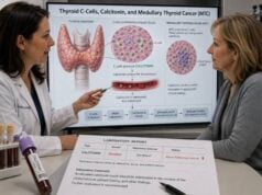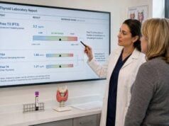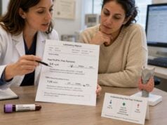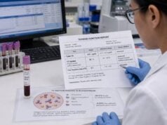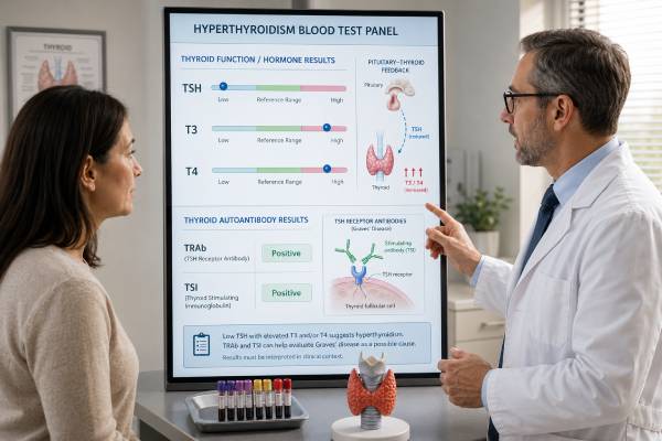
A hyperthyroidism blood test panel checks whether the body has too much thyroid hormone and helps identify why. The usual starting tests are thyroid-stimulating hormone (TSH) and free T4, followed by total or free T3 when the pattern is unclear or T3-predominant disease is possible. A very low TSH with high T4, high T3, or both usually confirms overt thyrotoxicosis. Antibody tests such as TSH receptor antibodies (TRAb) and thyroid-stimulating immunoglobulin (TSI) can then support Graves disease as the cause. Results still need context because pregnancy, severe illness, thyroid medication, iodine exposure, and supplements such as biotin can alter the numbers. The panel does not always distinguish hormone overproduction from hormone leakage caused by thyroiditis, so imaging or repeat testing may be needed. Symptoms, heart rate, medication history, examination findings, and the direction of change over time are all part of a safe interpretation.
- Low TSH with high free T4 and/or T3 usually indicates overt hyperthyroidism or another form of thyrotoxicosis.
- Low TSH with normal T4 and T3 is called subclinical hyperthyroidism and often requires confirmation before treatment.
- High T3 with normal free T4 can represent T3-predominant hyperthyroidism, especially early Graves disease or an autonomous nodule.
- Positive TRAb or TSI strongly supports Graves disease, but a negative result does not exclude every case.
- Biotin can create a false hyperthyroid pattern on some assays; many laboratories advise stopping it for at least 48 hours before testing.
- Chest pain, severe shortness of breath, fainting, confusion, or a very fast irregular heartbeat requires urgent medical assessment.
Table of Contents
- Tests Included in a Hyperthyroidism Panel
- How to Read Common Result Patterns
- Causes of Low TSH and High Thyroid Hormones
- Preparation, Timing, and Test Interference
- How Antibodies and Imaging Identify the Cause
- Using Blood Tests During Treatment
- Special Situations and Urgent Findings
Tests Included in a Hyperthyroidism Panel
A hyperthyroidism panel is not one fixed laboratory package. Clinicians usually order tests in stages, beginning with the markers most likely to confirm excess thyroid hormone and adding tests that explain the cause. A broad panel may include TSH, free T4, total T3, TRAb, TSI, and sometimes thyroid peroxidase antibodies (TPOAb). The results answer different questions, so no single value should be interpreted as the whole diagnosis.
TSH: the sensitive first signal
TSH comes from the pituitary gland. It tells the thyroid how strongly to make hormone. When T4 and T3 rise, the pituitary normally reduces TSH through a feedback system. For that reason, TSH is commonly the earliest value to fall in primary hyperthyroidism.
Many laboratories use an adult TSH reference interval near 0.4 to 4.0 mIU/L, but the exact limits vary. In overt hyperthyroidism, TSH is often below the assay’s measurable limit, such as less than 0.01 mIU/L. A mildly low result, for example 0.1 to 0.39 mIU/L, is less specific and can occur with temporary illness, medication effects, pregnancy, or subclinical hyperthyroidism.
A low TSH does not automatically prove that the thyroid gland is overproducing hormone. Pituitary disease, glucocorticoids, dopamine-related medicines, severe non-thyroid illness, and assay interference can also suppress it. The result becomes much more informative when viewed alongside free T4 results and T3.
Free T4: circulating unbound thyroxine
Free T4 measures the small portion of thyroxine that is not attached to transport proteins. It is generally more useful than total T4 when pregnancy, estrogen therapy, liver disease, or inherited binding-protein differences may change the amount of bound hormone.
An elevated free T4 with suppressed TSH supports overt thyrotoxicosis. A normal free T4 does not end the evaluation when symptoms are convincing and TSH is clearly low, because T3 may rise first. Laboratories use different free T4 methods and ranges; a typical interval may be about 0.8 to 1.8 ng/dL, but the printed laboratory interval should always control interpretation.
T3: severity and T3-predominant disease
T3 is the more active thyroid hormone. Total T3 is often preferred in hyperthyroidism because commonly available free T3 immunoassays can be less dependable. A total T3 test can detect cases in which TSH is suppressed and free T4 remains normal.
T3 may also help estimate disease severity and recognize T3-predominant Graves disease. Total T3, however, rises when thyroid-binding proteins increase, including during pregnancy or estrogen use. Severe illness can lower T3 even when thyroid physiology is abnormal, so an apparently normal value needs context.
TRAb and TSI: antibodies linked to Graves disease
TRAb tests detect antibodies that bind the TSH receptor. Depending on the assay, they may include stimulating, blocking, and neutral antibodies. TSI assays are designed to identify the stimulating activity most closely associated with Graves disease. Positive results can establish an autoimmune cause without a radioactive iodine uptake scan in many patients.
Neither antibody concentration directly measures how much hormone is circulating at that moment. TSH, free T4, and T3 describe thyroid function; TRAb and TSI help describe the cause and sometimes the chance of remission, relapse, or fetal exposure during pregnancy.
How to Read Common Result Patterns
The pattern across the panel is more reliable than an isolated high or low flag. The following combinations are common starting points, not final diagnoses.
| TSH | Free T4 | T3 | Common interpretation |
|---|---|---|---|
| Low or suppressed | High | High or normal | Overt thyrotoxicosis; determine whether the cause is Graves disease, autonomous nodules, thyroiditis, medication, or iodine |
| Low or suppressed | Normal | High | T3-predominant thyrotoxicosis |
| Low | Normal | Normal | Subclinical hyperthyroidism, temporary suppression, medication effect, pregnancy-related change, or assay interference |
| Low | Low | Low or normal | Not a standard primary hyperthyroid pattern; consider pituitary disease, severe illness, recent treatment, or drug effects |
| Normal or high | High | High | Discordant pattern requiring repeat testing and evaluation for interference, thyroid hormone resistance, or a rare TSH-secreting pituitary tumor |
Overt hyperthyroidism versus thyrotoxicosis
“Thyrotoxicosis” means that tissues are exposed to too much thyroid hormone. “Hyperthyroidism” specifically means the thyroid is actively producing too much. Graves disease and toxic nodules cause true hyperthyroidism. Thyroiditis can produce the same low-TSH, high-hormone blood pattern because stored hormone leaks from damaged thyroid cells, even though new hormone production is not increased.
That distinction changes treatment. Antithyroid drugs reduce new hormone synthesis and therefore work for Graves disease and autonomous nodules. They generally do not correct the hormone leakage of thyroiditis. Blood values confirm the biochemical state, while antibodies, imaging, tenderness, recent pregnancy, and medication history help classify it.
Subclinical hyperthyroidism
Subclinical hyperthyroidism means TSH is below range while free T4 and T3 remain within their ranges. The term refers to the laboratory pattern; some people still have palpitations, tremor, heat intolerance, anxiety, or reduced exercise tolerance.
A single mildly low TSH often needs repetition. Temporary suppression may follow acute illness, pregnancy, thyroiditis, steroid use, or a recent change in thyroid hormone medication. Persistent TSH below 0.1 mIU/L carries more concern than a result between 0.1 mIU/L and the lower reference limit, especially in adults over 65 or people with atrial fibrillation, heart disease, osteoporosis, or high fracture risk.
T3-predominant results
In early or recurrent Graves disease, T3 can rise before free T4. Autonomous nodules may also produce proportionally more T3. This is why testing only TSH and free T4 can miss some overt cases. A suppressed TSH plus high T3 and normal free T4 is not “almost normal”; it is a recognized biochemical form of thyrotoxicosis.
Conversely, T3 may be normal in an older or acutely ill patient with genuine hyperthyroidism because illness reduces conversion of T4 to T3. Symptoms, free T4, and the clinical setting prevent overreliance on one marker.
Causes of Low TSH and High Thyroid Hormones
The same hormone pattern can arise through several mechanisms. Identifying the mechanism prevents unnecessary or ineffective treatment.
Graves disease
Graves disease is an autoimmune disorder in which stimulating antibodies activate the TSH receptor. It often causes diffuse thyroid enlargement, smooth increased blood flow through the gland, and sometimes thyroid eye disease. TRAb or TSI is commonly positive. The hormone pattern may show elevations of both T3 and T4, with T3 disproportionately high in some cases.
The dedicated Graves disease blood test panel is especially useful when eye symptoms, a diffuse goiter, a personal or family autoimmune history, or relapse after antithyroid therapy raises suspicion.
Toxic adenoma and toxic multinodular goiter
A toxic adenoma is one thyroid nodule that produces hormone without responding to pituitary control. Toxic multinodular goiter contains several autonomous areas. These conditions are more common with advancing age and may develop gradually. TRAb and TSI are usually negative. A thyroid scan may show one “hot” focus or a patchy pattern of increased uptake.
Autonomous nodules rarely enter lasting remission from antithyroid medication alone. Medication can control hormone production, while radioactive iodine or surgery may provide definitive treatment when appropriate.
Thyroiditis
Subacute, painless, postpartum, and drug-induced thyroiditis can release stored T4 and T3. Subacute thyroiditis often causes neck pain and tenderness, sometimes after a respiratory infection. Painless and postpartum thyroiditis may have no neck discomfort. The thyrotoxic phase commonly lasts weeks, followed in some patients by a temporary hypothyroid phase.
Radioactive iodine uptake is low because the inflamed gland is not actively trapping iodine to make new hormone. TRAb is usually negative, although autoimmune markers such as TPOAb may be present in painless or postpartum disease. Beta blockers may reduce palpitations and tremor; antithyroid drugs usually offer little benefit in a purely destructive process.
Medication, supplements, and iodine
Taking too much levothyroxine or liothyronine can suppress TSH and raise thyroid hormones. The source may be an intentionally suppressive dose after thyroid cancer treatment, a dose that has become excessive after weight loss or aging, accidental duplication, or unsupervised use for weight loss.
Amiodarone can cause thyrotoxicosis through iodine-driven hormone synthesis, destructive thyroiditis, or a mixture of both. Iodinated contrast, kelp products, and high-dose iodine preparations can trigger excess hormone in susceptible glands. Some immune therapies and other medicines can inflame the thyroid.
Less common explanations
A high free T4 with a TSH that is not suppressed is not the usual feedback response. The laboratory should often repeat the measurements using another method before rare diagnoses are pursued. Heterophile antibodies, biotin, abnormal binding proteins, and anti-reagent antibodies can create misleading combinations. Genuine causes include resistance to thyroid hormone and a TSH-secreting pituitary adenoma, both of which need specialist evaluation.
Preparation, Timing, and Test Interference
Fasting is usually unnecessary for a hyperthyroidism panel. Consistency in medication timing and full disclosure of supplements are more important than an empty stomach.
Before the blood draw
Tell the ordering clinician and laboratory about:
- Biotin, including hair, skin, and nail products and high-dose neurologic formulations
- Levothyroxine, liothyronine, desiccated thyroid, or weight-loss products that may contain thyroid hormone
- Amiodarone, lithium, glucocorticoids, dopamine-related drugs, antiseizure medicines, and immune therapies
- Recent CT contrast, angiography, radioactive iodine, or high-iodine supplements
- Pregnancy, recent delivery, severe illness, hospitalization, and known pituitary disease
Biotin is a frequent source of false results because some immunoassays use biotin-streptavidin chemistry. It can make TSH appear falsely low and free T4, T3, or antibody results appear falsely high, imitating Graves disease. The American Thyroid Association advises avoiding biotin for at least two days before thyroid testing, but high doses or reduced kidney function may require a longer interval set by the clinician or laboratory. Prescription biotin should never be stopped without medical guidance.
Timing thyroid medicine
A dose of levothyroxine shortly before a blood draw can temporarily increase free T4. Liothyronine produces a more pronounced T3 peak after dosing. For serial monitoring, clinicians often prefer blood collected before the morning dose or at the same interval after every dose. The exact plan should be consistent and documented rather than improvised on test day.
Do not withhold an antithyroid drug, beta blocker, or thyroid hormone unless the treating clinician specifically instructs it. Missing doses can distort the result and may be unsafe.
Repeat testing when results conflict
Repeat testing is reasonable when the numbers do not fit symptoms, prior results, or one another. A useful verification process may include:
- Repeating TSH, free T4, and total T3 after removing a suspected interference.
- Asking the laboratory to use a different assay platform.
- Measuring total hormone or free hormone by a reference method when available.
- Reviewing medications, supplements, pregnancy status, and recent illness again.
- Comparing the current result with the person’s stable baseline rather than only the population interval.
Unexpected results should be investigated before permanent treatment such as radioactive iodine or surgery is chosen.
How Antibodies and Imaging Identify the Cause
Hormone tests show that thyrotoxicosis exists. Cause-finding tests show whether the gland is stimulated, autonomous, inflamed, or exposed to an outside hormone source.
TRAb versus TSI
A TRAb test commonly uses a binding method. It reports whether antibodies compete for the TSH receptor but may not identify whether each antibody stimulates or blocks the receptor. A TSI test is intended to detect stimulating activity. Modern versions of both tests perform well for diagnosing Graves disease, although cutoffs and reporting units differ.
A positive result in a person with suppressed TSH and elevated thyroid hormone often makes Graves disease the most likely explanation. A negative result lowers the probability but does not completely exclude early, mild, treated, or antibody-negative Graves disease. The clinician may then use a radioactive iodine uptake test or Doppler ultrasound.
Antibody numbers should not be compared directly between laboratories when platforms or units differ. A result reported as an index, IU/L, percentage, or times the upper limit of normal must be read against that assay’s cutoff.
Radioactive iodine uptake and scan
An uptake test measures how much tracer the thyroid collects over a set period. A scan shows its distribution.
- Diffusely high uptake supports Graves disease.
- One focal hot area supports a toxic adenoma.
- Patchy uptake supports toxic multinodular goiter.
- Very low uptake supports thyroiditis, recent iodine exposure, or outside thyroid hormone use.
Radioactive iodine testing is generally avoided during pregnancy and breastfeeding. Recent iodinated contrast or amiodarone can suppress uptake and make the study hard to interpret for weeks or longer.
Ultrasound with Doppler
Ultrasound evaluates thyroid size, texture, nodules, and blood flow. Markedly increased diffuse vascularity can support Graves disease, while nodules may point toward autonomous disease. Ultrasound does not measure hormone production by itself, and incidental nodules are common. A scan is often more direct when the question is whether a specific nodule is functioning.
TPOAb and thyroglobulin antibodies can indicate thyroid autoimmunity but are not specific for Graves disease. They may appear in Graves disease, Hashimoto thyroiditis, or people with normal thyroid function. A broad thyroid antibody panel should therefore be interpreted by antibody type, not simply labeled positive or negative as a group.
Using Blood Tests During Treatment
Monitoring intervals and target markers depend on treatment type. Early in therapy, free T4 and T3 often guide changes more safely than TSH because TSH can remain suppressed for weeks or months after hormone levels improve.
Antithyroid medication
Methimazole is commonly preferred outside certain pregnancy situations. Propylthiouracil is used in selected cases, including part of pregnancy management and thyroid storm. Before starting, clinicians may obtain a complete blood count and liver profile to establish a baseline and identify pre-existing abnormalities.
Thyroid tests are often repeated about every 4 to 6 weeks after starting or changing a dose, then less frequently once stable. If free T4 normalizes but T3 remains high, reducing treatment based only on free T4 could leave T3 thyrotoxicosis uncontrolled. If both fall low while TSH remains suppressed, continuing the same dose may cause treatment-induced hypothyroidism.
TRAb may be checked near the end of a planned antithyroid-drug course. A persistently elevated concentration suggests a higher chance of relapse, while a low or negative result supports—but does not guarantee—remission. Symptoms and hormone tests still determine current control.
Anyone taking methimazole or propylthiouracil should know the uncommon but serious warning signs. Fever or a severe sore throat may signal agranulocytosis, a dangerous drop in infection-fighting white blood cells. The medicine is generally held while urgent blood-count testing is arranged under medical instruction. Yellow skin or eyes, dark urine, severe itching, pale stools, or marked abdominal pain can indicate liver injury and also require prompt care.
Radioactive iodine
After radioactive iodine, hormone release may fluctuate before thyroid function declines. TSH, free T4, and sometimes T3 are checked at intervals such as every 4 to 6 weeks initially or according to the treatment protocol. Hypothyroidism is expected after treatment for Graves disease and can appear within months or later. Once replacement is stable, monitoring becomes similar to routine levothyroxine dose monitoring.
Surgery
Before thyroidectomy, clinicians aim to control hormone levels and heart-rate symptoms to reduce operative risk. After total thyroidectomy, free T4 falls and lifelong thyroid hormone replacement is required. TSH usually becomes the principal long-term marker once the pituitary has had time to respond to the new dose.
Special Situations and Urgent Findings
Pregnancy, older age, severe illness, and cardiac disease change the risks and the way results are interpreted.
Pregnancy and the postpartum period
Human chorionic gonadotropin can mildly stimulate the thyroid in early pregnancy and lower TSH, especially in the first trimester. Severe vomiting of pregnancy may produce temporary gestational thyrotoxicosis. Pregnancy-specific TSH and free T4 reference intervals are preferable because standard adult ranges can misclassify results.
Graves disease can also begin or recur during pregnancy. TRAb crosses the placenta, so a high maternal value can affect the fetal or newborn thyroid even after the mother has had surgery or radioactive iodine in the past. Medication selection and dose targets require obstetric and endocrine supervision. The related thyroid tests in pregnancy use trimester-aware interpretation rather than ordinary adult assumptions.
Postpartum thyroiditis commonly starts with a temporary thyrotoxic phase and may be followed by hypothyroidism. TRAb can help distinguish it from postpartum Graves disease, although imaging choices are limited during breastfeeding.
Older adults and heart risk
Older adults may lack classic tremor, heat intolerance, or obvious hyperactivity. They may instead develop fatigue, weight loss, weakness, depression, atrial fibrillation, or worsening heart failure. Persistent subclinical hyperthyroidism is also more important in this group because even modest hormone excess can affect rhythm and bone.
A new irregular pulse, resting tachycardia, exertional shortness of breath, or unexplained weight loss deserves prompt assessment. Clinicians may order an electrocardiogram and evaluate bone health in addition to thyroid tests.
Thyroid storm and other emergencies
Thyroid storm is a rare, life-threatening state of severe thyrotoxicosis with systemic decompensation. The hormone concentrations do not need to be higher than those seen in uncomplicated hyperthyroidism; the diagnosis depends heavily on clinical illness. Triggers include infection, surgery, trauma, childbirth, stopping antithyroid medicine, and acute iodine exposure.
Emergency features can include:
- High fever
- Very fast or irregular heartbeat
- Agitation, delirium, severe weakness, seizure, or coma
- Vomiting, diarrhea, abdominal pain, or jaundice
- Heart failure, chest pain, low blood pressure, or severe shortness of breath
These symptoms require emergency services rather than waiting for an outpatient panel. Treatment begins from clinical suspicion while blood tests are being processed.
For non-emergency abnormalities, the safest next step is usually confirmation of the pattern, a review of medicines and supplements, and a cause-focused test such as TRAb/TSI or imaging. Results that change in a coherent direction over time are more useful than repeated broad panels ordered without a clear question.
References
– Hyperthyroidism: A Review 2023 (Review) – 2022 Update on Clinical Management of Graves’ Disease and Thyroid Eye Disease 2022 (Review) – The Diagnosis and Management of Thyrotoxicosis in Adults 2024 (Guideline) – 2025 Korean Thyroid Association Management Guidelines for Radioactive Iodine Therapy in Patients with Hyperthyroidism 2025 (Guideline) – Thyroid Function Tests 2023 – Hyperthyroidism 2023
Disclaimer
This information is for education and does not diagnose or treat hyperthyroidism. Laboratory ranges, assay methods, pregnancy targets, and treatment plans vary, so review abnormal results with a qualified clinician. Seek urgent care for severe heart, breathing, neurologic, or fever symptoms.