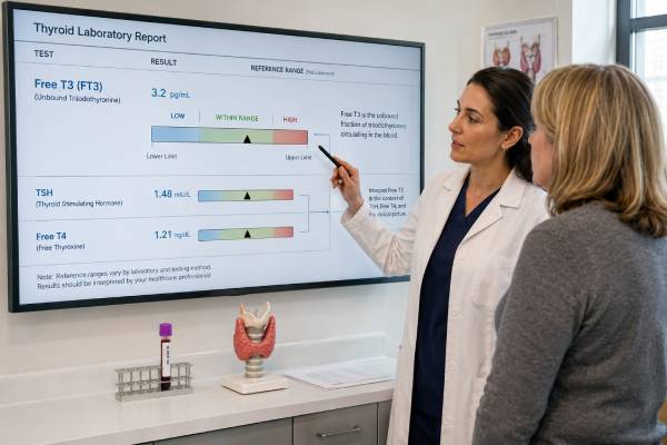
A free T3 test measures the small portion of triiodothyronine circulating without a binding protein. T3 is the thyroid hormone that acts most strongly in many tissues, but free T3 is not usually the first or only test used to assess thyroid function. It is most helpful when TSH is low and free T4 is normal, a pattern that can occur in T3-predominant hyperthyroidism. A high result may also reflect thyroid medicine, pregnancy-related changes, assay interference, or uncommon disorders rather than an overactive thyroid. A low result is common during serious illness, calorie restriction, and recovery from surgery and does not automatically mean hypothyroidism. Free T3 assays are technically difficult because the unbound amount is tiny and results differ among laboratory methods. For that reason, clinicians interpret free T3 with TSH, free T4, symptoms, medicines, and the laboratory’s own reference interval instead of using a universal “optimal” value.
- Free T3 measures unbound triiodothyronine, but TSH and free T4 usually provide the main framework for thyroid diagnosis.
- High free T3 with low TSH can identify T3 thyrotoxicosis even when free T4 remains normal.
- Low free T3 often reflects acute or chronic illness rather than primary hypothyroidism.
- Reference ranges vary by assay, age, pregnancy status, and laboratory; there is no universal ideal free T3 level.
- Unexpected results should be repeated and checked for biotin, thyroid medication timing, antibody interference, and method differences.
Table of Contents
- What Free T3 Measures
- When a Free T3 Test Is Useful
- Preparing for the Blood Test
- Normal Range and Result Patterns
- Causes of High Free T3
- Causes of Low Free T3
- Accuracy, Interference, and Assay Limits
- What to Do With an Abnormal Result
What Free T3 Measures
Triiodothyronine contains three iodine atoms, which is why it is called T3. The thyroid gland releases some T3 directly, but most circulating T3 is made outside the thyroid when enzymes remove one iodine atom from thyroxine, or T4. This conversion occurs in the liver, kidneys, muscles, brain, and other tissues. T3 then binds to nuclear receptors and changes the expression of genes involved in heat production, heart rate, digestion, muscle function, brain activity, and energy use.
Almost all T3 in blood is attached to proteins, mainly thyroxine-binding globulin, albumin, and transthyretin. Only about 0.2% is unbound. A free T3 assay attempts to measure that tiny unbound fraction, while a total T3 test measures both bound and unbound hormone.
The distinction matters when binding proteins change. Estrogen, pregnancy, liver disease, inherited binding-protein differences, and some medicines can alter total T3 without producing true hyperthyroidism or hypothyroidism. In theory, free T3 should be less affected. In practice, common free T3 immunoassays remain partly influenced by binding proteins and other substances in the sample, so they are not perfectly independent measurements.
TSH provides information about the pituitary gland’s response to circulating thyroid hormone. Free T4 shows the available pool of T4. Free T3 adds a narrower piece of information about the active T3 fraction. These tests are related but not interchangeable. The same free T3 result can mean different things when TSH is suppressed, normal, or elevated.
Free T3 is also different from reverse T3. Both are made from T4, but reverse T3 is biologically inactive. During severe illness, the body often decreases conversion to active T3 and increases production of reverse T3. That adaptive pattern can lower free T3 without indicating failure of the thyroid gland itself.
A result must be considered in the context of the hypothalamic-pituitary-thyroid axis. The hypothalamus releases thyrotropin-releasing hormone, the pituitary releases TSH, and TSH stimulates the thyroid to make T4 and T3. T4 and T3 then feed back to reduce TSH. Because this feedback is very sensitive, TSH often changes before free T3 leaves its reference interval.
When a Free T3 Test Is Useful
Free T3 has its clearest role in evaluating possible hyperthyroidism. It may be ordered when symptoms suggest excess thyroid hormone and the TSH result is low, especially if free T4 is normal or only slightly high.
Detecting T3 thyrotoxicosis
T3 thyrotoxicosis describes a pattern of suppressed TSH, elevated T3, and normal free T4. It can occur early in Graves disease, with an autonomously functioning thyroid nodule, or in toxic multinodular goiter. Without a T3 measurement, this form of hyperthyroidism may be missed.
Some laboratories and clinicians prefer total T3 for this purpose because free T3 assays show more method variation. Others use free T3 based on local assay performance. Either test should be interpreted with TSH, free T4, symptoms, and the clinical cause under consideration.
Defining the severity or pattern of hyperthyroidism
T3 can rise disproportionately in Graves disease because the stimulated thyroid produces and releases more T3 and converts T4 efficiently. In destructive thyroiditis, stored hormone leaks from damaged tissue and the T3-to-T4 pattern can differ. T3 results may support the distinction, but they do not replace TSH receptor antibodies, radioactive iodine uptake, or imaging when the cause remains uncertain.
During antithyroid drug treatment, T3 may stay elevated after free T4 improves. Clinicians sometimes monitor T3 along with free T4 in the early phase of treatment. TSH can remain suppressed for weeks or months and may not immediately reflect the current hormone state.
Evaluating unusual or conflicting results
A free T3 test may be used when symptoms and other laboratory values do not fit. Examples include an unexpectedly low TSH with normal free T4, a suspected assay problem, thyroid hormone resistance, a TSH-secreting pituitary tumor, or use of T3-containing medication. These situations require careful specialist interpretation because laboratory interference is often more common than a rare endocrine disorder.
Free T3 is not generally useful for diagnosing ordinary primary hypothyroidism. In early thyroid failure, TSH rises while free T4 and T3 can remain normal. As hypothyroidism progresses, the body may maintain T3 through increased conversion even after free T4 falls. A normal free T3 therefore does not rule out hypothyroidism, and a low result in a sick patient does not establish it.
Routine free T3 testing is also not needed in every wellness panel. More testing can create misleading flags when the pretest chance of thyroid disease is low. A focused thyroid function test panel should be chosen for the clinical question rather than ordered as an unrestricted list.
Preparing for the Blood Test
Free T3 is measured from a venous blood sample. Fasting is usually unnecessary unless another test on the same order requires it. The blood draw takes a few minutes and may cause brief discomfort, bruising, or lightheadedness.
Preparation matters most when a result will be compared with earlier values or used to adjust medication. Ask the ordering clinician or laboratory about these points:
- Biotin: High-dose biotin can interfere with many thyroid immunoassays, often creating a pattern that resembles hyperthyroidism. The laboratory may advise pausing nonessential biotin for a defined period. Do not stop medically prescribed biotin without guidance.
- Thyroid medicine: Liothyronine causes a rapid rise in serum T3 after a dose. Desiccated thyroid also contains T3. The time between the dose and blood collection can substantially change the result.
- Levothyroxine: T4 medication affects free T4 more directly, but timing can still influence the overall pattern and conversion to T3. Follow the clinician’s instructions consistently.
- Antithyroid drugs: Methimazole, carbimazole, and propylthiouracil lower thyroid hormone production. Propylthiouracil and high doses of some other medicines can also reduce T4-to-T3 conversion.
- Recent contrast or iodine exposure: Iodinated contrast, amiodarone, and large iodine loads can alter thyroid function in susceptible people.
- Acute illness: Hospitalization, infection, trauma, fasting, and surgery can lower T3 independently of thyroid disease.
- Pregnancy and estrogen: These change binding proteins and require pregnancy- and method-aware interpretation.
People taking T3-containing medication should not guess whether to skip the morning dose. Some clinicians want a trough sample before the dose; others use a standardized time after dosing. The correct approach depends on the treatment goal. Record the exact medication, dose, and time taken so the result can be interpreted accurately.
Testing at a similar time of day and with the same laboratory can reduce avoidable variation. T3 has biological fluctuation, and assay methods differ. A small change may not represent a clinically meaningful shift when collection conditions are different.
Tell the clinician about heparin, glucocorticoids, dopamine, anticonvulsants, amiodarone, lithium, estrogen, testosterone, and supplements. Some alter thyroid physiology, binding proteins, or assay behavior. No medicine should be stopped solely because it appears on a list of possible influences.
Normal Range and Result Patterns
Free T3 may be reported in picograms per milliliter (pg/mL) or picomoles per liter (pmol/L). A rough adult reference interval might be about 2.0–4.4 pg/mL, equivalent to approximately 3.1–6.8 pmol/L, but ranges differ substantially. The interval printed by the performing laboratory is the correct reference for that method.
Age affects interpretation. T3 concentrations tend to be higher in children and younger adults and may decline with age. Pregnancy changes binding proteins and thyroid physiology. Serious illness can shift values well outside a healthy outpatient reference interval. For these reasons, a number copied from another laboratory or website should not override the report’s population- and assay-specific range.
The relationship among TSH, free T4, and free T3 is more informative than free T3 alone.
| TSH | Free T4 | Free T3 | Pattern that may be considered |
|---|---|---|---|
| Low | High | High or normal | Overt hyperthyroidism, depending on symptoms and cause |
| Low | Normal | High | T3 thyrotoxicosis |
| Low | Normal | Normal | Subclinical hyperthyroidism, transient suppression, medicine effect, or assay issue |
| High | Low | Low or normal | Primary hypothyroidism; free T3 may remain normal until later |
| High | Normal | Usually normal | Subclinical hypothyroidism |
| Normal or low-normal | Low | Low | Nonthyroidal illness, central hypothyroidism, medicine effect, or assay limitation |
| Normal or high | High | High | Assay interference, thyroid hormone resistance, or a TSH-secreting pituitary tumor among possible causes |
This table describes possibilities, not diagnoses. The duration of the abnormality, symptoms, medication use, pregnancy, and pituitary status can change the meaning.
“Optimal” free T3 ranges promoted for fatigue, weight management, or wellness are not standardized clinical targets. Healthy individuals have personal set points, but current assays and outcome evidence do not support adjusting treatment to push every person into the upper portion of the range. For most people receiving levothyroxine for primary hypothyroidism, TSH remains the main biochemical treatment target unless pregnancy, pituitary disease, thyroid cancer, or another special circumstance changes the plan.
A result just outside the range may be due to ordinary analytical and biological variation. The farther the value is from the limit and the more clearly it matches TSH, free T4, symptoms, and repeated testing, the more confidence clinicians can place in it.
Causes of High Free T3
A high free T3 result usually requires a TSH result to establish whether the thyroid-pituitary feedback loop is suppressed.
Hyperthyroid conditions
Graves disease is a common cause. TSH receptor-stimulating antibodies drive the thyroid to make excess hormone, often with a strong T3 component. Symptoms can include palpitations, tremor, heat intolerance, sweating, anxiety, frequent bowel movements, muscle weakness, and unintentional weight loss. Eye irritation or eye prominence can occur in thyroid eye disease.
Toxic adenoma and toxic multinodular goiter produce hormone independently of normal TSH control. T3 may rise before free T4, particularly in milder or early disease. Thyroid ultrasound and radioactive iodine uptake can help identify the source.
Early thyroiditis can release stored T4 and T3 into the circulation. Causes include painless thyroiditis, postpartum thyroiditis, subacute painful thyroiditis, and immune-related drug effects. Because the gland is leaking hormone rather than actively overproducing it, treatment and uptake findings differ from Graves disease.
Medication and hormone exposure
Liothyronine, compounded T3, desiccated thyroid, and some nonprescription “thyroid support” products can raise free T3. Levels may peak two to four hours after oral liothyronine and fall later in the day, so a single sample can reflect dose timing more than the average tissue exposure. Excess dosing can cause atrial fibrillation, bone loss, anxiety, and other thyrotoxic effects.
Taking thyroid hormone without medical supervision can produce low TSH and high T3. In factitious thyrotoxicosis, thyroglobulin may be low because the thyroid gland itself is not releasing hormone. Clinicians may use several tests to distinguish this from endogenous hyperthyroidism.
Less common explanations
A TSH-secreting pituitary adenoma can cause high T3 and T4 with a TSH that is not suppressed. Thyroid hormone resistance can create a similar biochemical pattern but different clinical features. Both are rare and should be considered only after medication effects and assay interference are addressed.
Struma ovarii, an ovarian teratoma containing functioning thyroid tissue, can rarely produce thyroid hormone. Trophoblastic disease with very high human chorionic gonadotropin can stimulate the TSH receptor. These causes require specialist evaluation.
A high free T3 with normal TSH is often not true hyperthyroidism. Interference, method bias, or a binding-protein issue may be more likely, especially when the person has no symptoms and free T4 is normal. Repeating the sample with a different method can prevent unnecessary imaging or treatment.
Causes of Low Free T3
Low free T3 is less specific than high free T3. It commonly appears when the body changes thyroid hormone metabolism during illness.
Nonthyroidal illness syndrome
Serious infection, heart failure, kidney disease, liver disease, cancer, trauma, burns, surgery, and prolonged hospitalization can reduce conversion of T4 to T3. The earliest pattern is often low T3 with normal or low-normal free T4 and TSH. With severe or prolonged illness, free T4 and TSH may also fall. During recovery, TSH can temporarily rise.
This pattern is sometimes called euthyroid sick syndrome or low T3 syndrome. It does not necessarily mean the thyroid gland is failing. Routine thyroid hormone treatment has not shown clear benefit for most critically ill patients without established thyroid disease. Clinicians usually focus on the underlying illness and repeat thyroid tests after recovery when the diagnosis is uncertain.
Reduced calorie intake and physiological stress
Fasting, severe calorie restriction, eating disorders, and rapid weight loss can lower T3 as the body conserves energy. Overtraining and significant physiological stress may contribute. A low free T3 in this setting is not proof that thyroid medication is needed. Nutritional status, menstrual function, bone health, and the broader medical picture deserve attention.
Medicines
Glucocorticoids, amiodarone, propranolol at higher doses, and propylthiouracil can reduce T4-to-T3 conversion. Dopamine and some critical-care medicines suppress TSH. Antiseizure medicines and heparin can alter thyroid tests through other mechanisms. The expected pattern depends on dose, timing, illness, and assay.
Hypothyroidism and central disease
Free T3 can be low in overt primary hypothyroidism, but it often falls later than free T4. High TSH and low free T4 establish the common primary pattern more reliably. In central hypothyroidism caused by pituitary or hypothalamic disease, free T4 is low while TSH may be low, normal, or only mildly high. Free T3 alone cannot diagnose central hypothyroidism.
Older adults may have lower T3 values without symptomatic thyroid failure. Chronic kidney or liver disease can also lower T3 through altered conversion and protein binding. Interpretation should avoid labeling every age- or illness-related decline as a treatment target.
Accuracy, Interference, and Assay Limits
Free T3 is challenging to measure because the free fraction is extremely small and must remain in equilibrium with a much larger protein-bound pool. Most routine laboratories use automated competitive immunoassays. These are fast and practical, but their results can differ significantly among manufacturers.
Direct methods first separate free hormone by equilibrium dialysis or ultrafiltration and then measure it, often with liquid chromatography-tandem mass spectrometry. They are technically demanding and usually available only through reference laboratories. Even direct methods require careful control of temperature, dilution, membrane behavior, and sample handling.
Common reasons for a misleading free T3 result include:
- High-dose biotin in assays that use biotin-streptavidin chemistry
- Heterophile antibodies that react with assay components
- Anti-T3 autoantibodies
- Abnormal albumin or other binding proteins
- Heparin-related release of free fatty acids after the blood sample is drawn
- Severe nonthyroidal illness
- Pregnancy-related binding changes
- Differences in assay calibration and reference intervals
- Sample mix-up, handling problems, or transcription error
Interference should be suspected when the numbers conflict with physiology—for example, very high free T3 with normal TSH and no symptoms, or sharply different results from two laboratories. A clinician can ask the laboratory to repeat the test after dilution, use blocking reagents, check total T3, analyze the sample on another platform, or arrange a direct method.
A result can be analytically correct yet clinically misleading. During acute illness, the measured concentration may accurately reflect altered hormone metabolism, but treating the number as primary thyroid disease would still be inappropriate. Good interpretation separates analytical error from a real but adaptive biological change.
Because method differences are largest at the high and low ends, serial monitoring should ideally use the same assay. Numeric changes across platforms should not be treated as a continuous trend unless the methods have been compared.
What to Do With an Abnormal Result
The next step depends on the full pattern and the reason the test was ordered.
For a high free T3 with low TSH, clinicians commonly confirm free T4, review thyroid hormone exposure, and assess the cause of thyrotoxicosis. TSH receptor antibody testing, radioactive iodine uptake, or ultrasound with blood-flow assessment may follow. Treatment urgency depends on symptoms, heart rate, age, pregnancy, and complications.
For a low free T3 during acute illness, immediate thyroid treatment is usually not based on free T3 alone. Prior thyroid history, TSH, free T4, medications, and illness severity guide interpretation. Repeat testing after recovery may provide a clearer baseline.
For an isolated unexpected result, useful steps include:
- Verify the units and laboratory reference interval.
- Review biotin, supplements, thyroid medicine, and dose timing.
- Compare TSH and free T4 rather than interpreting free T3 alone.
- Repeat the test under standardized conditions when clinically appropriate.
- Ask the laboratory about interference or an alternative assay if results remain discordant.
- Seek endocrinology input for persistent, unexplained, or complex patterns.
Urgent assessment is appropriate for chest pain, fainting, severe shortness of breath, a resting heart rate that remains very fast, new confusion, high fever with marked hyperthyroid symptoms, or severe weakness. These can signal a cardiac complication or, rarely, thyroid storm. A laboratory result by itself does not establish an emergency, but serious symptoms should not wait for a routine appointment.
Questions worth asking include whether total T3 would be more reliable on the local platform, whether the sample timing reflects a T3 medicine peak, and whether a repeat should use the same or a different method. The purpose is not to chase a preferred number. It is to determine whether the result reflects true thyroid hormone excess or deficiency, an adaptive response to illness, medication timing, or a measurement problem.
References
- Limited Utility of Free Triiodothyronine Testing 2023
- Analysis of free, unbound thyroid hormones by liquid chromatography-tandem mass spectrometry: A mini-review of the medical rationale and analytical methods 2023 (Review)
- Large method differences for free thyroid hormone assays are not explained by differences in T4 and T3 binding proteins 2023
- Advances in Thyroid Function Tests: Precision Diagnostics and Clinical Applications 2023 (Review)
- Free thyroid hormone: Methods and standardization 2025 (Review)
- Assay of Thyroid Hormone and Related Substances 2025 (Review)
Disclaimer
Free T3 results vary by assay and must be interpreted with TSH, free T4, symptoms, medicines, and the clinical setting. Do not change thyroid medication or start T3 treatment based on one result without medical guidance. Seek prompt care for severe cardiac, neurological, or hyperthyroid symptoms.