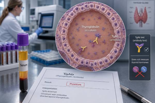
A thyroglobulin antibody test detects immune proteins directed against thyroglobulin, a protein made by thyroid follicular cells. TgAb is most often used for two reasons: supporting a diagnosis of autoimmune thyroid disease and checking whether antibodies may interfere with thyroglobulin testing after differentiated thyroid cancer. A positive result is common in Hashimoto thyroiditis and can also occur in Graves disease or in people whose thyroid function is currently normal. It does not show whether the thyroid is underactive or overactive; TSH and free T4 provide that information. In thyroid cancer follow-up, TgAb is especially important because it can make a thyroglobulin immunoassay appear falsely low. The antibody trend may then act as an indirect marker: falling values are generally reassuring, while persistent or rising values can prompt closer evaluation. Reference ranges and units vary greatly between assays, and results from different laboratories are not directly comparable. The clinical meaning depends on the reason for testing, hormone results, treatment history, and trend.
- Positive TgAb supports thyroid autoimmunity but does not by itself diagnose Hashimoto thyroiditis or require treatment.
- TSH and free T4 determine thyroid function; the TgAb concentration does not measure hormone deficiency.
- TPOAb is generally more sensitive for Hashimoto thyroiditis, while TgAb can add evidence when TPOAb is negative.
- Every thyroglobulin tumor-marker test should be paired with TgAb because antibodies can make Tg unreliable.
- After total thyroidectomy, falling TgAb is usually reassuring; a sustained rise may lead to ultrasound or other imaging.
- Compare serial TgAb values with the same assay because method changes can create a false trend.
Table of Contents
- What the TgAb Test Measures
- Why Thyroglobulin Antibodies Are Tested
- Positive, Negative, High, and Borderline Results
- TgAb in Hashimoto and Graves Disease
- TgAb During Thyroid Cancer Follow-Up
- Preparation, Assay Differences, and Interference
- Follow-Up and When More Testing Is Needed
What the TgAb Test Measures
Thyroglobulin is a large protein stored inside thyroid follicles. Iodine is attached to parts of this protein during the production of T4 and T3. When immune tolerance breaks down, B lymphocytes can make antibodies against thyroglobulin. The laboratory detects those circulating antibodies and reports them as TgAb, anti-Tg, or antithyroglobulin antibodies.
TgAb is a marker of immune recognition. It is not the same as thyroglobulin itself. The two tests answer different questions:
- Thyroglobulin (Tg): measures a thyroid-cell protein and is mainly used as a tumor marker after treatment for differentiated thyroid cancer.
- Thyroglobulin antibody (TgAb): detects antibodies that suggest autoimmunity and may interfere with Tg measurement.
The distinction matters because a report may list both side by side. A low Tg with positive TgAb cannot be interpreted as confidently as a low Tg with negative TgAb.
How results are reported
Laboratories commonly report TgAb in IU/mL, kIU/L, or another manufacturer-specific unit. The upper reference limit may be less than 1 IU/mL on one platform and less than 20 or 40 IU/mL on another. This does not mean one person’s antibody level changed twentyfold. The assays recognize different antibody populations and use different calibration.
Results may appear as:
- Negative or below the detection limit
- Borderline or equivocal
- Positive
- A numeric concentration above the laboratory cutoff
A positive result should be interpreted against that laboratory’s limit, not an internet “optimal” range. The degree of elevation does not map cleanly to the amount of thyroid damage or symptom severity.
Why antibodies form
Genetic susceptibility, sex hormones, iodine exposure, pregnancy-related immune shifts, infections, smoking patterns, and other environmental factors can influence thyroid autoimmunity. TgAb often occurs with TPOAb because both arise from the same autoimmune process. It can also occur alone.
The antibody itself may participate in inflammation, but the measured serum concentration is mainly used as a marker. Treatment decisions are not based on lowering TgAb to a specific number.
Why Thyroglobulin Antibodies Are Tested
The indication determines whether TgAb is a secondary autoimmune marker or a central part of cancer surveillance.
Suspected Hashimoto thyroiditis
When TSH is high, free T4 is low or normal, and autoimmune thyroiditis is suspected, clinicians usually order TPOAb first. TgAb may be included in a thyroid antibody panel or added when TPOAb is negative despite a strong clinical picture.
Evidence for Hashimoto thyroiditis becomes stronger when TgAb accompanies:
- Elevated TSH
- Positive TPOAb
- A firm or enlarged thyroid
- A heterogeneous, hypoechoic ultrasound pattern
- Another autoimmune condition
- A family history of autoimmune thyroid disease
A positive TgAb with normal TSH and free T4 means autoimmune susceptibility, not established hypothyroidism.
Graves disease
TgAb can be positive in Graves disease, but it does not cause the characteristic receptor stimulation and is not the preferred diagnostic antibody. TRAb or TSI provides much stronger evidence for Graves hyperthyroidism. TgAb may simply show that broader thyroid autoimmunity is present.
Differentiated thyroid cancer monitoring
Every serum thyroglobulin tumor-marker test should include TgAb. Standard immunometric Tg assays can read falsely low in the presence of antibodies. Detecting TgAb warns the clinician that an apparently undetectable Tg may not be reliable.
After total thyroidectomy and radioactive iodine, TgAb itself may be followed as an indirect marker. Antibody production often falls as normal and malignant thyroid tissue is removed. A rising trend can indicate renewed antigen exposure from persistent or recurrent tissue, although immune fluctuation and assay changes can also affect it.
Pregnancy and postpartum evaluation
Thyroid autoantibodies can identify people at higher risk of thyroid dysfunction during and after pregnancy. TPOAb is the more commonly used marker in pregnancy guidance, but TgAb may also be present. Thyroid hormone treatment decisions depend on TSH, free T4, gestational age, and clinical history—not TgAb alone.
The postpartum immune rebound can trigger painless thyroiditis. Antibodies may be positive before pregnancy, during pregnancy, or after delivery. Serial hormone tests determine whether the person is in a thyrotoxic, hypothyroid, or recovered phase.
Before selected treatments
Baseline thyroid antibodies may be measured before immune checkpoint inhibitors, interferon, amiodarone, or other treatments associated with thyroid dysfunction. Positive antibodies can identify increased risk in some populations, but they do not predict exactly who will develop disease. Scheduled TSH monitoring remains necessary.
Positive, Negative, High, and Borderline Results
TgAb is not interpreted like a hormone with a narrow physiologic target. The result establishes whether potentially relevant antibodies are detectable and whether the concentration is changing.
Positive TgAb with abnormal TSH
A positive result with high TSH and low free T4 strongly supports autoimmune primary hypothyroidism, especially if TPOAb is also positive. With high TSH and normal free T4, it supports early or subclinical autoimmune hypothyroidism and a higher chance of progression than the same TSH pattern without antibodies.
The hypothyroidism blood test panel determines whether treatment is needed. Antibody positivity can influence the threshold for closer monitoring, but the TgAb value itself is not a levothyroxine dose target.
Positive TgAb with low TSH
Low TSH with high free T4 or T3 indicates thyrotoxicosis. Positive TgAb does not identify the cause. Graves disease, thyroiditis, medication, or nodular disease still needs evaluation. TRAb or TSI is more specific for Graves disease, while low radioactive iodine uptake may support thyroiditis.
Positive TgAb with normal thyroid function
Some people have positive TgAb and completely normal TSH and free T4. They are euthyroid. No thyroid hormone is needed simply because antibodies exist. Periodic TSH checks may be reasonable, particularly with symptoms, goiter, pregnancy plans, another autoimmune disease, or a strong family history.
Testing every few weeks is unnecessary because autoimmune thyroid failure usually develops over months or years. A clinician may choose annual testing or a risk-based interval.
Very high TgAb
A value many times above the upper limit confirms antibody positivity but does not precisely predict how severe hypothyroidism will be. One person with a very high level can have normal TSH, while another with a modest positive result can have overt disease. Symptoms also do not rise in direct proportion to the antibody concentration.
High concentrations can increase the likelihood of interference with Tg tumor-marker assays. Even low-level antibodies below the manufacturer’s formal positive cutoff may interfere in some samples, which is why trends and assay comments matter in cancer care.
Borderline TgAb
A borderline result can reflect early autoimmunity, assay variation, low-level cross-reactivity, or a value near an arbitrary statistical cutoff. Useful steps include checking TSH, free T4, and TPOAb; reviewing prior values; and repeating only when the result will change management.
In thyroid cancer follow-up, a newly borderline result deserves attention because any detectable antibody may complicate Tg interpretation. The same laboratory should be used for confirmation.
Negative TgAb
A negative result reduces the likelihood of autoimmune thyroiditis but does not eliminate it. Some people with Hashimoto thyroiditis are antibody-negative, especially after substantial gland destruction or when antibodies remain below the assay cutoff. Ultrasound, persistent TSH elevation, and exclusion of other causes may still support the diagnosis.
In cancer follow-up, negative TgAb makes immunometric Tg more dependable, but heterophile antibodies and other assay problems can still occur. Structural disease can also be present with low Tg production.
TgAb in Hashimoto and Graves Disease
TgAb appears in both major autoimmune thyroid diseases but has different diagnostic weight.
Hashimoto thyroiditis
Hashimoto thyroiditis involves immune-mediated destruction of follicular cells. TPOAb is positive in most cases and is generally the most sensitive antibody. TgAb is also common and can be the only detectable antibody in a smaller group.
The dedicated Hashimoto panel combines:
- TSH to measure pituitary demand
- Free T4 to measure circulating hormone supply
- TPOAb as the principal autoimmune marker
- TgAb as additional evidence
Early disease can pass through stages. Antibodies may be positive while function is normal. TSH may then rise with normal free T4, followed later by low free T4. Some people remain stable indefinitely.
Graves disease
Graves disease is driven by antibodies against the TSH receptor. TgAb and TPOAb are frequently present because the immune response is not limited to one antigen. Their presence does not replace TRAb testing or TSI.
TgAb has been studied as a predictor of Graves outcomes, eye disease, or treatment response, but results are inconsistent and it is not a standard treatment target. TSH, free T4, T3, TRAb/TSI, symptoms, and treatment course provide the actionable information.
Switching between autoimmune patterns
A person can move from Graves hyperthyroidism to hypothyroidism after treatment or through natural autoimmune changes. Others experience a transient thyrotoxic phase of Hashimoto thyroiditis before becoming hypothyroid. TgAb cannot determine which direction function will move at a specific time. Repeated TSH and free T4 show the actual state.
Other autoimmune associations
Thyroid antibodies occur more often with type 1 diabetes, celiac disease, pernicious anemia, vitiligo, rheumatoid arthritis, lupus, and Addison disease. A positive TgAb does not diagnose those conditions, but the wider history may justify targeted testing when symptoms suggest them.
Diet and supplements
No supplement has proven ability to eliminate TgAb and prevent thyroid failure. Selenium can modestly reduce TPOAb in some studies, but clinical outcome benefits are uncertain, and excessive selenium is toxic. High-dose iodine can aggravate autoimmune thyroid dysfunction in susceptible people. Treatment should focus on adequate—not excessive—iodine, confirmed hormone deficiency, and specific nutritional deficiencies.
TgAb During Thyroid Cancer Follow-Up
In differentiated thyroid cancer, TgAb is both an interference marker and, when positive, a possible surrogate trend.
Why Tg becomes unreliable
Immunometric Tg assays use antibodies to capture and detect thyroglobulin. Patient TgAb can cover the protein’s binding sites or alter its clearance, leading most often to a falsely low Tg. A result reported as less than 0.1 ng/mL may therefore fail to reflect existing thyroid tissue.
Radioimmunoassay and mass-spectrometry methods can reduce some forms of interference, but neither guarantees detection of every recurrence. Imaging remains essential when risk, symptoms, or antibody trends are concerning.
Expected TgAb decline
After total thyroidectomy, TgAb may fall gradually over months to years as antigen disappears. The decline is not immediate because antibody-producing immune cells persist. A downward trend measured with the same assay is generally favorable.
The speed depends on the starting concentration, remaining normal thyroid tissue, radioactive iodine, autoimmune activity, and assay. A positive value one year after treatment does not automatically mean cancer remains if it is falling and imaging is negative.
Stable TgAb
Stable antibodies can represent persistent immune memory or remaining benign thyroid tissue. The original recurrence risk, Tg result, TSH, ultrasound, and duration of stability guide follow-up. Low-risk patients with long-term stable or declining TgAb and negative imaging may need less intensive surveillance than high-risk patients.
Rising or newly positive TgAb
A sustained rise on sequential same-assay tests raises concern. It can precede measurable Tg or visible disease and often prompts neck ultrasound. Cross-sectional imaging or other studies may be considered according to cancer type, original stage, magnitude of change, and symptoms.
One isolated rise should be verified. Assay change, laboratory variation, infection, autoimmune fluctuation, and TSH changes can affect results. A clinician looks for a reproducible trajectory rather than reacting to a single percentage difference.
After lobectomy
When a thyroid lobe remains, normal tissue continues to expose the immune system to thyroglobulin. TgAb may persist and is less specific as a recurrence marker. Ultrasound and pathology-based risk are more important. Total-thyroidectomy response thresholds should not be applied after lobectomy.
Pairing results correctly
Every cancer follow-up interpretation should state:
- Tg value and assay
- TgAb value and assay
- TSH at the same blood draw
- Surgery type
- Radioactive iodine history
- Prior Tg/TgAb trend
- Recent neck ultrasound or other imaging
Without those details, “positive antibodies” is incomplete information.
Preparation, Assay Differences, and Interference
Fasting is usually not required. The blood draw is routine, but consistent conditions improve serial interpretation.
Before testing
Tell the clinician about:
- Thyroid surgery and radioactive iodine dates
- Levothyroxine dose and TSH suppression target
- Pregnancy or recent delivery
- Biotin-containing supplements
- Immune therapies, amiodarone, lithium, or interferon
- Recent neck biopsy, ablation, or surgery
- Prior laboratory platform changes
Biotin can interfere with some immunoassays. The direction depends on the method, so laboratories often advise stopping non-prescription biotin for at least 48 hours. High-dose prescribed biotin requires individualized instructions.
Fasting and medication timing
Food does not directly determine TgAb. Levothyroxine can usually be taken as prescribed, although the timing may affect same-day free T4 and, over time, TSH. For serial cancer surveillance, use a consistent schedule relative to the morning dose.
Do not withdraw thyroid hormone unless the care team is deliberately performing stimulated testing. TgAb itself is not expected to change acutely with TSH stimulation, but Tg and TSH interpretation will.
Assay variability
TgAb assays have poor agreement across manufacturers because antibodies are heterogeneous. Two platforms may classify the same sample differently. Even when both use IU/mL, the numbers are not interchangeable.
For trends:
- Stay with the same laboratory when possible.
- Confirm whether the method or reference limit changed.
- Treat the first value after a platform switch as a new baseline.
- Avoid calculating percentage growth across different assays.
Other antibodies
Heterophile antibodies and human anti-animal antibodies can interfere with immunoassays. Macro-forms and nonspecific binding may also create unexpected values. The laboratory can investigate with dilution, blocking reagents, alternate platforms, or specialized testing when results conflict with the clinical picture.
Ultrasound does not measure antibodies
A thyroid ultrasound can show a heterogeneous autoimmune pattern, gland size, and nodules. It cannot prove whether TgAb is present or quantify immune activity. Conversely, positive TgAb does not mean an ultrasound is needed when the thyroid is not enlarged and there is no nodule or cancer history.
Follow-Up and When More Testing Is Needed
Follow-up should answer a specific question: Is thyroid function changing, or is thyroid cancer surveillance becoming more concerning?
Autoimmune thyroid disease follow-up
When TgAb is positive but TSH and free T4 are normal, periodic TSH is more useful than repeating antibodies. Retest sooner if symptoms develop, pregnancy occurs, a goiter enlarges, or a medicine known to affect the thyroid starts.
When hypothyroidism is treated, levothyroxine is adjusted by TSH and sometimes free T4. Falling TgAb is not required for successful treatment. Repeating the antibody every dose change adds little.
Cancer follow-up
Repeat Tg and TgAb together at the interval set by recurrence risk and response. Rising TgAb, new neck symptoms, abnormal examination, or suspicious ultrasound may lead to:
- Repeat high-resolution neck ultrasound
- Fine-needle aspiration of a lymph node, sometimes with Tg washout
- Contrast CT or MRI
- Radioactive iodine imaging
- FDG PET/CT in selected iodine-negative disease
The presence of TgAb does not specify where disease is located. Imaging provides that information.
When results deserve prompt review
Contact the clinician promptly when:
- TgAb becomes newly positive after being negative
- The concentration rises consistently on the same assay
- Tg is unexpectedly undetectable despite known disease
- TSH or free T4 becomes clearly abnormal
- A neck mass, persistent hoarseness, swallowing difficulty, or unexplained cough develops
- Pregnancy occurs in someone with known autoimmune thyroid disease
A positive TgAb result is usually not an emergency. It is a clue that gains meaning from thyroid function tests or cancer history. The most useful approach is to keep the test in its proper role: cause marker in autoimmunity, interference marker in Tg testing, and trend marker in selected post-treatment cancer surveillance.
References
– The Usefulness of Thyroid Antibodies in the Diagnostic Approach to Autoimmune Thyroid Disease 2023 (Review) – Revisiting Thyroglobulin Measurement: Current Methods and Emerging Alternative Approaches 2025 (Review) – Thyroglobulin (Tg) Assays 2026 – Thyroid Function Tests 2023 – Thyroid antibodies explained 2024 – Significance of thyroperoxidase and thyroglobulin antibodies in clinically and serologically confirmed Graves’ disease 2023
Disclaimer
This article is educational and cannot diagnose autoimmune thyroid disease or determine whether thyroid cancer has returned. TgAb assays vary substantially, and cancer interpretation requires Tg, TSH, treatment history, and imaging. Review positive or changing results with a qualified clinician.