
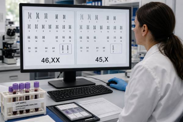
Turner syndrome genetic testing determines whether cells contain complete or partial loss of one sex chromosome in a person with a female phenotype. The best-known result is 45,X, often called monosomy X, but Turner syndrome also includes mosaic cell lines and structural X-chromosome changes. Testing may begin with a prenatal cell-free DNA screen, an ultrasound finding such as cystic hygroma, short stature in childhood, delayed puberty, or unexplained ovarian insufficiency. These starting points are not equivalent. Cell-free DNA is a placental screening test; chorionic villus sampling and amniocentesis are prenatal diagnostic procedures; and a postnatal blood karyotype directly examines the patient’s cells. A confirmed chromosome result establishes the genetic pattern but does not predict an exact phenotype. Heart, kidney, hearing, growth, endocrine, fertility, and learning outcomes vary widely, particularly in mosaic Turner syndrome. Results should therefore lead to condition-specific evaluation and long-term care rather than a one-time label.
- A high-chance monosomy X blood screen is not a fetal diagnosis. The signal can come from the fetus, placenta, or pregnant patient.
- Turner syndrome is broader than 45,X. Mosaicism, ring X, isochromosome Xq, and X-chromosome deletions can also be relevant.
- Ultrasound severity and karyotype are related imperfectly. Cystic hygroma or hydrops raises concern, but a normal ultrasound does not exclude Turner syndrome.
- Amniocentesis may clarify a positive screen better than placental testing in some cases. Confined placental mosaicism is an important source of discordance.
- Diagnosis should trigger lifelong surveillance. Cardiovascular risk, hearing, thyroid function, growth, puberty, and reproductive health require planned follow-up.
Table of Contents
- Which chromosome patterns are called Turner syndrome
- How Turner syndrome is suspected before birth
- Choosing and confirming a diagnostic test
- Understanding 45,X, mosaic, and structural results
- What prenatal findings can and cannot predict
- Testing after birth and lifelong medical meaning
- Result limitations, family questions, and next steps
Which chromosome patterns are called Turner syndrome
Turner syndrome is associated with complete or partial absence of the second sex chromosome in an individual with a female phenotype and compatible clinical features. A typical chromosome complement is written 46,XX. In classic monosomy X, the karyotype is 45,X: each tested cell has one X chromosome and no second sex chromosome.
The diagnosis is not limited to that one notation. Other patterns include:
- 45,X/46,XX mosaicism, with both monosomy X and typical XX cell lines;
- 45,X/46,XY mosaicism or another cell line containing Y-chromosome material;
- 46,X,i(Xq), in which one X is an isochromosome composed mainly of two long arms;
- 46,X,r(X), with a ring X chromosome;
- 46,X,del(Xp) or another X deletion, depending on the missing region;
- more complex mosaics or structural rearrangements involving X.
Not every small X-chromosome change is Turner syndrome. The location and size of a deletion, the genes involved, X-inactivation, age, phenotype, and proportion of abnormal cells all influence classification. A genetics professional should review structural findings rather than infer the diagnosis from the phrase “X chromosome deletion.”
The missing dosage of genes that escape X-inactivation contributes to Turner features. The SHOX gene in the pseudoautosomal region is especially relevant to short stature and skeletal findings. Other health effects arise from broader X-chromosome dosage, developmental differences, and associated congenital anomalies. The biology is more complex than simply having “half an X,” because one X chromosome is normally largely inactivated in 46,XX cells and different genes behave differently.
Turner syndrome occurs sporadically in most families. It is usually caused by a chromosome-separation or early cell-division event rather than an inherited variant or anything a parent did. Recurrence risk is generally low, although unusual structural rearrangements or parental chromosome findings can change counseling.
Many 45,X conceptions end in miscarriage, often before Turner syndrome is recognized. The liveborn population therefore does not represent all affected conceptions. This selection helps explain why severe prenatal findings and relatively mild postnatal diagnoses can both be associated with a Turner chromosome pattern.
How Turner syndrome is suspected before birth
There are two common prenatal routes to suspicion: a chromosome screening signal and an ultrasound phenotype. They provide different evidence.
Cell-free DNA screening may report high risk, positive, or increased chance for monosomy X. The blood sample comes from the pregnant patient, and most pregnancy-associated fragments arise from the placenta. The screen estimates whether X-chromosome representation is lower than expected; it does not directly count chromosomes in fetal cells.
Monosomy X has a lower positive predictive value than trisomy 21 screening. A positive result can represent fetal Turner syndrome, confined placental mosaicism, maternal X-chromosome mosaicism, a maternal structural X finding, a vanished co-twin, or technical variation. The laboratory’s overall accuracy statement should not be mistaken for the chance that this individual fetus has 45,X.
Ultrasound findings that may prompt diagnostic testing include markedly increased nuchal translucency, septated cystic hygroma, generalized edema or hydrops, pleural effusions, certain cardiac abnormalities, renal differences, growth restriction, or a combination. Coarctation of the aorta and left-sided heart abnormalities are particularly relevant, although not always visible early.
A cystic hygroma is not specific to Turner syndrome. Other chromosome conditions, copy-number changes, single-gene disorders, infections, and non-genetic causes can produce overlapping findings. Diagnostic testing may therefore include chromosomal microarray or additional sequencing depending on the ultrasound pattern and initial chromosome result.
A normal ultrasound does not rule out Turner syndrome. Many fetuses with mosaic or structural X findings have no distinctive prenatal anomaly, and XX/45,X mosaicism may be detected only because of screening. Conversely, a severe ultrasound pattern does not prove 45,X without chromosome testing.
When both cfDNA and ultrasound point toward Turner syndrome, the probability of a fetal diagnosis is higher than when the blood screen is positive and anatomy is unremarkable. Even then, confirmation is needed because the specific karyotype matters for counseling and medical planning.
Patients should be told before cfDNA screening that sex chromosome analysis may reveal information about them. A previously unrecognized maternal 45,X/46,XX mosaic result can have implications for cardiovascular and reproductive health. This possibility is one reason current guidance treats sex chromosome screening as a choice requiring explicit counseling rather than a trivial extension of fetal sex reporting. The broader sex chromosome aneuploidy test guide explains these discordant pathways.
Choosing and confirming a diagnostic test
A positive prenatal screen should lead to genetic counseling, targeted ultrasound, and an offer of diagnostic testing before decisions that depend on fetal chromosome status.
Chorionic villus sampling (CVS) obtains placental tissue, usually in the first trimester. It can give an earlier diagnosis and is appropriate when ultrasound findings are severe or early information is a priority. However, placenta and fetus may have different sex chromosome cell lines. A CVS result showing mosaic 45,X may require amniocentesis because confined placental mosaicism cannot always be excluded. Laboratories may compare direct villus analysis with cultured cells to understand which placental layers contain the finding.
Amniocentesis samples fetal-derived cells in amniotic fluid, generally from 15 weeks onward. When cfDNA is positive for monosomy X but ultrasound is normal, amniocentesis is often especially useful because it reduces the ambiguity created by testing a second placental sample. The timing, patient’s values, ultrasound findings, and local expertise determine which procedure is preferred.
The diagnostic laboratory strategy may include:
- Karyotype, the standard test for chromosome count, large X-structural changes, Y-chromosome material, and mosaic cell lines;
- Chromosomal microarray, which can define smaller X-chromosome gains or losses and detect other clinically significant copy-number changes;
- FISH, which may count X and Y signals in additional uncultured cells when low-level mosaicism is suspected;
- Targeted testing for Y material, when the karyotype, phenotype, or virilization raises concern;
- Additional molecular testing, when ultrasound findings are not adequately explained by the chromosome result.
A rapid test that reports one X signal may provide an initial answer but should not replace a complete chromosome analysis. Structural X variants and mosaic cell lines may be missed by a narrow assay. The full report should specify specimen type, number of cells examined, culture method, resolution, and any limitations.
After birth, peripheral blood karyotyping is recommended to confirm a prenatal diagnosis and establish a baseline chromosome result. If clinical suspicion remains high despite a normal blood karyotype, analysis of more cells or another tissue may be considered. Buccal cells, skin fibroblasts, or targeted FISH can sometimes reveal mosaicism not detected in routine blood testing.
A positive cfDNA result with normal fetal diagnostic testing may warrant maternal evaluation. Maternal karyotype or microarray is selected according to the screen pattern and counseling goals. Low-level loss of X in blood becomes more common with age and does not always equal Turner syndrome, so the maternal phenotype and percentage matter.
For an overview of what a prenatal chromosome study can and cannot show, see the prenatal karyotype test article.
Understanding 45,X, mosaic, and structural results
The karyotype notation is a map of the observed cells, not a severity score.
45,X in all analyzed cells supports nonmosaic monosomy X in that specimen. Prenatally, the report should still be interpreted alongside specimen source. A nonmosaic amniotic-fluid result is strong evidence of fetal 45,X. A nonmosaic villus result may be highly informative, especially with compatible ultrasound findings, but placental-fetal discordance remains biologically possible.
45,X/46,XX mosaicism means both cell lines were observed. A notation such as mos 45,X[10]/46,XX[20] states that 10 counted cells had 45,X and 20 had 46,XX under the laboratory conditions. It does not mean 33% of the person’s body has monosomy X. Different tissues can have different proportions, and cell culture can favor one line.
Mosaic 45,X/46,XX is often associated with milder average features than nonmosaic 45,X, but the ranges overlap. Some individuals have spontaneous puberty and fertility; others have significant cardiovascular, endocrine, or ovarian involvement. A low percentage in blood does not eliminate health risks.
45,X with Y-chromosome material requires precise characterization. Y material may be visible as a 46,XY line, a marker chromosome, or a molecular signal. Gonadal development can vary, and some Y-chromosome sequences increase gonadoblastoma risk in dysgenetic gonads. Evaluation should involve a multidisciplinary team experienced in Turner syndrome and differences of sex development. Gonadal management is individualized and should not be based on an incomplete rapid result.
Isochromosome Xq usually means loss of much of Xp with duplication of Xq material. It is associated with short stature and ovarian insufficiency and may have a different autoimmune profile from some other karyotypes. Ring X varies greatly because ring size, deleted material, X-inactivation, and mosaicism differ; a small ring that does not undergo normal X-inactivation can be associated with neurodevelopmental concerns not typical of most Turner syndrome.
X-chromosome deletions require breakpoint-level interpretation. Loss of Xp regions containing SHOX can contribute to short stature. Xq deletions may affect ovarian function depending on the region. Distal changes that do not produce a Turner phenotype should not be overclassified.
Normal 46,XX result lowers the likelihood of Turner syndrome in the tested tissue but may not end the evaluation if the phenotype is compelling. Low-level or tissue-limited mosaicism can fall below detection. The laboratory can advise whether more metaphases, interphase FISH, or another tissue is reasonable.
Variant or uncertain result means the laboratory cannot confidently connect a chromosome change with disease. It should not be treated as a confirmed explanation for every feature. Parental testing, higher-resolution analysis, or periodic reinterpretation may help.
What prenatal findings can and cannot predict
Prenatal prognosis depends on more than the chromosome notation. The strongest immediate predictors of pregnancy course are often the ultrasound findings, particularly persistent hydrops, large cystic hygroma, cardiac abnormalities, and severe growth concerns.
A fetus with 45,X and extensive hydrops has a high risk of miscarriage or stillbirth. Some hygromas and edema regress, and survival is possible, but the course cannot be guaranteed. Serial ultrasound helps assess fluid collections, growth, cardiac anatomy, kidneys, and placental function.
When Turner syndrome is confirmed, fetal echocardiography is generally recommended because aortic coarctation, bicuspid aortic valve, and other left-sided lesions can be subtle. Prenatal imaging may miss lesions later found after birth, so a normal fetal echocardiogram does not replace postnatal cardiac assessment.
Kidney anatomy should be reviewed, including the possibility of horseshoe kidney or collecting-system differences. Delivery planning depends on hydrops, growth, heart findings, obstetric factors, and neonatal resources rather than the karyotype alone.
A fetus with mosaic 45,X/46,XX and normal anatomy may have a favorable pregnancy course, but prenatal testing cannot predict final height, timing of ovarian failure, hearing, learning profile, or cardiovascular risk. The proportion of 45,X cells in amniotic fluid is not a validated conversion table for those outcomes.
Counseling should distinguish survival prognosis from life-course prognosis. Severe fetal hydrops concerns whether the pregnancy can continue safely. A liveborn child with Turner syndrome faces a separate spectrum of treatable or monitorable health issues and often has typical overall intelligence and meaningful quality of life.
Families need balanced descriptions of uncertainty. Historical prenatal series may overrepresent severe ultrasound cases, while postnatal clinic series may overrepresent people with health problems that led to diagnosis. Individuals diagnosed incidentally or through modern screening can be milder than either group suggests.
If the pregnancy continues, the team can prepare for neonatal examination, echocardiography, renal imaging, glucose and feeding support when needed, and confirmatory chromosome testing. If parents are considering pregnancy termination, adoption planning, or perinatal palliative care in a severe case, counseling should remain nondirective and consistent with local law and available services.
Testing after birth and lifelong medical meaning
Turner syndrome may first be suspected outside pregnancy. Newborn clues include lymphedema of the hands and feet, webbed neck, low hairline, heart or kidney findings, and poor growth. In childhood, unexplained short stature is a major reason to test. During adolescence, absent or incomplete puberty and lack of menstrual periods can prompt evaluation. Adults may be diagnosed during infertility workup, recurrent pregnancy loss, premature ovarian insufficiency, or cardiac imaging.
The first-line diagnostic test is usually a peripheral blood karyotype with enough cells counted to evaluate mosaicism. Chromosomal microarray can help define structural X abnormalities but may be less sensitive to very low-level mosaicism and does not replace karyotype for every question. Testing for occult Y-chromosome material is considered when the karyotype or clinical findings make it relevant.
A confirmed diagnosis should lead to a coordinated baseline assessment rather than testing only reproductive hormones. Key areas include:
- Cardiovascular health: echocardiography or cardiac MRI, blood pressure, aortic dimensions, congenital lesions, and lifelong risk-based surveillance;
- Growth: serial height and discussion of growth hormone when appropriate;
- Puberty and ovarian function: gonadotropins, estrogen status, induction of puberty when needed, and individualized hormone replacement;
- Hearing: baseline and repeated audiology because recurrent ear disease and progressive hearing loss are common;
- Kidneys and urinary tract: imaging and management of structural differences;
- Thyroid and metabolic health: thyroid function, glucose, lipids, liver enzymes, and weight-related risks;
- Bone health: calcium, vitamin D, physical activity, estrogen adequacy, and bone density when indicated;
- Learning and psychosocial support: attention to visuospatial, executive, social-cognitive, anxiety, and school needs while recognizing substantial individual variability.
Fertility ranges from spontaneous conception in some mosaics to ovarian insufficiency in most nonmosaic 45,X cases. Fertility preservation may be possible for selected children, adolescents, or adults with evidence of ovarian reserve, but timing is difficult and procedures are not guaranteed. Pregnancy using spontaneous conception or donor oocytes can carry major cardiovascular risk.
Before attempting pregnancy, a person with Turner syndrome needs specialized cardiovascular assessment, including aortic imaging and risk calculation. Certain aortic dimensions or cardiac conditions make pregnancy dangerously high risk or contraindicated. Assisted reproduction does not remove those maternal risks.
Diagnosis can also affect relatives only in selected structural cases. Most 45,X and common mosaics are not inherited. Genetic counseling can determine whether parental karyotypes are useful or whether routine family testing would add little.
Result limitations, family questions, and next steps
Every Turner syndrome result has three boundaries: the assay’s detection limit, the tissue sampled, and the uncertain relationship between karyotype and phenotype.
A cell-free DNA screen can be wrong about the fetus because it measures a mixed maternal and placental signal. CVS can be discordant because it samples placenta. Amniocentesis better reflects fetal cells but samples only certain fetal-derived tissues. A postnatal blood test can miss mosaicism concentrated elsewhere. Culture may alter the apparent proportion of cell lines.
Karyotype has limited resolution for small deletions. Microarray may define copy-number changes but can miss balanced rearrangements or very low-level mosaicism. FISH is targeted and cannot provide a complete genome-wide chromosome assessment. No test predicts which medical features will occur with certainty.
Questions to ask after a prenatal high-chance result include:
- What is the condition-specific positive predictive value, not just sensitivity?
- Is the ultrasound normal, or are there Turner-associated findings?
- Would CVS answer the question now, or is amniocentesis more likely to distinguish fetus from placenta?
- Which diagnostic methods will be performed, and can they detect mosaicism and structural X changes?
- If fetal testing is normal, should the pregnant patient be evaluated for an X-chromosome finding?
Questions after a diagnostic result include:
- What is the exact karyotype and specimen source?
- How many cells were examined, and was mosaicism found?
- Is any Y-chromosome material present or suspected?
- Which fetal or postnatal heart and kidney evaluations are recommended?
- What can the ultrasound predict, and what remains unknowable before birth?
- Which Turner syndrome center or multidisciplinary clinicians will coordinate care?
Keep the complete cytogenetic report. A shorthand phrase such as “Turner positive” omits the cell lines, counts, breakpoints, and method that future clinicians need. For prenatal diagnoses, keep cfDNA, ultrasound, CVS or amniocentesis, and postnatal confirmation reports together.
Turner syndrome information should be delivered without equating disability with tragedy or minimizing serious medical risks. Short stature, ovarian insufficiency, heart disease, hearing loss, and learning differences deserve candid discussion. So do the possibilities of typical intelligence, education, work, relationships, and a full adult life. The result is most useful when it starts accurate surveillance and individualized support rather than ending the conversation with a chromosome label.
References
- Clinical practice guidelines for the care of girls and women with Turner syndrome (2024, international guideline)
- Society for Maternal-Fetal Medicine Consult Series #74: Cell-free DNA screening for aneuploidies (2025, guideline)
- American College of Obstetricians and Gynecologists: Screening for Fetal Chromosomal Abnormalities (2026, practice advisory)
- Cell-free DNA screening positive for monosomy X: clinical evaluation and management (2022, expert review)
- Comparison of prenatal cell-free DNA screen positive for monosomy X with maternal and fetal outcomes (2025, research article)
- NHS Genomics Education Programme: Turner syndrome (2024, clinical resource)
Disclaimer
This article is for general education and cannot replace individualized prenatal diagnosis, endocrine care, cardiology assessment, or genetic counseling. The meaning of a Turner syndrome result depends on the exact karyotype, specimen, testing method, ultrasound findings, and patient’s health. Discuss personal results with clinicians experienced in Turner syndrome.





