
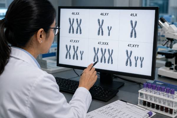
A sex chromosome aneuploidy test looks for an unexpected number of X or Y chromosomes. Depending on the setting, it may be a prenatal blood screen, a diagnostic test on chorionic villi or amniotic fluid, a postnatal blood karyotype, or a targeted analysis prompted by infertility, growth, puberty, or developmental concerns. The most commonly discussed results are 45,X; 47,XXY; 47,XXX; and 47,XYY, but mosaic and structural findings can be more complicated. A prenatal cell-free DNA result is not a fetal diagnosis because most analyzed DNA comes from the placenta and the signal may also reflect the pregnant patient. Confirmation is especially important for sex chromosome findings, which have lower and more variable positive predictive values than screening for trisomy 21. Even after diagnosis, chromosome notation does not predict an individual’s exact health, learning profile, fertility, gender identity, or life experience. Interpretation should combine the testing method, specimen source, ultrasound findings, and condition-specific counseling.
- “Positive” means different things for screening and diagnosis. A positive cell-free DNA screen raises probability; a diagnostic karyotype or microarray directly examines sampled pregnancy cells.
- Each sex chromosome condition has a broad spectrum. Some people have significant medical needs, while others have mild features or are diagnosed only in adulthood.
- Monosomy X screening has particularly important false-positive pathways. Placental mosaicism and maternal X-chromosome mosaicism can create discordant results.
- Mosaicism changes interpretation but does not provide a simple severity percentage. The cells tested may not represent every tissue in the body.
- A result should never be used as a stand-alone prediction of identity or ability. Chromosomes are medically relevant, but they do not determine a person’s full development or future.
Table of Contents
- What sex chromosome aneuploidy testing can find
- The main X and Y chromosome results
- How prenatal screening estimates chance
- How a screening result is confirmed
- Reading positive, negative, mosaic, and no-result reports
- What a confirmed result may mean
- Counseling limits and practical next steps
What sex chromosome aneuploidy testing can find
Most human cells contain 46 chromosomes, including two sex chromosomes. A typical chromosome result may be written 46,XX or 46,XY. Sex chromosome aneuploidy means that one or more X or Y chromosomes are missing or extra. Structural changes—such as a deletion, duplication, ring X, or isochromosome—can also alter sex chromosome function even when the total chromosome count is 46.
Testing may occur in several contexts:
- Prenatal screening with cell-free DNA estimates whether the placental DNA pattern is more consistent with an X or Y chromosome aneuploidy.
- Prenatal diagnosis uses chorionic villus sampling or amniocentesis followed by karyotype, chromosomal microarray, rapid aneuploidy testing, or a combination.
- Newborn or childhood testing may be prompted by swelling, heart findings, growth differences, genital findings, developmental concerns, or an unexpected prenatal result.
- Adolescent or adult testing may follow delayed puberty, short stature, tall stature, primary ovarian insufficiency, low testosterone, infertility, or recurrent pregnancy loss.
A karyotype displays whole chromosomes and is particularly useful for chromosome number, large structural changes, and mosaic cell lines. A chromosomal microarray can detect smaller gains and losses but may not characterize every balanced structural change. FISH can rapidly count targeted chromosomes in selected cells, while PCR-based rapid aneuploidy assays answer narrower questions. The laboratory may recommend more than one method when the first result is mosaic or structurally complex.
The words “sex chromosome test” can also refer to fetal sex prediction, carrier testing for an X-linked condition, Y-chromosome microdeletion testing, or analysis of differences of sex development. Those are not interchangeable with aneuploidy testing. The order and report should name the condition or chromosome question being assessed.
A sex chromosome result is a medical chromosome description, not a complete description of sex. Gonadal development, internal and external anatomy, hormones, gene expression, health, gender identity, and lived experience are related in complex ways. Respectful counseling should use the language preferred by the patient or family while explaining the relevant biology accurately.
The main X and Y chromosome results
The four most frequently screened sex chromosome aneuploidies have different natural histories and cannot be summarized by one prognosis.
45,X, associated with Turner syndrome, means that sampled cells contain one X chromosome and no second sex chromosome. Many conceptions with full 45,X do not survive to birth. Among liveborn individuals, possible features include short stature, ovarian insufficiency, congenital heart or kidney differences, hearing issues, thyroid disease, and specific learning or visuospatial challenges. Intelligence is usually in the typical range. Mosaic forms and structural X abnormalities can have different presentations. More detail is available in the Turner syndrome genetic test guide.
47,XXY, associated with Klinefelter syndrome, means an extra X chromosome in an individual with Y-chromosome material. Many pregnancies have no distinctive ultrasound findings. Later features may include tall stature, small testes, lower testosterone, reduced facial or body hair, gynecomastia, language or learning differences, and infertility. Some individuals are diagnosed only during fertility evaluation, and some remain undiagnosed.
47,XXX, often called trisomy X or triple X syndrome, means an extra X chromosome in an individual with no Y chromosome detected. Many have few obvious physical findings. Possible features include tall stature, low muscle tone, speech-language delay, learning differences, anxiety or attention concerns, and, in a subset, ovarian insufficiency. Fertility is often preserved.
47,XYY means an extra Y chromosome. Possible features include tall stature, speech-language or motor delay, learning differences, attention or behavioral concerns, and low muscle tone. Puberty and fertility are often typical. XYY does not predict aggression, criminality, or a particular personality; historical stereotypes were based on flawed interpretation.
Mosaic results contain two or more cell lines, such as 45,X/46,XX or 46,XY/47,XXY. Mosaicism may arise early in development or be limited to the placenta or another tissue. The percentage reported in a blood, villus, or amniotic-fluid sample is the percentage observed in that specimen under that laboratory method. It is not a direct percentage of the whole body and does not translate linearly into symptom severity.
Structural findings can include deletion of part of Xp or Xq, an isochromosome Xq, ring X, an X-autosome translocation, or rearranged Y chromosome. Their implications depend on the genes and regulatory regions involved, whether X-inactivation is balanced, mosaicism, and the presence of clinically important Y-chromosome material. These results require individualized genetics review rather than assignment to a broad aneuploidy label.
How prenatal screening estimates chance
Prenatal cell-free DNA screening analyzes short DNA fragments in maternal plasma. Most fragments considered “fetal” are released by the placenta, not directly by fetal organs. The laboratory compares representation of X and Y chromosome sequences with an expected pattern and reports a high-chance, low-chance, or sometimes condition-specific probability result.
Sex chromosome screening is technically and biologically more challenging than screening for trisomy 21. The X and Y chromosomes differ in size and sequence composition; X-chromosome dosage is influenced by X-inactivation; and the pregnant patient contributes most of the circulating DNA. These factors create more opportunities for an unexpected signal.
A high-chance screen can reflect:
- a fetal sex chromosome aneuploidy;
- confined placental mosaicism, in which the placenta differs from the fetus;
- maternal mosaic or constitutional sex chromosome variation;
- a vanished co-twin with a different chromosome complement;
- maternal organ transplant from a donor with different sex chromosomes;
- rarely, a maternal medical condition that alters cell-free DNA patterns;
- technical or statistical variation.
The positive predictive value, or PPV, is the chance that a positive screen is confirmed in the fetus. PPV is not the same as sensitivity or specificity. It depends on the condition, test platform, population, fetal fraction, ultrasound findings, and starting prevalence. Published studies generally find that PPV varies considerably among sex chromosome conditions and is often lowest for monosomy X. A laboratory-wide “sex chromosome PPV” may obscure those condition-specific differences.
Professional recommendations are not completely uniform. The American College of Medical Genetics and Genomics has recommended offering sex chromosome screening with informed counseling. Updated Society for Maternal-Fetal Medicine guidance recommends that sex chromosome aneuploidy screening be optional, or “opt in,” rather than automatically included, because false positives and unexpected maternal findings are more common and the conditions have variable phenotypes. The practical point is shared decision-making before the blood draw.
Pretest counseling should clarify whether the panel includes monosomy X, XXY, XXX, and XYY; whether fetal sex will be reported; how no-call results are handled; and what the patient would do with a positive result. Some laboratories bundle sex chromosome analysis into a default panel, while others require a separate selection.
Traditional first- or second-trimester serum screening is designed mainly for common autosomal trisomies and does not reliably screen for most sex chromosome aneuploidies. Ultrasound can raise suspicion for Turner syndrome when cystic hygroma, hydrops, or certain heart findings are present, but XXY, XXX, and XYY often have no specific prenatal ultrasound pattern. A normal ultrasound therefore cannot rule them out.
How a screening result is confirmed
A positive cell-free DNA screen should be followed by genetic counseling, detailed ultrasound review, and an offer of diagnostic testing before irreversible pregnancy decisions. Repeating the same screening test is generally not diagnostic and may reproduce the same placental or maternal signal.
Chorionic villus sampling (CVS) is usually performed in the first trimester and samples placental villi. It can provide an earlier result, but its placental source is especially relevant after a sex chromosome screen. If CVS shows mosaicism or a finding that could be confined to the placenta, amniocentesis may be needed to clarify fetal status. Laboratory teams may analyze direct and cultured villus preparations because the cell populations can differ.
Amniocentesis is generally performed from 15 weeks onward and samples fetal-derived cells in amniotic fluid. For a positive monosomy X screen with no major ultrasound abnormality, many specialists favor amniocentesis because it is less vulnerable than CVS to a placenta-only 45,X cell line. The optimal procedure depends on gestational age, ultrasound findings, the specific screen result, and how quickly a patient needs information.
Diagnostic laboratory options include:
- Karyotype, which can show 45,X, 47,XXY, 47,XXX, 47,XYY, many structural changes, and mosaic cell lines at the assay’s detection level;
- Chromosomal microarray, which can detect extra or missing chromosome material at higher resolution but may have limitations for balanced rearrangements and low-level mosaicism;
- Rapid FISH or QF-PCR, which can provide an early targeted answer but may require confirmation with a full chromosome study;
- Targeted molecular testing, when a structural Y or X finding raises a specific gene-level question.
A discordant prenatal result may require testing the pregnant patient. For example, a cell-free DNA pattern suggesting monosomy X could arise from age-related or constitutional maternal X-chromosome mosaicism. Maternal blood karyotype or microarray may be offered after counseling. A maternal finding can have implications for cardiac, endocrine, or reproductive health, but low-level mosaicism in blood must be interpreted cautiously.
A diagnostic sample can itself be mosaic. The laboratory may count additional cells, analyze a second culture, use FISH on uncultured cells, or recommend postnatal confirmation. When ultrasound anatomy and the chromosome result conflict, a clinical geneticist and cytogenetics laboratory should review specimen source and method before conclusions are made.
Reading positive, negative, mosaic, and no-result reports
The first line of a report is rarely enough. Interpretation starts by identifying whether the test was screening or diagnostic, which specimen was tested, and the exact chromosome notation.
High chance, positive, or screen detected means the algorithm found an X or Y chromosome pattern above its reporting threshold. It does not state that the fetus has the condition. Ask for the condition-specific PPV, fetal fraction, laboratory limitations, and recommended diagnostic pathway. A generic statement that the test is “more than 99% accurate” usually does not apply to every sex chromosome condition or to PPV.
Low chance, negative, or screen not detected means the assay did not identify the targeted pattern. It lowers the chance of the included conditions but does not eliminate it. The test may miss low-level mosaicism, unusual structural changes, samples with borderline signal, or conditions outside the panel. It does not evaluate all genetic disorders or birth defects.
No result, no call, or nonreportable may reflect low fetal fraction, assay quality limits, unusual sex chromosome data, early gestational age, higher maternal weight, placental factors, or sample problems. A no-call is not a negative result. The next step can be redraw, ultrasound, diagnostic testing, or a different strategy based on gestational age and the reason supplied by the laboratory. When the no-call is specifically due to atypical X or Y findings, repeating may be less useful than diagnostic and maternal evaluation.
Atypical finding or suspected maternal origin means the pattern does not fit the laboratory’s standard fetal categories. The report may be unable to predict the chromosome complement. This wording should prompt direct discussion with the laboratory or a genetics professional rather than conversion into a presumed fetal diagnosis.
Confirmed 45,X, 47,XXY, 47,XXX, or 47,XYY on a diagnostic karyotype describes the cells analyzed. The report should state the number of cells counted and whether mosaicism was observed. A nonmosaic prenatal result may still be confirmed after birth, especially when medical management depends on it.
Mosaic notation, such as mos 45,X[8]/46,XX[22], records the cell lines and the number observed under specified conditions. Culture can favor one cell line, and tissue distribution varies. The bracketed counts are laboratory observations, not a forecast of the child’s clinical severity.
Structural or marker chromosome result may require parental studies or additional methods. A small marker chromosome that contains X or Y material can affect gonadal development or other health features depending on its content. The phrase “variant of uncertain significance” should not be treated as equivalent to a known syndrome.
What a confirmed result may mean
A confirmed diagnosis gives a name to a chromosome pattern, but prenatal counseling must avoid both false reassurance and worst-case prediction. Ascertainment matters: adults diagnosed because of infertility or medical problems may appear more affected than people identified prenatally or incidentally, while prenatal cohorts may be too young to show later features.
For Turner syndrome, pregnancy care may include detailed fetal anatomy, fetal echocardiography, and monitoring for hydrops or growth concerns. After birth, evaluation commonly includes heart and kidney imaging, hearing assessment, growth monitoring, thyroid screening, and referral to a Turner-focused team. Growth hormone and estrogen replacement may be considered at developmentally appropriate times. The exact plan depends on karyotype and clinical findings.
For XXY, prenatal ultrasound is often unremarkable. Postnatal plans may include developmental surveillance, speech-language support, endocrine assessment around puberty, and fertility counseling later. Testosterone treatment is individualized rather than assumed from the prenatal karyotype alone.
For XXX and XYY, early development may be typical, but awareness can support prompt evaluation of speech, motor, learning, attention, or emotional concerns. Many individuals attend mainstream school, have relationships and employment, and live independently. A diagnosis is useful when it opens access to support, not when it is used to impose a predetermined future.
Fertility implications vary. Ovarian insufficiency is common in Turner syndrome but not universal in all mosaics; some individuals with XXX experience reduced ovarian reserve; XXY commonly affects sperm production; and XYY fertility is often typical. Fertility preservation discussions should be age-appropriate and led by specialists familiar with the condition.
Most sex chromosome aneuploidies occur as sporadic chromosome-separation events. Recurrence in a future pregnancy is generally low and is not usually explained by something either parent did. A structural rearrangement or parental mosaic finding may change that assessment, which is why parental testing is sometimes recommended.
A chromosome result also does not dictate gender identity or sexual orientation. Families should receive medically accurate information without assumptions about how a child will identify. When Y-chromosome material is detected in a fetus or child with atypical gonadal or genital development, management may involve specialists in differences of sex development and requires more specific analysis than an aneuploidy label.
Counseling limits and practical next steps
Sex chromosome testing has several layers of uncertainty: whether a screen reflects the fetus, whether a sampled tissue reflects the whole body, and how a confirmed chromosome pattern will express clinically. Good counseling names each layer instead of blending them into one percentage.
Before prenatal screening, ask:
- Is sex chromosome analysis included automatically or only if I opt in?
- Which conditions does the laboratory report, and does it report fetal sex separately?
- What are the condition-specific positive predictive values in a population like mine?
- Could the test reveal a chromosome finding in me rather than the fetus?
- Would I pursue CVS, amniocentesis, or neither after a positive screen?
After a positive result, obtain the complete report and arrange counseling before choosing a procedure. Confirm whether ultrasound shows cystic hygroma, hydrops, heart findings, growth differences, or no relevant abnormality. Ask whether CVS or amniocentesis better addresses the specific source of possible discordance. A general prenatal screening overview is available in the prenatal genetic screening article, while the prenatal karyotype test article explains diagnostic chromosome notation.
After a confirmed result, request condition-specific counseling that includes both medical risks and the range of real-world outcomes. Balanced information may come from a clinical geneticist, maternal-fetal medicine specialist, pediatric endocrinologist, developmental specialist, and reputable condition-specific organizations. Families often benefit from speaking with people living with the chromosome condition, provided personal experiences are not presented as universal predictions.
Testing limitations should remain visible. Cell-free DNA does not directly sample the fetus. CVS samples placenta. Amniocentesis samples fetal-derived cells but still cannot predict every tissue’s mosaic distribution. Karyotype has limited resolution, and microarray may not characterize all structural arrangements. Neither test predicts personality, intelligence, fertility, or health with certainty.
Medical terminology should also be used carefully. “Abnormal chromosome” can feel stigmatizing and may imply that a person is reducible to a laboratory finding. “Sex chromosome aneuploidy,” “chromosome variation,” or the specific karyotype is often clearer. At the same time, avoiding difficult medical information is not respectful; patients need candid discussion of heart disease, gonadal function, development, pregnancy loss, and other condition-specific risks.
The most useful result interpretation is a four-part statement: what method was used, whose cells or DNA probably produced the signal, what chromosome finding is supported, and what remains uncertain. That framework prevents a screening probability from becoming a fetal diagnosis and prevents a confirmed karyotype from becoming an overly narrow forecast of a person’s life.
References
- Society for Maternal-Fetal Medicine Consult Series #74: Cell-free DNA screening for aneuploidies (2025, guideline)
- American College of Obstetricians and Gynecologists: Screening for Fetal Chromosomal Abnormalities (2026, practice advisory)
- ACMG evidence-based guideline for noninvasive prenatal screening (2023, guideline)
- Accuracy of prenatal cell-free DNA screening for sex chromosome abnormalities (2023, systematic review and meta-analysis)
- Clinical evaluation of noninvasive prenatal testing for sex chromosome aneuploidies (2024, research article)
- Prenatal Diagnosis of Sex Chromosome Aneuploidies (2026, research article)
Disclaimer
This article is for general education and does not replace prenatal diagnosis, pediatric care, or individualized genetic counseling. Screening performance and confirmatory strategies vary by condition, laboratory, specimen, and pregnancy findings. Discuss any result with clinicians who can review the complete report and your specific circumstances.





