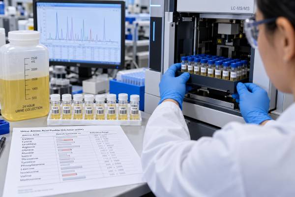
A 24-hour urine amino acids test measures how much of many individual amino acids are excreted over a full day. Amino acids are the building blocks of proteins, and the kidneys normally filter and then reabsorb most of them. An unusual urine pattern can therefore point to an inherited amino acid transport disorder, a metabolic disease that raises amino acids in blood, or a broader problem with the kidney’s proximal tubules. The test is not interpreted as one single “high” or “low” result because a panel may contain dozens of amino acids, each with age- and method-specific reference ranges. In many cases, urine findings must be compared with plasma amino acids, urine organic acids, ammonia, acid-base measurements, and the person’s symptoms. A complete 24-hour collection can quantify daily excretion, but urgent metabolic evaluations often use faster spot urine and blood tests rather than waiting a full day.
- The test looks for patterns, not one universal cutoff: Each amino acid has its own reference interval, and the combination of abnormalities often matters more than one value.
- High urine amino acids can come from two main mechanisms: blood levels may be high enough to exceed kidney reabsorption, or the kidney tubules may fail to reclaim amino acids normally.
- Cystinuria is a classic transport disorder: Excess urinary cystine and dibasic amino acids can help support the diagnosis and guide stone prevention.
- Generalized aminoaciduria can suggest proximal tubular dysfunction: Fanconi syndrome can cause urinary loss of amino acids along with glucose, phosphate, bicarbonate, uric acid, and other solutes.
- A normal 24-hour result does not exclude every metabolic disease: Timing, diet, age, treatment, kidney function, and the specific disorder can all affect the pattern.
Table of Contents
- What a 24-hour urine amino acids test measures
- Why the test is ordered
- How aminoaciduria happens
- How to collect the 24-hour sample
- How amino acid results are interpreted
- Disorders linked to abnormal urine amino acids
- Follow-up and next tests
What a 24-hour urine amino acids test measures
Amino acids circulate in blood after proteins are digested or after the body breaks down and rebuilds its own proteins. The kidneys filter many of these small molecules at the glomerulus. Specialized transporters in the proximal tubule then reclaim most filtered amino acids so they can return to the bloodstream.
A urine amino acid panel measures what escapes that reabsorption process. Depending on the laboratory, the panel may quantify alanine, glycine, glutamine, glutamate, serine, threonine, valine, leucine, isoleucine, phenylalanine, tyrosine, methionine, lysine, arginine, ornithine, citrulline, cystine, and many others. Some laboratories include dozens of compounds and report them as micromoles per 24 hours, milligrams per day, or values normalized to creatinine.
The 24-hour format answers a specific question: how much of each analyte was excreted over a complete day. A spot urine test, by contrast, is often expressed relative to urine creatinine. Spot testing is easier and may be preferable in infants, children, or urgent evaluations. A 24-hour collection can be useful when total daily loss is clinically important or when a specialist wants quantitative follow-up.
There is no meaningful single “normal urine amino acid level.” Reference intervals vary by amino acid, age, diet, collection type, and analytic method. Children may excrete different amounts than adults because kidney transport systems and metabolism change with age. Laboratories therefore provide analyte-specific ranges rather than one panel-wide cutoff.
The test is also different from plasma amino acid analysis. Plasma shows what is circulating in blood at the time of sampling, while urine reflects the interaction between blood concentration, kidney filtration, and tubular reabsorption over time. Comparing the two can help distinguish an overflow problem from a renal transport problem.
Because amino acids share several transport systems, the pattern can be more informative than the size of any one increase. A clinician may recognize a cluster of related compounds that points toward one transporter, while a broad rise across many unrelated amino acids suggests a more generalized tubular problem.
Sample timing can also affect pattern recognition. A collection obtained during acute illness may show catabolic changes that are absent when the person is well, while dietary treatment can suppress a previously obvious abnormality. When the diagnostic question is inherited disease, laboratories often want to know whether the person was fasting, acutely ill, receiving special formula, or taking amino-acid supplements during the specimen period.
Why the test is ordered
A 24-hour urine amino acid test may be ordered when an inherited metabolic disorder or renal amino acid transport defect is suspected. The exact reason depends heavily on age and symptoms.
In children, clinicians may investigate poor growth, developmental concerns, unexplained vomiting, recurrent episodes of lethargy, unusual odors, kidney stones, low blood sugar, high ammonia, or an abnormal newborn screening follow-up. In adults, testing may be used for recurrent cystine stones, suspected proximal tubular dysfunction, unexplained aminoaciduria, or follow-up of a known inherited disorder.
The test is rarely the only diagnostic study. If a person is acutely ill and an inborn error of metabolism is possible, clinicians usually prioritize tests that can be obtained quickly. These may include plasma amino acids, ammonia, glucose, blood gas or bicarbonate, lactate, ketones, acylcarnitines, and urine organic acids. Waiting 24 hours for a collection should not delay treatment of a metabolic emergency.
For kidney transport disorders, urine patterns can be especially informative. Cystinuria causes excessive excretion of cystine together with the dibasic amino acids ornithine, lysine, and arginine. A urine cystine test is often more directly focused on stone risk, while a broader amino acid profile can demonstrate the characteristic transport pattern.
Generalized loss of many amino acids can raise concern for proximal tubular dysfunction. In that setting, clinicians look for additional urine losses such as glucose despite normal blood glucose, phosphate, bicarbonate, uric acid, and low-molecular-weight proteins. The amino acid result is one piece of a larger tubular-function assessment.
How aminoaciduria happens
“Aminoaciduria” simply means increased amino acids in urine. It can develop through several mechanisms, and separating them is central to interpretation.
Overflow aminoaciduria occurs when one or more amino acids become very high in blood. The kidney’s transporters have a limited reabsorptive capacity. Once that capacity is exceeded, more of the amino acid spills into urine. Metabolic blocks that cause specific amino acids or related compounds to accumulate can produce this pattern.
Renal aminoaciduria occurs when blood levels may be normal or only mildly changed, but kidney transporters do not reabsorb amino acids properly. Inherited examples include cystinuria and Hartnup disorder. The pattern often reflects which transporter is affected rather than a generalized rise in all amino acids.
Generalized proximal tubular dysfunction can cause broad aminoaciduria. Fanconi syndrome is the classic example. Because the proximal tubule normally reabsorbs many filtered substances, Fanconi syndrome can cause urinary losses of amino acids, phosphate, glucose, bicarbonate, uric acid, potassium, and other solutes. It can be inherited or acquired from certain medicines, toxins, monoclonal protein disorders, or systemic diseases.
Diet and physiology can also influence excretion. A high protein intake can increase the filtered amino acid load. Fasting, catabolic illness, pregnancy, age, and nutritional status can change metabolism and urine patterns. These effects are one reason a mildly abnormal amino acid value should not be interpreted in isolation.
Kidney function adds another layer. Reduced filtration may alter the amount delivered to the tubules, while tubular injury can impair reabsorption. A clinician interpreting aminoaciduria therefore considers both glomerular filtration and tubular function rather than assuming every abnormality is a primary metabolic disease.
How to collect the 24-hour sample
The laboratory should provide the correct container and instructions because amino acid stability and preservative requirements can differ by method. Some laboratories require refrigeration throughout the collection, while others may provide a preservative or specify rapid return after completion.
A standard 24-hour urine collection usually follows these steps:
- At the chosen start time, empty the bladder into the toilet and record the time.
- Collect every urine sample after that for the next 24 hours.
- Store the container exactly as directed, usually refrigerated or kept cold.
- At the same time the next day, collect one final urine sample and add it to the container.
- Return the complete specimen with the requested start and stop times and total volume information.
The first void is discarded because it represents urine formed before the timed period. The final void is included because it contains urine produced during the last part of the 24 hours. The same principle applies to other timed urine collections.
Missing a void can make daily amino acid excretion appear lower than it really is. Collecting for too long can raise the reported total. If a sample is missed or spilled, contact the laboratory; restarting is often more reliable than submitting a known incomplete collection.
Diet instructions are test-specific. Some metabolic studies are intended to reflect the person’s usual intake, while others require fasting or controlled conditions. Do not impose a low-protein or high-protein diet unless instructed because changing intake may alter the pattern the clinician is trying to evaluate.
Medicines and supplements should be documented. Some can affect amino acid metabolism, kidney transport, or analytic measurements. A patient should not stop essential medication without medical guidance. If the test is part of treatment monitoring, the clinician may specifically want the sample collected while the usual therapy continues.
For children who cannot reliably complete a 24-hour collection, a spot urine amino acid profile may be more practical. The laboratory can normalize results to creatinine, although age-specific interpretation remains important. The best specimen is the one that answers the clinical question and can be collected accurately.
How amino acid results are interpreted
Interpretation starts with the pattern. A single mildly elevated amino acid is very different from a coordinated increase in a group that shares a transporter or metabolic pathway.
Laboratories flag values against age- and method-specific reference intervals. A specialist then asks several questions: Which amino acids are high? Are any low? Is the pattern selective or generalized? Are plasma levels also abnormal? Does the person have kidney dysfunction, stones, neurologic symptoms, liver disease, malnutrition, or recent acute illness?
The relationship between plasma and urine can be particularly useful. If an amino acid is very high in both blood and urine, overflow from a metabolic block is possible. If urine excretion is high while plasma concentration is normal, a renal reabsorption defect becomes more likely. This is a simplified framework, not a rule, because transport systems overlap and disease states can affect several processes at once.
Urine creatinine may be measured to assess collection plausibility or to normalize a spot result. A low 24-hour creatinine total can suggest undercollection, but it can also reflect low muscle mass, small body size, older age, or low meat intake. The 24-hour urine creatinine test should therefore be used as a quality clue rather than a rigid completeness standard.
A result within range does not exclude every inherited metabolic disorder. Some abnormalities appear mainly during illness, fasting, or catabolic stress. Treatment may also normalize a previously abnormal profile. In addition, some diseases are better detected with plasma amino acids, acylcarnitines, organic acids, enzyme testing, or molecular genetics.
Small isolated abnormalities often need confirmation before they are given diagnostic weight. Pre-analytic factors, diet, medication, age, and specimen handling can produce minor changes. A metabolic specialist may repeat testing under more standardized conditions or compare it with a targeted blood panel.
Disorders linked to abnormal urine amino acids
Several inherited disorders produce recognizable urine amino acid patterns, but laboratory findings must be linked to clinical features and confirmatory testing.
Cystinuria is caused by impaired renal reabsorption of cystine and dibasic amino acids. Cystine is poorly soluble, especially in acidic or concentrated urine, so affected people can develop recurrent kidney stones from childhood or young adulthood. Hexagonal cystine crystals in urine are a classic clue, but they are not always present. Quantitative cystine measurement and genetic testing may help confirm and manage the disorder.
Hartnup disorder affects transport of neutral amino acids in the kidney and intestine. Many people remain asymptomatic, but some can develop photosensitive rash, neurologic symptoms, or pellagra-like features related to reduced tryptophan availability. Urine testing can show increased neutral amino acids.
Lysinuric protein intolerance involves defective transport of cationic amino acids, including lysine, arginine, and ornithine. It can cause feeding problems, poor growth, episodes of high ammonia after protein intake, and systemic complications. Urinary loss of these amino acids is one part of the diagnostic pattern.
Fanconi syndrome causes broad proximal-tubule wasting rather than a single transporter defect. Generalized aminoaciduria may appear with glucosuria, phosphate wasting, bicarbonate loss, and other abnormalities. The cause may be inherited, medication-related, toxin-related, or associated with plasma-cell disorders in adults.
Metabolic pathway disorders can also change urine amino acids through overflow. Conditions affecting phenylalanine, tyrosine, branched-chain amino acids, methionine, or the urea cycle may produce characteristic blood and urine changes. However, modern diagnosis usually relies on combined biochemical and genetic testing rather than a 24-hour urine amino acid profile alone.
Because the same amino acid can be abnormal for different reasons, pattern recognition is essential. For example, high cystine in urine has different implications when it appears with lysine, arginine, and ornithine than when it is part of generalized tubular wasting.
Follow-up and next tests
An abnormal panel usually leads to targeted confirmation rather than immediate treatment based on the urine result alone. The next tests depend on the pattern and the person’s symptoms.
For suspected inherited metabolic disease, clinicians may order quantitative plasma amino acids, urine organic acids, acylcarnitines, ammonia, lactate, glucose, electrolytes, liver tests, and genetic testing. Enzyme assays or specialized metabolite studies are used for some conditions. A metabolic geneticist or biochemical genetics service may coordinate the workup.
For suspected renal transport disease, follow-up can include serum electrolytes, bicarbonate, phosphate, uric acid, glucose, creatinine, urinalysis, and measurements of urine glucose or phosphate handling. Recurrent stone formers may need a broader 24-hour kidney stone risk profile to assess urine volume, calcium, citrate, oxalate, uric acid, sodium, pH, and other factors.
Urgent evaluation is important when abnormal amino acid testing accompanies confusion, unusual sleepiness, repeated vomiting, seizures, severe weakness, rapidly worsening illness, or suspected high ammonia. In infants and children, poor feeding and sudden lethargy can be signs of metabolic decompensation. These situations require prompt medical care rather than waiting for outpatient 24-hour results.
Long-term monitoring depends on the diagnosis. A person with cystinuria may have repeated urine cystine and stone-risk testing. Someone with Fanconi syndrome may need monitoring of electrolytes, phosphate, bicarbonate, bone health, and kidney function. An inherited amino acid metabolism disorder may require diet therapy, medications, supplements, or emergency sick-day plans supervised by a metabolic team.
The practical value of a 24-hour urine amino acids test comes from matching the specimen to the question. It can quantify daily excretion and reveal useful transport patterns, but it is most informative when interpreted with plasma results, kidney function, diet, treatment status, and the patient’s clinical story.
References
- Mechanisms involved in aminoacidurias: impacts of genetic and environmental factors 2025 (Review)
- Inborn errors of amino acid metabolism – from underlying pathophysiology to therapeutic advances 2023 (Review)
- Cystinuria in children: diagnosis and treatment 2025 (Review)
- Inborn Errors of Amino Acid Metabolism Revisited: Clinical Implications and Insights into Current Therapies 2026 (Review)
- Plasma calprotectin is extremely high in patients with lysinuric protein intolerance 2023 (Research Article)
- Cystinuria: An Overview of Diagnosis and Medical Management 2022 (Review)
Disclaimer
This article is for general education and does not diagnose an inherited metabolic or kidney transport disorder. Urine amino acid results require age-specific laboratory ranges and should be interpreted with plasma tests, kidney function, symptoms, diet, and treatment status. Severe vomiting, confusion, seizures, marked lethargy, or concern for high ammonia requires urgent medical evaluation.