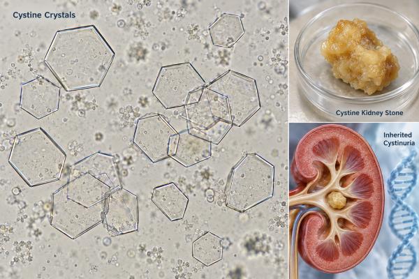
Cystine crystals in urine are an important finding because their classic hexagonal shape strongly points to cystinuria, an inherited disorder that causes excess cystine to remain in the urine. Cystine is poorly soluble, especially in acidic to neutral urine, so it can crystallize and form recurrent kidney stones. Unlike many common urine crystals, true cystine crystals are not usually dismissed as a harmless incidental finding. They should lead to confirmation of the diagnosis, assessment of stone burden, and a prevention plan that lowers urinary cystine concentration and raises cystine solubility. Cystinuria can present in childhood, adolescence, or adulthood, and stone episodes may recur throughout life. Treatment often requires unusually high fluid intake, lower sodium intake, urine alkalinization, and sometimes cystine-binding thiol medicines such as tiopronin. Because recurrent obstruction and procedures can damage kidneys over time, long-term monitoring matters even when a person currently feels well.
- Cystine crystals are typically colorless hexagonal plates and are highly characteristic of cystinuria.
- Cystinuria is a genetic transport disorder involving the SLC3A1 or SLC7A9 genes and causes recurrent cystine kidney stones.
- Cystine becomes more soluble as urine pH rises, so treatment often aims for alkaline urine under medical guidance.
- Prevention usually requires high fluid intake, sodium restriction, and sometimes tiopronin or another cystine-binding thiol drug.
- A first cystine stone or convincing cystine crystals should prompt metabolic evaluation because recurrence risk is high.
Table of Contents
- What Cystine Crystals in Urine Mean
- Appearance and Identification of Cystine Crystals
- Cystinuria and Genetic Risk
- Why Cystine Stones Form and Recur
- Diagnosis and Testing for Cystinuria
- Treatment and Prevention of Cystine Stones
- Monitoring and When to Seek Urgent Care
What Cystine Crystals in Urine Mean
True cystine crystals are a strong clue to cystinuria and deserve follow-up. Cystinuria is an inherited disorder in which the kidney tubules do not reabsorb cystine and several related dibasic amino acids normally. Cystine then reaches high concentrations in urine, where its poor solubility allows crystals and stones to form.
This finding differs from common amorphous urates or phosphates, which may appear in otherwise healthy urine. Cystine crystals are much more specific. When a laboratory confidently identifies their classic morphology, clinicians usually look for cystinuria even if the person has never had a known kidney stone.
The crystal itself does not show how many stones are present or whether a stone is currently blocking urine flow. A person can have cystinuria without visible crystals in every sample, because crystallization changes with urine concentration, pH, and timing. Likewise, a single sample may contain many crystals during a period of dehydration and fewer after heavy fluid intake.
A complete urine microscopy examination can also show blood from stone irritation, white blood cells, bacteria, or other crystals. Those findings help identify complications such as infection or suggest that more than one process is present.
The practical meaning is therefore twofold: cystine crystals can help establish the underlying diagnosis, and their presence suggests that urinary cystine has exceeded its solubility under current conditions. Long-term care focuses on changing those conditions so cystine stays dissolved.
Appearance and Identification of Cystine Crystals
Cystine crystals classically look like colorless, flat, six-sided plates. Their regular hexagonal shape is distinctive and may appear as single plates, overlapping groups, or stacks. The edges are usually well defined, although crystal size and thickness can vary.
Because rare crystals can be missed or confused with other forms, fresh urine microscopy is useful when cystinuria is suspected. Crystal formation may be more obvious in concentrated or acidic urine, while aggressive hydration and alkalinization can reduce the number seen.
The laboratory finding should be distinguished from the stone itself. Cystine stones are often yellowish and can have a waxy appearance, but visual inspection is not enough for reliable composition. If a stone is passed or removed, formal kidney stone analysis provides stronger confirmation.
Older screening methods may use the cyanide-nitroprusside reaction to detect excess urinary cystine and homocystine-related sulfhydryl compounds after chemical processing. A positive screen can support the diagnosis, but quantitative testing is more useful for long-term management and may be preferred where available.
Microscopy has an important limitation: absence of hexagons does not rule out cystinuria. Crystal visibility depends on urinary supersaturation at that moment. Someone who drinks large amounts of water or takes alkali may have cystinuria with few visible crystals.
When the morphology is uncertain, laboratories can repeat microscopy or use additional methods rather than relying on an atypical crystal. A rare genetic stone disease has lifelong implications, so confirmation should rest on the total clinical picture, stone composition, and urine testing.
Cystinuria and Genetic Risk
Cystinuria is caused by inherited defects in the renal transport system for cystine and the dibasic amino acids ornithine, lysine, and arginine. The main genes are SLC3A1 and SLC7A9, which encode parts of the transporter in the proximal renal tubule.
Cystine is the amino acid that creates the stone problem because it is much less soluble in urine than the others. The transport defect raises urinary concentrations, but only cystine commonly precipitates into stones.
Inheritance is often described as autosomal recessive, but genotype and clinical expression can be more complex, especially with SLC7A9-related disease. Some people with one pathogenic variant can have increased urinary amino acids or stone risk. This is one reason family patterns do not always look like a simple textbook recessive pedigree.
Genetic testing can confirm the molecular cause, help clarify uncertain cases, and support family counseling. It is not always necessary to begin treatment when the clinical diagnosis is already clear from cystine stones, characteristic crystals, and quantitative urine findings. A cystine stone and genetic risk evaluation may be particularly useful for children, unusual presentations, or families with several affected members.
First-degree relatives may benefit from discussion with a clinician if cystinuria is confirmed in the family, especially when there is a history of stones at a young age. The goal is not simply to label genetic risk; it is to identify people who may benefit from prevention before recurrent obstruction or surgery occurs.
Cystinuria accounts for only a small proportion of all kidney stones in adults but a larger share of stones in children. Because it often begins early and recurs frequently, lifetime stone burden can be substantial. Repeated obstruction, infections, and procedures may contribute to chronic kidney disease, making prevention especially important.
Why Cystine Stones Form and Recur
Cystine stones form when urine contains more cystine than can remain dissolved. Three factors are especially important: the amount of cystine excreted, urine volume, and urine pH.
Cystine solubility is limited around ordinary urinary pH and rises as urine becomes more alkaline. This is why concentrated, acidic urine creates a particularly favorable environment for crystallization. Drinking more fluid lowers cystine concentration, while alkalinization increases how much cystine can remain in solution.
Supersaturation can change across the day. A person may drink well during waking hours but become relatively dehydrated overnight, allowing urine to become concentrated for many hours. Prevention plans therefore often spread fluid intake throughout the day and include water before bed and sometimes during the night when clinically practical.
Sodium intake also matters. Higher sodium intake tends to increase urinary cystine excretion, so sodium restriction can reduce the cystine load that must be kept soluble. Very high animal-protein intake may add acid load and make urinary alkalinization harder, although the strongest routine dietary emphasis is often on fluid and sodium.
Cystine stones can grow quickly and recur despite treatment because the underlying transport defect is lifelong. They may form in both kidneys and can become large. Stone fragments left after a procedure can act as sites for additional growth, which is one reason follow-up imaging and complete clearance are important goals.
The high recurrence risk means a person should not wait for pain before thinking about prevention. A urine cystine test can quantify the abnormal excretion, while measures such as cystine capacity or supersaturation can help estimate whether urine conditions favor crystal growth.
Cystinuria is therefore best viewed as a chronic metabolic stone disorder rather than a series of unrelated stone attacks.
Diagnosis and Testing for Cystinuria
Diagnosis combines stone composition, urine findings, and quantitative metabolic testing. The exact set of tests depends on whether the patient presents with a first stone, recurrent stones, childhood disease, or a family history.
Useful components include:
- Urinalysis and microscopy. Characteristic hexagonal cystine crystals can provide an immediate clue. Blood may appear during a stone episode, while white blood cells or bacteria raise concern for infection.
- Stone analysis. A confirmed cystine-containing stone strongly supports cystinuria and can distinguish pure cystine from mixed composition.
- Quantitative urine cystine. A timed 24-hour collection estimates daily cystine excretion and provides a baseline for therapy.
- Full 24-hour stone profile. Volume, sodium, creatinine, calcium, oxalate, citrate, uric acid, and pH can reveal additional stone risks and show whether the collection was adequate.
- Cystine capacity or related monitoring assays. These can help assess whether urine can dissolve additional cystine or is supersaturated, especially in patients taking cystine-binding drugs where direct cystine measurement can be difficult to interpret.
- Imaging. Ultrasound or CT can determine stone burden, obstruction, and changes over time.
- Genetic testing when useful. Molecular testing can identify variants in SLC3A1 or SLC7A9 and help with counseling or atypical cases.
A cystine capacity test can be valuable during treatment because thiol drugs chemically bind cystine. Depending on the laboratory method, ordinary cystine measurements may not fully reflect the stone-forming potential once these medicines are used.
A broader 24-hour urine kidney stone risk test is still useful because a patient with cystinuria can also have low volume, high sodium, hypercalciuria, or low citrate.
Children and young adults with recurrent stones warrant especially careful evaluation. A cystine stone at a young age is not simply a dietary event; it points to a lifelong transport disorder that benefits from early preventive care.
Collection quality matters because cystine management often depends on 24-hour results. A collection that misses several hours can underestimate cystine and sodium excretion, while overcollection can do the opposite. Laboratories commonly use urine creatinine and the recorded collection duration as plausibility checks. When a result does not fit the clinical picture, repeating the collection is often more useful than making a major treatment change from a questionable sample.
Treatment and Prevention of Cystine Stones
Treatment aims to keep cystine dissolved by diluting the urine, reducing cystine excretion, increasing urine pH, and binding cystine when needed. Most patients require lifelong prevention.
High fluid intake
Fluid is the foundation of treatment. Adults with cystinuria are often advised to produce more than about 3 liters of urine per day, which may require drinking substantially more than 3 liters depending on climate, activity, and other losses. Pediatric targets are adjusted for body size.
The key is urine output, not a fixed number of glasses. Fluid should be distributed through the day because long dry intervals allow cystine concentration to rise.
Sodium restriction
Reducing sodium intake can lower urinary cystine excretion. Many specialist recommendations aim for roughly less than 2 g of sodium per day in adults, equivalent to about 5 g of table salt, while individual needs may differ. Reading food labels matters because processed foods often supply most dietary sodium.
Urine alkalinization
Potassium citrate is commonly used to raise urine pH because cystine becomes more soluble in alkaline urine. Specialist guidance often targets a pH above 7.0 and, in some European recommendations, above 7.5 for cystine prevention. The target must be individualized because very high pH can increase calcium phosphate stone risk.
Frequent home urine pH testing can help guide dosing. One isolated pH reading is less useful than a pattern across the day.
Cystine-binding thiol drugs
If high fluid intake, sodium restriction, and alkalinization do not adequately control stones, a clinician may prescribe a cystine-binding thiol drug. Tiopronin is commonly used; D-penicillamine is another option. These medicines react with cysteine to form more soluble compounds, lowering free cystine available for crystals.
Thiol drugs can cause significant adverse effects and require monitoring. Protein in the urine, blood counts, liver tests, and other safety checks may be used depending on the medicine and treatment plan. Dose decisions should be made by a clinician experienced in cystinuria.
Treatment targets are also dynamic. Hot weather, heavy exercise, fever, vomiting, or travel can sharply reduce urine volume even in someone who normally meets a fluid goal. During these periods, patients often need a deliberate hydration plan and closer attention to urine color or measured output. If illness makes it impossible to maintain fluids or medications, clinicians may need to adjust the plan temporarily rather than allowing prolonged concentrated urine.
Monitoring and When to Seek Urgent Care
Long-term monitoring is essential because cystinuria remains active even between stone attacks. Follow-up typically combines urine chemistry, imaging, kidney function, and treatment-safety checks.
A clinician may repeat 24-hour urine testing after major changes in fluid, diet, alkali, or thiol therapy. The goal is to confirm that urine volume is high, sodium intake is controlled, pH is in the intended range, and cystine supersaturation or capacity has improved. Home pH logs can reveal periods when urine becomes too acidic or excessively alkaline.
Imaging frequency depends on stone burden, age, symptoms, and prior procedures. Ultrasound can monitor many patients without radiation, while low-dose noncontrast CT may be used when greater detail is needed. Serum creatinine and estimated GFR help track kidney function over time.
Seek urgent medical care for fever or chills with flank pain, severe persistent pain, repeated vomiting, inability to pass urine, or markedly reduced urine output. A blocked urinary tract with infection can become life-threatening and may require urgent drainage rather than waiting for the stone to pass.
Prompt evaluation is also important with pain in a person who has only one functioning kidney, is pregnant, or has known advanced kidney disease. Children with severe pain, dehydration, or suspected obstruction need timely assessment.
For routine care, repeated stone passage despite good adherence is a reason to reassess the prevention plan rather than simply tolerating another episode. Medication doses, pH targets, sodium intake, and cystine capacity may need adjustment, and residual stones may need urologic treatment.
The central goal is not to eliminate every visible crystal on a microscope slide. It is to lower cystine supersaturation enough to prevent new stones, slow growth of existing stones, preserve kidney function, and reduce the need for repeated procedures.
Procedures may still be necessary when stones cause obstruction, recurrent infection, persistent pain, or progressive growth. The choice among ureteroscopy, shock-wave treatment, and percutaneous approaches depends on stone size, location, density, anatomy, and local expertise. Cystine stones can be relatively resistant to some fragmentation methods, so urologic planning should account for composition rather than treating every stone as mechanically identical.
References
- Cystinuria: Diagnosis, Medical and Surgical Management, and Emerging Therapies. A Narrative Review. 2026 (Review)
- Cystine Renal Calculi: New Aspects Related to Their Formation and Development. 2024
- A Summary of Current Guidelines and Future Directions for Medical Management and Monitoring of Patients with Cystinuria. 2023 (Review)
- Cystinuria: An Overview of Diagnosis and Medical Management. 2022 (Review)
- Metabolic Evaluation and Recurrence Prevention for Urinary Stone Patients: An EAU Guidelines Update 2024 (Guideline Update)
Disclaimer
This article provides general education about cystine crystals, cystinuria, and cystine stones and is not a personal diagnosis or treatment plan. Fluid, sodium, urine pH, and medication targets should be individualized, especially for children, pregnancy, kidney disease, or people taking cystine-binding drugs. Seek urgent care for fever with flank pain, inability to urinate, or symptoms of urinary obstruction.