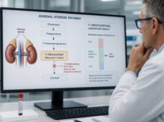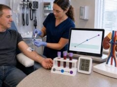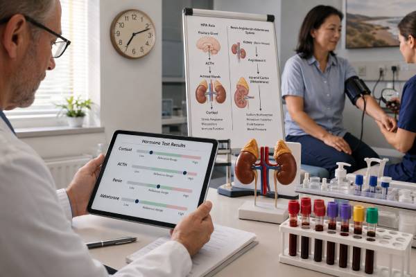
An adrenal insufficiency test panel looks for inadequate cortisol and then determines whether the problem begins in the adrenal glands or in the pituitary-hypothalamic signaling system. The usual starting measurements are an 8–9 a.m. serum cortisol and plasma ACTH drawn at the same time. Renin, aldosterone, sodium, and potassium show whether mineralocorticoid function is also impaired. A cosyntropin stimulation test often confirms limited cortisol reserve.
The pattern matters more than any isolated flag. Low cortisol with clearly high ACTH, high renin, and low or inappropriately normal aldosterone supports primary adrenal insufficiency. Low cortisol with low or normal ACTH and preserved aldosterone suggests central adrenal insufficiency, often related to pituitary disease or glucocorticoid suppression. Normal values do not always settle an early or partially treated disorder, and assay-specific cutoffs are essential. In a sick or unstable person, testing must not delay treatment. Adrenal crisis can cause vomiting, dehydration, low blood pressure, low glucose, confusion, and shock; clinicians draw cortisol and ACTH first only when doing so will not postpone hydrocortisone and intravenous fluids.
- The initial panel usually pairs 8–9 a.m. cortisol with ACTH and adds sodium, potassium, renin, and aldosterone when primary disease is possible.
- Low cortisol with high ACTH points to primary adrenal failure; low cortisol with low or normal ACTH points to a central cause.
- High renin with low aldosterone supports mineralocorticoid deficiency, which is typical of primary but not central adrenal insufficiency.
- An intermediate morning cortisol usually requires cosyntropin stimulation using an assay-specific peak cutoff.
- Suspected adrenal crisis is treated immediately; a complete diagnostic panel can be interpreted after stabilization.
Table of Contents
- Types and Causes of Adrenal Insufficiency
- Who Needs Testing
- Tests in the Panel
- Preparation and Timing
- Interpreting Result Patterns
- Confirming the Diagnosis and Cause
- Urgent Care and Long-Term Follow-Up
Types and Causes of Adrenal Insufficiency
Adrenal insufficiency means cortisol production is inadequate for ordinary needs, physiologic stress, or both. The location of the defect determines the accompanying hormone pattern.
Primary adrenal insufficiency
Primary disease begins in the adrenal cortex. Cortisol falls, feedback to the pituitary is lost, and ACTH rises. Because the outer adrenal zone is often damaged too, aldosterone falls and renin rises. Addison disease commonly refers to chronic primary adrenal insufficiency.
Autoimmune adrenalitis is a leading cause in many countries. Other causes include tuberculosis or fungal infection, bilateral adrenal hemorrhage, metastatic or infiltrative disease, bilateral adrenal removal, congenital adrenal hyperplasia, adrenoleukodystrophy, and medicines that inhibit cortisol synthesis. Autoimmune disease may coexist with autoimmune thyroid disease, type 1 diabetes, pernicious anemia, or other conditions.
Secondary and tertiary adrenal insufficiency
Secondary disease results from inadequate pituitary ACTH. Tertiary disease reflects reduced hypothalamic CRH, most commonly after prolonged glucocorticoid exposure. Clinicians often group them as central adrenal insufficiency because their routine laboratory patterns overlap.
Pituitary tumors, surgery, radiation, apoplexy, head trauma, inflammation, or congenital pituitary disorders can reduce ACTH. Glucocorticoid tablets, repeated injections, high-dose inhalers, potent topical products, and certain drug interactions can suppress the entire HPA axis. Recovery after withdrawal may take months.
Aldosterone is usually preserved in central disease because renin and angiotensin II, rather than ACTH, provide its main control. Hyperkalemia and salt wasting are therefore less typical, although low sodium can occur from impaired water excretion.
| Feature | Primary | Central |
|---|---|---|
| ACTH | High | Low or inappropriately normal |
| Aldosterone | Often low | Usually preserved |
| Renin | Often high | Usually not elevated from adrenal failure |
| Potassium | May be high | Usually normal |
| Hyperpigmentation | May occur | Not expected |
Who Needs Testing
Symptoms are often gradual and nonspecific. Testing becomes more important when fatigue occurs with objective clues or a strong risk factor.
Possible features include:
- unintentional weight loss or reduced appetite;
- persistent nausea, abdominal pain, vomiting, or diarrhea;
- low blood pressure or dizziness on standing;
- salt craving, dehydration, or recurrent low sodium;
- high potassium or low glucose;
- progressive skin darkening, including gums or skin creases;
- loss of underarm or pubic hair in some people with primary disease;
- poor growth or recurrent low glucose in a child; and
- worsening during infection, injury, or surgery.
Risk is higher after pituitary or adrenal surgery, cranial radiation, bilateral adrenal hemorrhage, immune checkpoint inhibitor treatment, tuberculosis, known autoimmune disease, or long-term glucocorticoid use. A person tapering steroids may experience fatigue, aches, and nausea from withdrawal, disease flare, or true adrenal insufficiency; a structured plan separates these possibilities.
One-sided adrenal masses rarely cause adrenal insufficiency because the other gland has reserve. Bilateral disease, hemorrhage, infiltration, or removal is more concerning. Pituitary symptoms such as headache, visual-field change, menstrual disturbance, low libido, or other hormone deficits suggest a central cause.
Pregnancy complicates recognition because nausea, fatigue, and lower blood pressure can be normal. Persistent vomiting, weight loss, salt craving, marked weakness, or unusual electrolyte changes warrant assessment, especially with autoimmune disease or steroid exposure. Cortisol-binding globulin rises during pregnancy, so nonpregnant total-cortisol cutoffs are unreliable. Endocrine and obstetric teams should coordinate testing and treatment.
Older adults may present without classic hyperpigmentation or salt craving. Falls, delirium, poor appetite, weight loss, or unexplained low sodium can dominate. Medicines and chronic disease broaden the differential, but age should not be used to dismiss possible adrenal insufficiency when objective clues are present.
Nonspecific tiredness alone does not prove adrenal disease. An adrenal fatigue test is not a validated substitute for the recognized panel. Sleep, anemia, thyroid, medication, mood, infection, and cardiopulmonary causes should be evaluated as indicated.
Tests in the Panel
Morning cortisol
Cortisol normally peaks around waking for daytime sleepers. An 8–9 a.m. total serum cortisol is the most useful baseline screen in a stable outpatient. A very low value supports deficiency, a clearly robust value makes it unlikely, and an intermediate result needs dynamic testing. Thresholds differ by assay and guideline.
Estrogen and pregnancy raise cortisol-binding globulin and total cortisol. Low albumin, liver disease, nephrotic protein loss, or critical illness can lower total cortisol. Free cortisol may help in selected binding-protein disorders, but availability and validated cutoffs vary.
ACTH
ACTH should be drawn with the baseline cortisol before cosyntropin or glucocorticoid treatment when possible. It is unstable and needs chilled EDTA plasma with prompt processing. A delayed, warm sample can look falsely low and incorrectly suggest central disease.
High ACTH is an appropriate response to low cortisol and supports primary failure. A normal-range ACTH can still be abnormal if cortisol is low, because the expected response is a clear rise.
Renin and aldosterone
These tests evaluate mineralocorticoid function. Aldosterone retains sodium and promotes potassium excretion; renin rises when the kidneys sense reduced effective volume or sodium delivery. In untreated primary adrenal insufficiency, aldosterone is low or inappropriate and renin is high.
Posture, sodium intake, potassium, dehydration, kidney disease, and many blood-pressure drugs affect both. Their interpretation in suspected deficiency differs from the aldosterone-to-renin ratio used to screen for primary aldosteronism.
Electrolytes, glucose, and supporting tests
Sodium, potassium, bicarbonate, glucose, and kidney function show physiologic impact. Primary disease may cause low sodium, high potassium, and volume depletion. Central disease can cause low sodium without high potassium. Normal electrolytes do not exclude either type.
DHEA-S may be low because adrenal androgen production is ACTH-dependent, but it declines with age and is nonspecific. Eosinophils or calcium can be abnormal but are not diagnostic. A broad panel adds context; it does not replace cortisol reserve testing.
Twenty-one-hydroxylase antibodies support autoimmune primary adrenal insufficiency, but they are a cause test rather than a reserve test. A positive antibody does not state how much cortisol can be produced today, and a negative result does not exclude other forms of primary disease. Similarly, adrenal imaging shows structure but cannot prove adequate hormone production.
Thyroid testing deserves careful sequencing in autoimmune or pituitary presentations. Hypothyroidism can overlap with fatigue and low sodium. If both thyroid and cortisol deficiencies are possible, clinicians secure glucocorticoid coverage before starting or increasing thyroid hormone because faster cortisol metabolism can unmask or worsen adrenal crisis.
Preparation and Timing
Arrange baseline collection between 8 and 9 a.m. when possible and record the exact time. Shift workers need an endocrinology plan because clock time may not match biologic morning. Acute illness can change cortisol, yet testing should proceed urgently when adrenal insufficiency is possible.
Give the clinician a complete list of:
- hydrocortisone, prednisone, prednisolone, methylprednisolone, dexamethasone, and other steroid tablets;
- steroid injections into joints, spine, muscle, or skin;
- inhaled, nasal, topical, eye, ear, and rectal glucocorticoids;
- oral estrogen, contraception, fertility treatment, or pregnancy;
- opioids and medicines that alter steroid metabolism;
- blood-pressure medicines and diuretics affecting renin or aldosterone;
- DHEA, biotin, adrenal glandular, or other supplements; and
- recent surgery, fever, trauma, vomiting, sleep change, or hospitalization.
Do not stop prescribed glucocorticoids on your own. Hydrocortisone is cortisol and will be measured by the assay. Prednisolone cross-reacts with some immunoassays. Dexamethasone generally does not cross-react much but suppresses ACTH. The clinician may time the draw after a safe pause or choose a different strategy.
For renin and aldosterone, follow posture and salt instructions. Dehydration can raise renin. Correct severe potassium abnormalities. Medication changes must be supervised because stopping antihypertensives can be dangerous.
If adrenal crisis is possible, collect cortisol, ACTH, renin, and aldosterone before hydrocortisone only if the tubes can be drawn immediately. Treatment has priority. A pre-treatment sample can later provide valuable diagnostic evidence even if the full stimulation test is deferred.
Interpreting Result Patterns
Morning cortisol cutoffs are not universal. NICE 2024 guidance uses modern immunoassay decision bands and directs laboratories and clinicians to local methods. Endotext notes that values below about 3 mcg/dL strongly suggest deficiency and values above about 15 mcg/dL suggest adequate reserve in many settings, but these are broad guideposts, not portable rules.
| Pattern | Interpretation | Next step |
|---|---|---|
| Low cortisol, high ACTH, high renin, low aldosterone | Strongly supports primary adrenal insufficiency | Treat as needed and identify the adrenal cause |
| Low cortisol, low/normal ACTH, preserved aldosterone | Supports central adrenal insufficiency | Review steroids and evaluate pituitary function |
| Intermediate cortisol, uncertain ACTH | Baseline testing is nondiagnostic | Cosyntropin stimulation or specialist-directed testing |
| Normal cortisol during major stress | May be inappropriately low for the illness | Critical-care interpretation; do not use outpatient cutoffs |
Aldosterone does not need to be below the printed range to be inappropriate. If the person is volume depleted and renin is high, aldosterone should rise. A low-normal value can therefore support mineralocorticoid failure. Conversely, a diuretic can raise renin without adrenal disease.
Early autoimmune primary insufficiency can evolve. Renin or ACTH may rise before basal cortisol becomes clearly low. Serial standardized testing and adrenal antibodies can identify progression. Treatment decisions still depend on reserve and symptoms.
Recent glucocorticoid or pituitary suppression creates the opposite pattern: ACTH and DHEA-S may be low, cortisol may be low or confounded by medication, and aldosterone remains intact. Exact dose history is essential.
Hydrocortisone taken before the draw can make cortisol appear reassuring because the assay measures the medication as cortisol. Prednisolone may cross-react on some platforms. A steroid injection may suppress the axis long after local pain relief fades. The most useful history lists the drug name, dose, route, date, and time, including non-oral products.
Results obtained during acute illness require a different lens. Cortisol should rise during major physiologic stress, so a value that is normal for a quiet morning may be inadequate for septic shock. Binding proteins and metabolism also change in critical illness. No single outpatient cutoff diagnoses critical illness-related corticosteroid dysfunction; clinicians use the overall hemodynamic situation and critical-care guidance.
Confirming the Diagnosis and Cause
The standard cosyntropin stimulation test gives 250 mcg of synthetic ACTH and measures cortisol at baseline and 30 and/or 60 minutes. A peak that remains below the assay-specific cutoff supports adrenal insufficiency. Many newer assays use thresholds around 14–15 mcg/dL rather than the historical 18–20 mcg/dL.
The test is highly useful for established primary disease. It can miss very recent central insufficiency because the adrenal cortex may still respond to a strong external ACTH signal. In a high-risk recent pituitary event, an insulin tolerance test, metyrapone test, repeat testing later, or temporary clinical protection may be appropriate.
The insulin tolerance test examines the full HPA axis by inducing controlled hypoglycemia. It can be informative for central disease but carries seizure and cardiovascular risks and must be performed in an experienced unit. Metyrapone blocks cortisol synthesis and measures the resulting ACTH and 11-deoxycortisol response. Neither is a casual alternative to cosyntropin; patient safety and local expertise determine the choice.
Assay changes can alter interpretation without any change in the patient. Modern monoclonal immunoassays and LC-MS/MS often read lower than older polyclonal assays. The endocrinologist should know the laboratory platform and time-specific cutoff. A portal’s generic “normal range” may not include the dynamic decision limit.
After primary disease is supported, common cause-directed tests include 21-hydroxylase antibodies, adrenal CT when antibodies are negative or structural disease is suspected, and testing for infection, hemorrhage, adrenoleukodystrophy, or genetic disorders based on age and history.
After central disease is supported, evaluate other pituitary axes before starting thyroid hormone if both cortisol and thyroid deficiencies are possible; giving thyroid hormone without cortisol coverage can precipitate crisis. Tests may include free T4, TSH, prolactin, LH, FSH, sex hormones, IGF-1, and pituitary MRI.
Children need pediatric endocrinology input. Congenital adrenal hyperplasia is a major pediatric cause, and newborns can deteriorate rapidly with salt wasting. Diagnostic steroids, electrolytes, glucose, renin, and genetic testing are selected by the presentation.
Urgent Care and Long-Term Follow-Up
Adrenal crisis is a time-critical emergency. Warning signs include repeated vomiting or diarrhea, severe weakness, abdominal or back pain, dehydration, low blood pressure, fainting, confusion, fever, low glucose, or shock. Infection, surgery, trauma, missed replacement doses, and inability to absorb tablets are common triggers.
Emergency treatment uses parenteral hydrocortisone and rapid isotonic saline, with glucose and electrolyte treatment as needed. Clinicians should not wait for cortisol results. Blood drawn before treatment can help later, but a delayed sample is less important than survival.
Confirmed adrenal insufficiency requires individualized glucocorticoid replacement. Primary disease with aldosterone deficiency also needs fludrocortisone and sometimes salt guidance. Monitoring includes symptoms, sitting and standing blood pressure, sodium, potassium, and renin. ACTH is not used as a target to drive glucocorticoid doses to normal because doing so can cause overtreatment.
Patients and caregivers need written sick-day rules, an emergency injection kit where prescribed, training, medical identification, and a plan for surgery or dental procedures. Vomiting that prevents oral doses is a reason to use the emergency plan and seek care.
Long-term monitoring aims to replace hormones without chronic excess. Too little glucocorticoid causes fatigue, weight loss, and crisis risk; too much causes weight gain, diabetes, high blood pressure, thin skin, infection risk, and bone loss. In primary disease, fludrocortisone is adjusted using symptoms, posture-related pressure, electrolytes, and renin. ACTH is not normalized by escalating glucocorticoids because that approach can overtreat.
Recovery can occur after glucocorticoid-induced or some postoperative central insufficiency. Patients should remain protected until repeat morning cortisol or dynamic testing documents recovery. A failed test early in recovery does not automatically mean lifelong replacement, and a few symptom-free days do not prove adequate stress reserve.
Ask the clinician:
- Were cortisol and ACTH drawn before steroid treatment and at the same time?
- Was ACTH handled in a chilled tube?
- Is the cortisol cutoff validated for this assay?
- Does renin–aldosterone physiology support primary or central disease?
- Could a medicine or binding-protein change explain the result?
- Which confirmatory test is needed, and do I need temporary steroid protection now?
The panel should lead to a clear safety plan even while the cause is being investigated. Recognizing the hormone pattern, confirming reserve when stable, and treating crisis immediately prevent the most serious complications.
Keep copies of pre-treatment laboratory values because later replacement changes every hormone in the panel. The original ACTH, cortisol, renin, aldosterone, electrolytes, collection time, and medication record may be the best evidence for classifying the disorder. If those values were not obtained during an emergency, diagnosis can still be revisited safely under endocrine supervision; treatment should never be withheld merely to recreate an untreated state.
Share the emergency plan with family, school, workplace, and travel companions when appropriate. Replacement medicine and injection supplies should remain accessible, within date, and accompanied by written instructions. Testing establishes the diagnosis, but preparation for illness is what prevents a stable chronic condition from becoming a crisis.
Review the plan periodically, especially after a dose change, pregnancy, new diagnosis, or move to a different health system. Emergency instructions must remain current and practical.
Turning Results Into a Safety Plan
When testing confirms adrenal insufficiency, the discussion should extend beyond a prescription. Patients need written instructions for ordinary dosing, fever or illness, vomiting, procedures, intense physical stress, and emergency injection use when prescribed. A medical alert card, bracelet, or phone record can tell emergency teams that glucocorticoid treatment is time-sensitive. Close family members or caregivers should know where the injection kit is stored, how to use it, and when to call emergency services.
Laboratory follow-up differs by type. In primary adrenal insufficiency, renin, potassium, sodium, blood pressure, salt craving, and postural symptoms can help assess mineralocorticoid replacement. ACTH may remain elevated despite adequate treatment and is generally not a target for escalating glucocorticoid doses by itself. In central adrenal insufficiency, aldosterone is often preserved, but other pituitary hormones may need evaluation. Day-to-day glucocorticoid adjustment relies more on symptoms, weight, blood pressure, energy pattern, and signs of over- or under-replacement than on repeatedly measuring a random serum cortisol.
Equivocal panels need a defined next step. The clinician may repeat a properly timed morning cortisol and ACTH, perform dynamic testing, review recent steroid exposure, or investigate pituitary and adrenal causes. Results drawn after emergency hydrocortisone can be hard to interpret, so a pretreatment sample is helpful when it can be obtained without delaying care. Persistent vomiting, severe diarrhea, confusion, profound weakness, low blood pressure, or collapse can signal adrenal crisis. Emergency treatment should begin on clinical suspicion; completing an outpatient diagnostic sequence is secondary to stabilizing the patient.
References
- Adrenal insufficiency: identification and management 2024 (Guideline)
- A Contemporary Approach to the Diagnosis and Management of Adrenal Insufficiency 2024 (Review)
- Adrenal Crisis 2025 (Review)
- Adrenal Insufficiency in Children 2024 (Review)
- Adrenal Insufficiency 2022 (Review)
Disclaimer
This article is educational and does not diagnose adrenal insufficiency or replace an emergency plan. Cortisol thresholds and stimulation cutoffs vary by assay and clinical setting. Suspected adrenal crisis requires immediate hydrocortisone and medical care, not delayed testing.