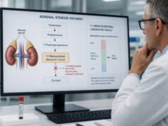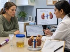
A 24-hour urine metanephrines test measures breakdown products of adrenaline and noradrenaline across a full day. It is a recommended biochemical test for pheochromocytomas and paragangliomas, rare neuroendocrine tumors that can produce catecholamines. Pheochromocytomas arise in the adrenal glands; related paragangliomas arise outside them.
The laboratory usually reports metanephrine, normetanephrine, and a combined total. These metabolites are often more reliable than catecholamines themselves because tumor cells produce them continuously, even when symptoms come in episodes. Accurate collection is essential: every urine void after the discarded start sample through the final 24-hour sample must go into the container. Stress, acute illness, nicotine, caffeine, intense exercise, and several medicines can raise results without a tumor. A value several times above the upper limit is more concerning than a slight increase. Biochemical confirmation generally comes before imaging, because incidental adrenal nodules are common and a scan alone cannot show whether a mass releases catecholamines.
- Urine fractionated metanephrines are a first-line option for detecting catecholamine-producing pheochromocytomas and paragangliomas.
- Collect every urine sample for exactly 24 hours; a missed void can produce a falsely low daily result.
- Metanephrine mainly reflects epinephrine metabolism, while normetanephrine mainly reflects norepinephrine metabolism.
- Results more than two to three times the upper limit are much more suspicious than borderline elevations.
- Chest pain, severe headache with very high blood pressure, fainting, weakness on one side, or breathing difficulty requires emergency care.
Table of Contents
- What Urine Metanephrines Show
- Reasons for Testing
- Preparation Before Collection
- Collecting Urine for 24 Hours
- Understanding the Results
- False Results and Repeat Testing
- Confirmation and Next Steps
What Urine Metanephrines Show
The adrenal medulla and sympathetic nerves make catecholamines to support the fight-or-flight response. Epinephrine, also called adrenaline, raises heart rate and energy availability. Norepinephrine, or noradrenaline, tightens blood vessels and helps maintain blood pressure. Dopamine has several roles in the nervous and endocrine systems.
Enzymes continuously convert these short-lived chemicals into more stable metabolites:
- epinephrine becomes metanephrine;
- norepinephrine becomes normetanephrine; and
- dopamine becomes 3-methoxytyramine.
Pheochromocytoma and paraganglioma cells can metabolize catecholamines inside the tumor even between symptomatic surges. For that reason, fractionated metanephrines offer better sensitivity than a one-time catecholamine measurement. Some specialized panels add 3-methoxytyramine when a dopamine-producing tumor, head-and-neck paraganglioma, metastatic disease, or a particular hereditary syndrome is suspected.
“Fractionated” means the laboratory reports metanephrine and normetanephrine separately instead of only a combined value. The pattern can provide clues about tumor biology, but it does not identify a tumor’s location with certainty. An adrenal pheochromocytoma may produce epinephrine and raise metanephrine; many extra-adrenal sympathetic paragangliomas predominantly raise normetanephrine. Exceptions are common enough that imaging and genetics cannot be chosen from the pattern alone.
The 24-hour measurement reports how much metabolite was excreted over the collection period, typically in micrograms per 24 hours or nanomoles per 24 hours. A concentration without a verified total volume is not equivalent. Laboratories often measure urine creatinine as a check on collection completeness.
Liquid chromatography–tandem mass spectrometry, or LC-MS/MS, is preferred because it separates closely related molecules and reduces analytical interference. Method-specific reference ranges remain necessary. A value from an older colorimetric or immunoassay method should not be judged against an LC-MS/MS interval.
Urine metanephrines and plasma free metanephrines are both accepted initial tests. Plasma testing is highly sensitive when the patient rests flat in a quiet setting before sampling, while urine testing integrates production over a day and may be more practical when correct supine plasma collection is unavailable. The choice also depends on pretest probability, kidney function, age, and ability to complete a 24-hour collection.
Neither specimen is automatically superior in every situation. A plasma sample is convenient and avoids missed urine, but posture, recent activity, and an inappropriate seated reference interval can reduce specificity. A urine collection smooths short-term variation and can be completed at home, but it depends on memory, storage, and exact timing. In a patient at high inherited risk or with a suspicious mass, clinicians often favor the most sensitive controlled protocol. In a lower-risk outpatient with a borderline plasma result collected under poor conditions, a complete urine collection can provide an independent check.
Reasons for Testing
Pheochromocytomas and paragangliomas are uncommon, so testing works best when symptoms, imaging, or inherited risk creates a reasonable suspicion. Ordering the test for isolated common symptoms increases the chance that a mild false positive will outnumber true tumors.
Clinicians may test when a person has spells that combine several of these features:
- pounding headache;
- racing, forceful, or irregular heartbeat;
- heavy sweating;
- pallor, shaking, or intense anxiety-like feelings;
- sudden high blood pressure or large pressure swings;
- chest or abdominal pain;
- nausea, weakness, or unexplained weight loss; or
- symptoms triggered by anesthesia, surgery, exercise, urination, certain foods, or particular medicines.
The traditional trio of headache, palpitations, and sweating is suggestive but not universal. Some tumors cause sustained hypertension without dramatic spells; others are found on imaging before symptoms develop. A minority are biochemically quiet or produce dopamine rather than epinephrine or norepinephrine.
Other reasons include an adrenal incidentaloma with imaging characteristics that do not clearly establish a benign lipid-rich adenoma, a known paraganglioma, or follow-up after treatment. Biochemical exclusion is important before biopsy or surgery on a suspicious mass because manipulating an unrecognized catecholamine-producing tumor can provoke a dangerous hypertensive crisis.
Inherited susceptibility is common enough that family history matters. Screening may be recommended for people with pathogenic variants or syndromes involving genes such as RET, VHL, NF1, SDHB, SDHD, SDHC, SDHA, MAX, or TMEM127. The starting age, interval, and choice of blood, urine, and imaging tests depend on the gene and family pattern. A relative’s result should guide genetic counseling, not informal self-testing.
Children with PPGL may present with sustained rather than episodic hypertension, headaches, sweating, visual symptoms, poor weight gain, or reduced exercise tolerance. The proportion with hereditary disease is higher than in unselected older adults, so pediatric evaluation usually includes genetics expertise. Age-adjusted reference intervals are essential. A child should not be judged by an adult urine limit, and a technically incomplete collection may be replaced by controlled plasma testing.
A pheochromocytoma and paraganglioma panel may combine metanephrines with selected catecholamines or 3-methoxytyramine. Vanillylmandelic acid is less sensitive and is not the preferred stand-alone screen for these tumors.
Preparation Before Collection
Preanalytical conditions are a major source of mild elevations. Ask the ordering clinician and laboratory for a personalized plan several days before collection. Do not stop a prescribed medicine simply because it appears on an interference list; the risk of stopping may be greater than the benefit, and many results can be interpreted with the exposure documented.
Medicines that may raise metanephrines through physiologic or analytical effects include some tricyclic and serotonin-norepinephrine reuptake inhibitor antidepressants, monoamine oxidase inhibitors, sympathomimetic decongestants, stimulants, levodopa, labetalol, and sotalol. Acetaminophen interferes with some older methods. Withdrawal from clonidine, alcohol, benzodiazepines, opioids, or other sedatives can also activate the sympathetic system.
Provide a complete list of:
- prescriptions and over-the-counter medicines;
- nicotine, vaping products, and cannabis;
- caffeine, energy drinks, and pre-workout products;
- supplements for weight loss, focus, or athletic performance;
- recent anesthesia, procedures, or contrast imaging;
- acute illness, pain, low blood sugar, or sleep loss; and
- known kidney or heart disease.
Many laboratories ask patients to avoid caffeine, nicotine, alcohol, and vigorous exercise for a defined period before and during collection, often 24 hours. Dietary restrictions differ by test method. If 3-methoxytyramine is included, fasting and avoidance of catecholamine-rich foods may be more important. Follow the exact laboratory sheet rather than a generic list from the internet.
Choose a calm, typical day if possible. Acute infection, severe pain, untreated sleep apnea, hypoglycemia, strenuous exercise, and major emotional stress can increase sympathetic activity. Testing should not be postponed when the clinician believes delay is unsafe, but the context must accompany the result.
Kidney impairment can alter urinary excretion and collection reliability. Severe renal disease may favor a carefully collected plasma test. Young children and people with incontinence may not be able to produce a valid 24-hour sample without specialized collection support.
Collecting Urine for 24 Hours
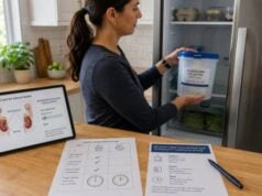
Use only the container and collection aid supplied or approved by the laboratory. The main jug may contain acid or another preservative. Avoid skin contact, do not pour it out, and do not urinate directly into a container when splashing is possible. Keep it upright and away from children. Ask whether it must remain refrigerated or on ice.
The standard collection proceeds as follows:
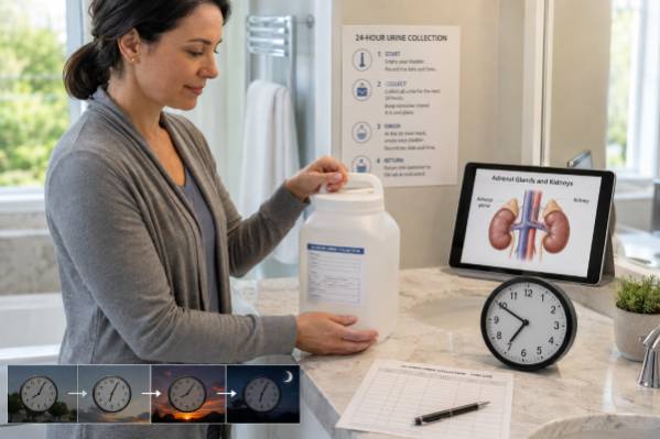
- Start with an empty bladder. On waking, urinate into the toilet and discard that urine. Record the exact date and time as the start.
- Save every later void. Collect all urine for the next 24 hours, including daytime, evening, nighttime, and urine passed with a bowel movement. Transfer it carefully into the large container.
- Store it correctly. Keep the lid tight and maintain the temperature required by the laboratory. Do not add water, tissue, stool, or household preservative.
- Include the ending void. Exactly 24 hours after the start, urinate one final time and add it to the collection. Record the end time.
- Return all containers promptly. If more than one jug was needed, label and return each as part of the same collection.
If any urine is missed or spilled, tell the laboratory. A partial collection can make excretion appear normal when it is not. Most laboratories will recommend restarting rather than estimating the missing amount. If a container fills early, obtain a second one and keep collecting; do not end the test early or discard urine.
Urine creatinine provides a rough completeness check. Low daily creatinine relative to the person’s age and muscle mass can suggest undercollection. A value above expectations may suggest an overly long collection. Creatinine is not perfect because diet, muscle mass, and kidney function influence it.
Menstrual blood or stool contamination can compromise a sample. Ask whether to delay or restart. For a child, use the pediatric collection system recommended by the laboratory. Urine extracted from absorbent diapers cannot provide an accurate 24-hour volume.
Record significant symptoms during the collection, including time, blood pressure if safely available, medicines taken, caffeine or nicotine exposure, and unusual stress. The record does not change the laboratory number, but it can help the endocrinologist explain a borderline result.
Understanding the Results
The report should list metanephrine, normetanephrine, and often total metanephrines. Reference intervals vary by age, sex, blood pressure population, method, and units. For example, one LC-MS/MS laboratory reports adult urine metanephrine intervals that differ for males and females and age-stratified normetanephrine intervals. Use the limits printed on the report rather than copying a number from another laboratory.
Interpret each analyte as a multiple of its upper limit of normal, or ULN:
| Pattern | Possible meaning | Common response |
|---|---|---|
| Both within range | A secreting tumor is unlikely after a complete, well-prepared collection | Reassess only if risk remains high |
| One value less than 2 times ULN | Borderline; false-positive factors are common | Review conditions and repeat or confirm with plasma testing |
| One value 2–3 times ULN | Increasing concern for PPGL | Prompt endocrine review and confirmatory strategy |
| One value more than 3 times ULN | Strong biochemical evidence when interference is excluded | Proceed to tumor localization under specialist care |
Exact thresholds are not absolute. Tumor size, genetic background, analyte measured, and sampling conditions affect the degree of elevation. A small tumor found during hereditary surveillance may produce a modest increase. Conversely, medications or severe illness can sometimes cause substantial abnormalities.
An isolated normetanephrine rise is common because norepinephrine increases with sympathetic activation. Sleep apnea, panic, pain, withdrawal, heart failure, and several drugs can contribute. A metanephrine-predominant pattern may be more suggestive of an adrenal epinephrine-producing tumor, but it still requires confirmation.
Total metanephrine can conceal useful detail. A normal total with an elevated fraction, or an elevated total driven by one metabolite, should be interpreted from the component results. Creatinine and total volume should be reviewed at the same time.
Normal results have high negative value for a typical secreting tumor when pretest conditions and collection are adequate. A normal urine test may not exclude a very small tumor, a dopamine-producing tumor, a nonsecretory head-and-neck paraganglioma, or disease detected in a high-risk gene carrier. Such cases require specialist surveillance rather than repeated unsupervised testing.
False Results and Repeat Testing
Most borderline positives do not lead to a tumor diagnosis, especially in low-risk patients. A careful review can prevent unnecessary scans and anxiety.
Common causes of false or nonspecific elevation include:
- antidepressants that block norepinephrine reuptake;
- stimulant or decongestant use;
- labetalol or method-specific analytical interference;
- caffeine, nicotine, and vigorous exercise;
- severe pain, anxiety, hypoglycemia, or acute illness;
- alcohol or sedative withdrawal;
- untreated obstructive sleep apnea;
- an overlong collection or incorrect total volume; and
- reference ranges not matched to age or method.
False-low results can occur with missed urine, a shortened collection, very small tumors, intermittent or dopamine-only biochemistry, and some nonsecretory paragangliomas. Improper storage can also compromise the specimen.
For a mild elevation, the clinician may correct reversible factors and repeat 24-hour urine testing. Another option is plasma free metanephrines collected under controlled conditions: fasting if required, resting supine in a quiet room for about 20–30 minutes, and using appropriate reference intervals. Drawing plasma while seated immediately after walking into the clinic can raise normetanephrine and create another borderline result.
The clonidine suppression test is reserved for selected persistent normetanephrine elevations and specialist use. It tests whether sympathetic norepinephrine production suppresses; tumor-related production may not. It is not suitable for every patient and can interact with medicines or cause low blood pressure.
Repeat testing should not become an endless cycle without reconsidering pretest probability. If symptoms are nonspecific, imaging is benign, and two high-quality biochemical assessments are normal, a PPGL is unlikely. If a pathogenic susceptibility gene, suspicious mass, or classic spells create high risk, normal or equivocal results deserve expert review.
Confirmation and Next Steps
Biochemical evidence usually precedes imaging. Once metanephrine elevation is convincing, CT or MRI is used to locate an adrenal or extra-adrenal tumor. Functional imaging may be chosen for multifocal, metastatic, recurrent, or genetically defined disease. Imaging before biochemical assessment can expose patients to incidental findings and, in rare cases, dangerous procedures.
Never biopsy a possible pheochromocytoma until the diagnosis has been considered and catecholamine excess appropriately excluded. Needle manipulation can trigger catecholamine release, severe hypertension, arrhythmia, stroke, or cardiovascular collapse.
If a PPGL is confirmed, the care team evaluates cardiovascular risk and prepares for surgery. Alpha-adrenergic blockade is generally established before surgery; a beta blocker, if needed for tachycardia, is added only after adequate alpha blockade. Starting beta blockade first can leave alpha-mediated vasoconstriction unopposed and cause a crisis. This treatment must be managed by experienced clinicians.
Genetic counseling and testing are commonly offered because a substantial proportion of PPGLs have a hereditary basis. Results can influence imaging, long-term surveillance, and testing for relatives. Pathology cannot always classify a tumor as harmless; all PPGLs have some metastatic potential, and follow-up is usually long term.
After surgery, metanephrines are rechecked to document biochemical response. A normal postoperative value supports complete removal of a secreting tumor but does not end surveillance. Recurrence or a new tumor can appear years later, particularly with hereditary disease, a paraganglioma, a large primary tumor, or an SDHB-related condition. Follow-up plans may include annual biochemical testing and periodic imaging tailored to risk. Using the same method and comparable preparation makes trends easier to interpret.
Ask the clinician:
- Was the 24-hour creatinine consistent with a complete collection?
- Which fraction was elevated, and by how many times the ULN?
- Could a medicine or collection-day stressor explain it?
- Should the result be repeated in urine or confirmed with controlled plasma sampling?
- Is 3-methoxytyramine appropriate?
- When is imaging justified?
- Does the personal or family history support genetic counseling?
While awaiting evaluation, do not deliberately provoke symptoms and do not change blood pressure medicines without advice. Seek emergency care for severe chest pain, neurologic symptoms, fainting, breathing difficulty, or a sudden extreme headache with very high blood pressure. Tell emergency clinicians that a catecholamine-producing tumor is being considered.
Home blood-pressure readings can be useful when taken safely, but repeated checking during a frightening spell may increase distress and delay care. Record the time, pulse, symptoms, and any likely trigger, then follow the emergency plan provided by the clinician. A normal pressure between spells does not exclude a tumor, and a high pressure alone does not establish one. Most hypertension has more common causes.
A reliable 24-hour result combines a complete collection, careful preparation, fraction-specific interpretation, and clinical risk. Marked abnormalities warrant rapid specialist follow-up; borderline results usually warrant better-controlled confirmation before imaging.
When repeating the test, preserve the first report and collection notes. Comparing methods, units, total volume, creatinine, medicines, and symptoms is more informative than comparing two isolated portal flags.
Making Sense of a Borderline Elevation
Small metanephrine elevations are much more common than pheochromocytoma or paraganglioma. Acute pain, severe illness, strenuous exercise, obstructive sleep apnea, nicotine, and several medicines can raise sympathetic activity or interfere with measurement. The exact pattern is informative: an isolated, slight increase close to the laboratory cutoff is handled differently from a result several times the upper limit or a consistent rise in both metanephrine fractions. The clinician should compare the result with symptoms, inherited-risk history, imaging findings already known, and the conditions under which the urine was collected.
For a borderline result, the next step may be a repeat 24-hour collection after correcting avoidable problems. Another option is plasma free metanephrine testing under controlled conditions, often after the patient has rested quietly while lying down. Medication review needs clinical judgment because abruptly stopping antidepressants, blood-pressure drugs, or other prescriptions can cause harm. The laboratory or specialist can identify true analytical interferences and decide which changes, if any, are safe.
A clearly marked and reproducible biochemical elevation usually leads to tumor localization with imaging and specialist referral. Imaging alone should not overrule weak biochemical evidence because adrenal nodules are common and many do not produce catecholamines. Conversely, severe spells with very high blood pressure, chest pain, fainting, neurologic symptoms, or shortness of breath require urgent assessment regardless of whether outpatient testing is complete. Results are best resolved through the combination of biochemical magnitude, collection quality, clinical risk, and confirmatory evaluation.
References
- Metanephrines, Fractionated, 24 Hour, Urine 2026
- Pheochromocytoma – Paraganglioma 2025
- A Laboratory Medicine Perspective on the Investigation of Phaeochromocytoma and Paraganglioma 2023 (Review)
- Hereditary Paraganglioma-Pheochromocytoma Syndromes 2023 (Review)
- Pheochromocytoma and Paraganglioma: An Endocrine Society Clinical Practice Guideline 2014 (Guideline)
Disclaimer
This information explains urine metanephrine testing and does not diagnose or exclude a tumor. Reference intervals, preparation, and medication restrictions are laboratory-specific; never stop prescribed medicine without medical advice. Emergency symptoms such as chest pain, fainting, stroke-like signs, or breathing difficulty need immediate care.