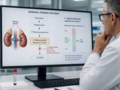
An ACTH blood test measures adrenocorticotropic hormone, the pituitary signal that tells the adrenal glands to make cortisol. Its value is not whether ACTH is simply high or low, but whether that level is appropriate for the cortisol measured at the same time. Low cortisol with high ACTH points toward primary adrenal failure. Low cortisol with low or inappropriately normal ACTH suggests a pituitary, hypothalamic, or glucocorticoid-related cause. After cortisol excess is confirmed, low ACTH supports an adrenal source, while measurable or high ACTH supports a pituitary or ectopic source.
ACTH changes in pulses and follows a strong daily rhythm, so clinicians usually collect it with cortisol between 7 and 10 a.m. The sample is fragile: it requires an ice-cooled EDTA tube and prompt processing according to the laboratory protocol. Stress, illness, sleep timing, pregnancy, and steroid medicines can change the result. A normal-range ACTH is not always physiologically normal; when cortisol is very low, the pituitary should be responding with a clear rise. Interpretation therefore relies on the paired hormone pattern, symptoms, timing, and often a dynamic test.
- ACTH should usually be interpreted with a cortisol sample drawn at the same time.
- Low cortisol plus high ACTH suggests primary adrenal insufficiency; low cortisol plus low or normal ACTH suggests central suppression.
- Confirmed high cortisol plus low ACTH suggests adrenal cortisol production; high cortisol plus measurable or high ACTH suggests an ACTH-dependent cause.
- Morning collection and rapid chilled handling are critical because ACTH is unstable after blood is drawn.
- Severe vomiting, dehydration, confusion, fainting, or very low blood pressure may signal adrenal crisis and needs emergency treatment.
Table of Contents
- What ACTH Does
- Why an ACTH Test Is Ordered
- Preparation and Sample Handling
- Normal Range and Paired Results
- Causes of High ACTH
- Causes of Low ACTH
- Follow-Up and Next Steps
What ACTH Does
ACTH is a peptide hormone made by corticotroph cells in the anterior pituitary gland. The hypothalamus releases corticotropin-releasing hormone, or CRH, in response to the body’s daily clock and physical stress. CRH prompts the pituitary to release ACTH. ACTH then binds receptors in the adrenal cortex and stimulates production of cortisol and adrenal androgens.
This linked system is called the hypothalamic-pituitary-adrenal, or HPA, axis. Cortisol feeds back to the hypothalamus and pituitary. When enough cortisol is present, ACTH signaling falls; when cortisol is low, ACTH should rise. That reciprocal relationship is why a paired result carries more information than either hormone alone.
ACTH and cortisol peak in the early morning for people who sleep at night and fall toward a late-evening low point. Secretion occurs in pulses, so two samples taken close together may differ. Night-shift work, jet lag, fragmented sleep, critical illness, and acute stress can shift or flatten the rhythm.
ACTH is produced from a larger precursor called proopiomelanocortin, or POMC. POMC processing also yields melanocyte-stimulating peptides. Very high ACTH can therefore darken skin, especially in creases, scars, gums, knuckles, and areas exposed to friction. Hyperpigmentation is a classic clue to primary adrenal insufficiency, although its absence does not exclude disease.
The blood test measures ACTH concentration in plasma, commonly in picograms per milliliter (pg/mL) or picomoles per liter (pmol/L). It does not show whether the adrenal glands can mount an adequate cortisol response under stress. A cosyntropin stimulation test is often used for that question.
An ACTH blood test is also different from receiving ACTH as a test medicine. In a baseline blood test, the laboratory measures the person’s own hormone. In stimulation testing, synthetic ACTH is administered and cortisol is measured afterward.
Why an ACTH Test Is Ordered
Clinicians order ACTH to identify the level of the HPA axis at which a cortisol problem begins. Common situations include suspected adrenal insufficiency, confirmed endogenous Cushing syndrome, a pituitary or adrenal mass, follow-up after pituitary surgery, and selected congenital adrenal disorders.
Suspected low cortisol
Symptoms of adrenal insufficiency can include persistent fatigue, muscle weakness, loss of appetite, weight loss, nausea, abdominal discomfort, dizziness on standing, low blood pressure, low sodium, or recurrent low glucose. Primary adrenal insufficiency may also cause salt craving, high potassium, and hyperpigmentation. Central adrenal insufficiency often preserves aldosterone, so high potassium and salt craving are less typical.
Risk factors shape the evaluation. Primary adrenal damage can result from autoimmune adrenalitis, infection, bleeding, infiltrative disease, bilateral adrenal surgery, metastatic disease, or inherited disorders. Central causes include pituitary tumors or surgery, cranial radiation, head injury, hypothalamic disease, and suppression after prolonged glucocorticoid exposure.
The most common acquired cause of central suppression is external glucocorticoid treatment. Tablets are obvious, but injections, high-dose inhalers, potent skin creams, and some drug interactions can also suppress ACTH. A 2024 joint guideline on glucocorticoid-induced adrenal insufficiency emphasizes planned tapering and morning cortisol assessment rather than abrupt withdrawal.
Confirmed high cortisol
ACTH is not the first screening test for Cushing syndrome. Hypercortisolism should first be demonstrated with urine free cortisol, late-night salivary cortisol, or dexamethasone suppression. After confirmation, ACTH divides the disorder into two broad groups:
- ACTH-independent: An adrenal adenoma, carcinoma, or nodular adrenal disease makes cortisol without pituitary stimulation, suppressing ACTH.
- ACTH-dependent: A pituitary corticotroph tumor causes Cushing disease, or a nonpituitary tumor produces ACTH or, rarely, CRH.
That distinction determines subsequent imaging and testing. Finding an adrenal or pituitary nodule before biochemical classification can mislead because small incidental lesions are common.
ACTH may also be part of a Cushing syndrome test panel, but its role is source classification after cortisol excess is established.
Monitoring and special situations
After surgery for an ACTH-producing pituitary tumor, low cortisol and ACTH can support remission, while later recovery or recurrence requires serial endocrine assessment. In congenital adrenal hyperplasia, ACTH may reflect inadequate cortisol feedback, but treatment monitoring relies more directly on specific steroid precursors, androgens, growth, and symptoms.
ACTH is sometimes included in adrenal incidentaloma evaluation or suspected ectopic ACTH syndrome. Severe low potassium, infections, marked muscle weakness, rapid onset of diabetes, or very high cortisol can make an ectopic source more urgent.
Preparation and Sample Handling
Unless the clinical situation requires immediate testing, ACTH and cortisol are usually drawn together in the early morning, often between 7 and 10 a.m. Record the exact collection time. A reference interval labeled for morning specimens should not be applied to an evening sample.
Fasting is not universally required, but the laboratory may request it when other tests are ordered. Avoid intense exercise immediately before the draw and sit quietly if possible. Acute pain and anxiety can transiently activate ACTH. Do not delay emergency evaluation merely to obtain ideal resting conditions.
Tell the clinician about:
- oral, injected, inhaled, nasal, topical, eye, ear, or rectal steroids;
- the time and dose of the most recent glucocorticoid;
- estrogen therapy, oral contraceptives, or pregnancy;
- opioid medicines and other drugs that affect pituitary signaling;
- supplements containing biotin, DHEA, pregnenolone, or hidden steroid ingredients;
- shift work, recent travel across time zones, or major sleep disruption; and
- fever, surgery, trauma, hospitalization, or severe emotional stress.
High-dose biotin can interfere with some immunoassays. One major laboratory asks patients to avoid biotin-containing supplements for 12 hours before its ACTH assay. Follow the local instruction because platform design differs. Do not stop prescribed steroids or opioids without a medical plan.
Why handling matters
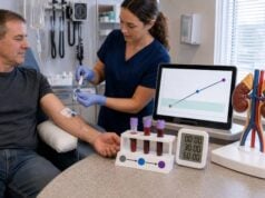
ACTH degrades in warm, unprocessed blood. A typical protocol uses an ice-cooled lavender-top EDTA tube, immediate mixing, rapid transport on ice, prompt centrifugation, and separation or freezing of plasma. Exact requirements vary. If the tube sits at room temperature or processing is delayed, the result can be falsely low.
Preanalytical error should be suspected when ACTH is unexpectedly low, conflicts with cortisol and symptoms, or was collected in an unsuitable tube. Repeating the paired sample at a laboratory experienced with ACTH may be more useful than building a diagnosis on a questionable specimen.
ACTH immunoassays can also differ. Rare antibodies or peptide fragments may cause spurious results. When a striking ACTH value does not match the cortisol pattern or clinical picture, the endocrinologist can consult the laboratory about dilution studies, an alternate assay, or repeat sampling.
Normal Range and Paired Results
There is no universal ACTH range. One commonly used morning plasma assay reports 7.2–63 pg/mL for samples collected between 7 and 10 a.m., but another laboratory may use different limits. Use the interval, units, and collection window printed on the report.
The physiologic relationship with cortisol is more informative:
| ACTH | Cortisol | Pattern suggested |
|---|---|---|
| High | Low | Primary adrenal insufficiency or impaired adrenal steroid synthesis |
| Low or inappropriately normal | Low | Secondary or tertiary adrenal insufficiency, often including glucocorticoid suppression |
| Low | High after hypercortisolism is confirmed | ACTH-independent adrenal cortisol production |
| Normal-high or high | High after hypercortisolism is confirmed | ACTH-dependent Cushing disease or ectopic ACTH production |
“Inappropriately normal” is important. If cortisol is dangerously low, a mid-range ACTH is not an adequate response. The pituitary should be driving ACTH upward. Conversely, a low ACTH can be expected when cortisol is genuinely high because negative feedback should suppress it.
ACTH values near a decision boundary may need repetition. In confirmed Cushing syndrome, some clinicians use approximate bands—suppressed, indeterminate, and clearly measurable—rather than one rigid threshold. Assay variation and episodic secretion limit precision. Indeterminate results may lead to repeat ACTH, DHEA-S, adrenal imaging context, or specialized dynamic testing.
Units can also create confusion. A laboratory may report pg/mL, ng/L, or pmol/L. The first two are numerically equivalent for ACTH, while pmol/L requires conversion. Never compare a result with an online range until both units match. The report’s flag reflects its own platform; it does not account for whether the paired cortisol makes the ACTH response appropriate.
A random afternoon ACTH cannot be compared fairly with a morning interval. Nor should a value obtained during critical illness be interpreted like a stable outpatient sample. Critical illness changes cortisol binding, metabolism, and feedback, and treatment decisions may precede full testing.
Causes of High ACTH
High ACTH means the pituitary is releasing a strong signal or an ectopic source is producing ACTH-like hormone. Cortisol shows whether the signal is compensatory or excessive.
With low cortisol, causes include:
- autoimmune Addison disease;
- adrenal infection, hemorrhage, infiltration, or metastatic destruction;
- bilateral adrenal surgery;
- congenital adrenal hyperplasia or another steroid synthesis defect;
- adrenoleukodystrophy or selected genetic disorders; and
- medicines that block cortisol synthesis.
In primary adrenal insufficiency, aldosterone deficiency may raise renin and potassium and lower sodium. Testing often includes electrolytes, renin, aldosterone, and 21-hydroxylase antibodies. Depending on the context, imaging or infectious, genetic, and metabolic evaluation may follow.
With high cortisol, ACTH-dependent causes include a pituitary corticotroph adenoma, called Cushing disease, and ectopic ACTH secretion. Ectopic sources can include neuroendocrine tumors of the lung, thymus, or pancreas and small-cell lung cancer. CRH-producing tumors are rare. Pituitary and ectopic values overlap; the ACTH number alone cannot identify the source.
High ACTH with apparently normal cortisol may occur early in primary adrenal insufficiency, during recovery from glucocorticoid suppression, with physiologic stress, or because of assay interference. Repeat paired morning testing and dynamic assessment help determine whether the finding is real.
Very high ACTH after both adrenal glands are removed can accompany growth of a corticotroph tumor, historically called Nelson syndrome. Progressive hyperpigmentation, rising ACTH, headaches, or visual symptoms require pituitary evaluation.
Congenital adrenal hyperplasia creates another high-ACTH pattern. An inherited enzyme block limits cortisol synthesis, so ACTH chronically stimulates the adrenal cortex and raises steroids before the blocked step. The companion markers vary by enzyme: 17-hydroxyprogesterone is prominent in 21-hydroxylase deficiency, while 11-deoxycortisol and deoxycorticosterone rise in 11β-hydroxylase deficiency. ACTH supports the feedback explanation but cannot name the enzyme defect.
Causes of Low ACTH
Low ACTH with low cortisol suggests insufficient hypothalamic or pituitary drive. Causes include:
- current or recent glucocorticoid treatment;
- a pituitary tumor compressing normal tissue;
- pituitary surgery or radiation;
- hypothalamic disease;
- traumatic brain injury, pituitary apoplexy, or postpartum pituitary injury;
- opioid-related HPA suppression; and
- congenital pituitary hormone deficiencies.
Central adrenal insufficiency can occur with low thyroid hormone, sex hormones, growth hormone, or prolactin abnormalities if several pituitary axes are affected. Headache, visual field loss, menstrual changes, low libido, or unexplained low sodium may prompt a broader pituitary hormone panel and MRI.
Low ACTH with confirmed cortisol excess points toward an adrenal source. Possibilities include a cortisol-producing adrenal adenoma, adrenocortical carcinoma, primary bilateral macronodular adrenal hyperplasia, or primary pigmented nodular adrenocortical disease. Exogenous glucocorticoids can produce Cushing features while suppressing both ACTH and endogenous cortisol, so medication history remains central.
Sample degradation can mimic low ACTH. If cortisol is low but the clinical picture strongly suggests primary adrenal insufficiency—especially hyperpigmentation, high potassium, or high renin—repeat a correctly handled ACTH sample while proceeding with necessary care.
ACTH can remain suppressed after a steroid injection even when the person no longer feels a medication effect. Long-acting preparations and repeated injections may suppress the axis for weeks. Drug interactions that slow steroid metabolism can extend exposure. For example, some strong CYP3A inhibitors can increase the systemic effect of certain inhaled or injected glucocorticoids. The exact product, dose, date, and route are therefore more useful than saying only “I took a steroid.”
A low ACTH result is not itself a reason to take an “ACTH booster” supplement. No over-the-counter product safely corrects pituitary-adrenal signaling, and hormone-containing supplements can make suppression worse.
Follow-Up and Next Steps
When low cortisol is suspected, the clinician may confirm adrenal reserve with cosyntropin. Cortisol is measured before and 30 or 60 minutes after synthetic ACTH. Peak cutoffs are assay-specific; newer assays often use lower thresholds than the historical 18–20 mcg/dL. A recent-onset central deficiency can sometimes retain a normal response because the adrenal glands have not yet atrophied, so test timing and clinical history matter.
For primary adrenal insufficiency, additional workup may include 21-hydroxylase antibodies, renin, aldosterone, electrolytes, adrenal imaging, or testing for infection and genetic causes. For central disease, other pituitary hormones and MRI are considered. Treatment should not wait in an unstable patient.
If high cortisol has already been confirmed, repeat ACTH may solidify source classification. Suppressed ACTH leads toward adrenal evaluation. Measurable or high ACTH leads toward pituitary MRI and sometimes CRH or desmopressin testing. When MRI is negative or shows a small lesion that may be incidental, bilateral inferior petrosal sinus sampling can distinguish pituitary from ectopic ACTH production at an expert center.
Questions to ask about a result include:
- Were ACTH and cortisol drawn together between 7 and 10 a.m.?
- Was the ACTH tube chilled and processed promptly?
- Is ACTH appropriate for the measured cortisol, even if it falls inside the printed range?
- Could any steroid route, supplement, opioid, biotin, illness, or sleep schedule affect the pair?
- Is a cosyntropin test needed, and which assay-specific cortisol cutoff will be used?
- Should renin, aldosterone, adrenal antibodies, or other pituitary hormones be measured?
Adrenal crisis is a clinical emergency. Severe weakness, repeated vomiting, abdominal pain, dehydration, confusion, fainting, low blood pressure, low glucose, or shock warrants immediate care and glucocorticoid treatment when suspected. Blood for cortisol and ACTH can be collected before treatment if this causes no delay, but lifesaving therapy takes priority.
For people using long-term glucocorticoids, recovery of ACTH and cortisol can take weeks to many months. Tapering should follow a clinician-designed plan. Symptoms during withdrawal may overlap with inflammatory disease flare and glucocorticoid withdrawal syndrome, so laboratory and clinical reassessment are often needed.
Pregnancy raises cortisol-binding globulin and total serum cortisol, while ACTH physiology also changes across gestation. Standard nonpregnant cortisol thresholds may not apply. Persistent vomiting, weight loss, low blood pressure, or unusual electrolyte findings in pregnancy deserve specialist assessment; common pregnancy symptoms should not automatically be assumed to explain them. Testing and replacement doses require obstetric and endocrine coordination.
In children, ACTH must be interpreted with pediatric cortisol methods and the growth pattern. Poor weight gain, recurrent low glucose, unexplained fatigue, or early androgen signs can prompt evaluation. Pediatric adrenal insufficiency may deteriorate quickly during gastroenteritis, so families with a confirmed disorder need written stress-dose and injection instructions.
The ACTH result becomes clear when read as part of a feedback pair. Timing, specimen integrity, cortisol, medication exposure, and the clinical setting determine whether a high, low, or normal-range value is appropriate.
Keep a copy of the report showing collection time, units, cortisol value, and assay notes. Those details allow an endocrinologist to compare future results accurately. A standalone screenshot of the ACTH number without its paired cortisol can hide the most important information. If repeat testing is planned, use similar timing and document medication doses so a true trend can be separated from changing conditions.
A written interpretation should state whether ACTH was appropriate for cortisol, not merely whether it was inside the interval. That single sentence often prevents future clinicians from misreading the result.
Resolving an ACTH and Cortisol Mismatch
ACTH should rarely be interpreted without a cortisol result obtained at about the same time. A high ACTH with low cortisol points toward impaired adrenal cortisol production, while low or inappropriately normal ACTH with low cortisol raises concern about pituitary or hypothalamic suppression. A low ACTH beside high cortisol can support ACTH-independent cortisol excess. These patterns guide testing, but they are not absolute diagnoses because timing, acute illness, intermittent hormone secretion, and assay differences can blur the expected relationship.
Preanalytical handling deserves special attention when the result does not fit the clinical picture. ACTH is unstable in blood after collection, so the correct tube, prompt cooling or processing, and timely analysis are important. A delayed or mishandled specimen may produce a falsely low value. Cortisol-binding changes from oral estrogen, pregnancy, severe liver disease, or low blood protein can alter total cortisol without changing the biologically active fraction in the same way.
The medication history can explain many apparent contradictions. Oral, injected, inhaled, topical, or intra-articular glucocorticoids may suppress ACTH, and some synthetic steroids are not detected as cortisol by every assay. Opioids and certain other drugs may also suppress the hypothalamic-pituitary-adrenal axis. Clinicians may repeat paired morning measurements, use an ACTH stimulation test, or select testing for cortisol excess depending on the pattern. Urgent symptoms—such as persistent vomiting, severe weakness, confusion, low blood pressure, or collapse—need immediate care rather than routine retesting.
References
- Adrenocorticotropic Hormone, Plasma 2026
- Physiology, Adrenocorticotropic Hormone (ACTH) 2025 (Review)
- Glucocorticoid-Induced Adrenal Insufficiency 2024 (Guideline)
- Diagnosis and management of adrenal insufficiency 2024 (Review)
- Consensus on Diagnosis and Management of Cushing’s Disease: A Guideline Update 2021 (Consensus Guideline)
Disclaimer
This article is educational and cannot diagnose a pituitary or adrenal disorder. ACTH ranges and cortisol cutoffs vary by assay, timing, and clinical setting. Seek emergency care for suspected adrenal crisis, severe low blood pressure, confusion, or rapid deterioration.