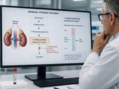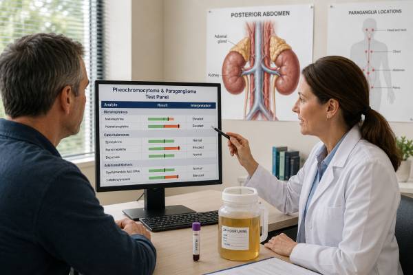
A pheochromocytoma and paraganglioma test panel looks for biochemical evidence of tumors that produce catecholamines or their metabolites. Pheochromocytomas arise in the adrenal medulla, while paragangliomas arise from related nerve-associated tissue outside the adrenal glands. The preferred first tests are plasma free metanephrines or 24-hour urine fractionated metanephrines because tumors continuously convert catecholamines into metanephrine and normetanephrine, even when symptoms are intermittent. Catecholamines may be added in selected situations, but they are less sensitive as a primary screen. Accurate testing depends on preparation: posture, rest, stress, exercise, caffeine, nicotine, illness, sleep apnea, and medicines can all produce false elevations. A result several times the upper limit is more concerning than a borderline rise. Mild abnormalities are usually repeated under controlled conditions before imaging. Once biochemical evidence is convincing, imaging, genetic assessment, and specialist management follow.
- Plasma free or 24-hour urine fractionated metanephrines are the preferred biochemical screening tests.
- Normetanephrine reflects norepinephrine metabolism; metanephrine reflects epinephrine metabolism.
- Values more than about three times the upper limit are strongly concerning, while small elevations often need repeat testing.
- Supine rest before plasma collection improves specificity and reduces posture-related false positives.
- Imaging usually follows convincing biochemical evidence, because incidental adrenal findings are common.
Table of Contents
- What the Panel Measures
- Who Should Be Tested
- Plasma Versus Urine Testing
- Preparation for Accurate Results
- How to Interpret Metanephrine Patterns
- Borderline Elevations and False Positives
- Imaging, Genetics, and Confirmation
- Urgent Symptoms and Next Steps
What the Panel Measures
Catecholamines are signaling chemicals made from tyrosine. The main ones are norepinephrine, epinephrine, and dopamine. They help regulate blood pressure, heart rate, alertness, and the response to danger or physical stress. Pheochromocytomas and sympathetic paragangliomas can produce one or more of these chemicals.
The body and the tumor convert catecholamines into more stable metabolites. Norepinephrine becomes normetanephrine, epinephrine becomes metanephrine, and dopamine becomes 3-methoxytyramine. Because this metabolism can continue inside tumor cells between symptomatic episodes, metanephrines are usually more sensitive than direct catecholamine measurements.
A comprehensive panel may include:
- Plasma free metanephrine and normetanephrine.
- 24-hour urine fractionated metanephrines.
- Plasma or urine 3-methoxytyramine in selected patients.
- Plasma or urine catecholamines: epinephrine, norepinephrine, and dopamine.
- Urine creatinine to assess completeness of a 24-hour collection.
Not every patient needs every analyte. Using both plasma and urine as routine duplicates can increase false positives without adding much when one high-quality test is clearly normal. The clinician selects the specimen according to risk, age, kidney function, ability to rest supine, and likelihood of completing a reliable urine collection.
Pheochromocytoma refers specifically to an adrenal medullary tumor. Paragangliomas can occur along sympathetic chains in the chest, abdomen, or pelvis and may secrete catecholamines. Head-and-neck paragangliomas are often parasympathetic and may be biochemically silent, although some produce dopamine-related markers.
The panel does not diagnose every adrenal mass and does not evaluate cortisol or aldosterone secretion. An adrenal incidentaloma may require a broader hormone workup, including cortisol suppression testing and aldosterone-renin testing when hypertension or low potassium is present.
Who Should Be Tested
Testing is appropriate when symptoms, imaging, or family history create a meaningful possibility of pheochromocytoma or paraganglioma. Classic spells include pounding headache, sweating, palpitations, tremor, pallor, anxiety, chest discomfort, and sudden high blood pressure. Episodes may last minutes to hours and can be triggered by exertion, anesthesia, surgery, certain medicines, abdominal pressure, urination in bladder tumors, or no clear trigger.
Many patients do not have the classic triad. Some have sustained hypertension, variable blood pressure, orthostatic dizziness, unexplained cardiomyopathy, weight loss, high glucose, or an incidental adrenal mass. Others are diagnosed through surveillance for an inherited syndrome.
Common indications include:
- An adrenal mass with imaging features that are not clearly benign.
- Paroxysmal or resistant hypertension with suggestive spells.
- A previous pheochromocytoma or paraganglioma requiring surveillance.
- A pathogenic variant or syndrome associated with these tumors.
- A first-degree relative with hereditary PPGL.
- An unexplained hyperadrenergic episode during surgery or anesthesia.
- A head-and-neck paraganglioma that may warrant dopamine-related testing.
Hereditary associations include variants in SDHx genes, VHL, RET, NF1, MAX, TMEM127, and others. A substantial proportion of patients carry a germline susceptibility variant, including some without an obvious family history. Genetic counseling and testing are therefore considered broadly after a tumor is diagnosed.
Screening everyone with common anxiety, palpitations, or mild hypertension is not helpful. These symptoms are frequent, while PPGL is rare. Poor patient selection increases the number of borderline results caused by stress, posture, medicines, or sleep disorders.
A biochemical panel may be bypassed only in specific circumstances, such as an emergency or a known nonsecretory hereditary lesion under specialist management. In ordinary evaluation, biochemical evidence should precede extensive tumor imaging.
Plasma Versus Urine Testing
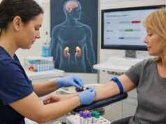
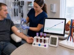
Plasma free metanephrines provide high sensitivity and are convenient when collected correctly. The most specific protocol has the patient fast when required, rest quietly while lying down for 20–30 minutes, and remain supine during blood collection. Reference intervals should match the collection posture. A seated sample compared with a supine range can create a false elevation, especially for normetanephrine.
The plasma free metanephrines test is often preferred in people at high pretest risk, those with a known hereditary syndrome, children, or patients who cannot complete a 24-hour collection. It may also be more practical in significant kidney impairment, although advanced illness and sympathetic activation still complicate interpretation.
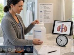
A 24-hour urine test measures fractionated metanephrines excreted over a full day. It is useful for outpatients who cannot undergo a controlled supine draw and can reliably collect every void. The collection begins after discarding the first morning urine, includes all urine for the next 24 hours, and ends with the first urine at the completion time. The container must be stored as instructed.
Urine creatinine helps assess completeness, but it cannot detect every collection error. Missing one void can lower results, while collecting longer than 24 hours can raise them. Kidney dysfunction can also affect excretion.
| Method | Advantages | Main limitations |
|---|---|---|
| Plasma free metanephrines | High sensitivity; one blood draw | Requires controlled posture and rest for best specificity |
| 24-hour urine fractionated metanephrines | Integrated collection; practical for many outpatients | Collection errors and kidney-function effects |
| Plasma catecholamines | Can characterize active secretion | Highly sensitive to stress and short-term fluctuations |
| Urine catecholamines | Captures daily output | Less sensitive than fractionated metanephrines |
LC-MS/MS is generally preferred because it separates analytes more specifically than some older methods. The report should identify the specimen, assay, units, and reference interval.
Preparation for Accurate Results
Preparation aims to reduce normal sympathetic activation. For plasma testing, the patient should ideally have an intravenous catheter placed and rest quietly in a dim, comfortable setting before the draw. Talking, scrolling through stressful messages, walking into the room immediately before collection, or a difficult needle insertion can raise norepinephrine and normetanephrine.
Instructions vary, but common measures include avoiding vigorous exercise, nicotine, and caffeine for a specified period, often at least several hours and sometimes 24 hours. Alcohol and certain foods may matter more when dopamine or 3-methoxytyramine is measured. Fasting overnight may be requested.
Medication review is essential. Drugs that can raise results or interfere analytically include some antidepressants, especially norepinephrine reuptake blockers and tricyclics; sympathomimetics; stimulant medicines; decongestants; levodopa; and several other agents. Abrupt withdrawal from clonidine, alcohol, benzodiazepines, or opioids can also activate the sympathetic system.
Do not stop prescription medicines on your own. The risk of withdrawal or loss of disease control may exceed the benefit of a cleaner test. The clinician can decide whether to continue and interpret, switch safely, or repeat later.
Acute illness, pain, hypoglycemia, heart failure, severe anxiety, obstructive sleep apnea, and intensive-care conditions can elevate normetanephrine. Nonurgent testing is often postponed until the person is stable. Untreated sleep apnea is a notable source of repeated mild elevations.
For urine collection, obtain written start and stop instructions. Record missed specimens, unusually heavy exercise, severe symptoms, and all medicines. A collection known to be incomplete should usually be repeated rather than interpreted as normal.
Samples should be handled promptly and according to laboratory temperature requirements. The laboratory may reject a specimen if the tube, preservative, volume, or timing is wrong.
How to Interpret Metanephrine Patterns
Interpretation begins with the degree of elevation. A result more than three times the upper reference limit is rarely caused by ordinary posture or mild stress and strongly raises concern for PPGL. Values between one and two times the limit are common in people without tumors and require careful review.
Metanephrine and normetanephrine patterns can suggest tumor biology. Predominant metanephrine elevation points toward epinephrine production, which is most often associated with adrenal pheochromocytoma. Predominant normetanephrine elevation reflects norepinephrine metabolism and can occur in adrenal or extra-adrenal sympathetic tumors. Elevated 3-methoxytyramine suggests dopamine production and may occur in some head-and-neck, SDHx-related, or metastatic tumors.
| Biochemical pattern | Possible implication | Interpretive caution |
|---|---|---|
| Marked metanephrine elevation | Epinephrine-secreting adrenal tumor more likely | Confirm specimen quality and assay |
| Marked normetanephrine elevation | Norepinephrine-secreting adrenal or extra-adrenal tumor | Stress and posture especially affect mild elevations |
| Elevated 3-methoxytyramine | Dopamine-related phenotype | Diet and levodopa can interfere |
| Normal metanephrines | Secretory PPGL unlikely with a high-quality test | Very small, dopamine-only, or nonsecretory tumors can be exceptions |
The upper reference limit is not a tumor threshold. Age affects normetanephrine, and some laboratories use age-adjusted ranges. Children need pediatric intervals. Results from plasma and urine cannot be directly compared by number because units and matrices differ.
A normal high-quality test usually excludes a catecholamine-producing PPGL in a symptomatic patient. Exceptions include very small tumors, nonfunctional head-and-neck paragangliomas, and rare dopamine-predominant lesions. Imaging characteristics and inherited risk can justify further evaluation despite normal routine metanephrines.
Borderline Elevations and False Positives
Borderline normetanephrine elevation is the most common difficult result. The clinician should ask whether the sample was drawn seated, immediately after walking, during pain, after caffeine or nicotine, or while taking a norepinephrine reuptake-blocking medicine. Repeating the test after 20–30 minutes of supine rest often resolves the issue.
False-positive causes include:
- Acute physical or emotional stress.
- Obstructive sleep apnea.
- Heart failure, severe illness, or hypoglycemia.
- Tricyclic or serotonin-norepinephrine reuptake inhibitor antidepressants.
- Stimulants, decongestants, nicotine, and heavy caffeine use.
- Levodopa or dopamine-related drugs.
- Withdrawal from clonidine, alcohol, sedatives, or opioids.
- Incorrect posture-specific reference intervals.
- Analytical interference with older assays.
A specialist may consider a clonidine suppression test for persistent isolated mild plasma normetanephrine elevation. Clonidine reduces sympathetic nerve release of norepinephrine in people without a tumor, while autonomous tumor production may remain elevated. The test has medication and safety limitations and is not used for metanephrine elevation or as a general screening test.
False-negative results are less common with properly collected metanephrines. They can occur with very small tumors, some nonsecretory lesions, or dopamine-predominant tumors when 3-methoxytyramine is not measured. A strong hereditary or imaging context therefore still matters.
The safest approach to a mild result is staged. Correct obvious confounders, repeat under optimal conditions, compare the magnitude and pattern, and only then decide whether imaging is justified. Moving directly from a 1.1-times elevation to whole-body scans commonly uncovers unrelated abnormalities.
Imaging, Genetics, and Confirmation
When biochemical evidence is convincing, CT or MRI is used to locate the tumor. The abdomen and pelvis are common starting regions because most secretory tumors occur in the adrenal glands or nearby sympathetic tissue. MRI may be preferred in children, pregnancy, or when radiation should be minimized.
Functional imaging is selected according to tumor location, biochemical phenotype, metastatic risk, and genetic background. Options include radiotracers targeting somatostatin receptors, norepinephrine transport, or glucose metabolism. The choice belongs to a specialized multidisciplinary team.
Imaging alone cannot prove secretion. Adrenal nodules are common, especially with age. A small benign adenoma may coexist with false-positive metanephrines, while an extra-adrenal tumor is missed if attention stops at the adrenal finding.
Genetic counseling is recommended broadly for confirmed PPGL because inherited variants influence tumor location, recurrence, metastatic risk, family screening, and the best functional imaging. Testing can be important even without a family history or young age.
Pathology confirms tumor type after surgery, but biopsy is generally avoided before biochemical exclusion of pheochromocytoma. Needle biopsy of an unrecognized catecholamine-producing tumor can provoke a dangerous hypertensive crisis and often cannot distinguish benign from malignant biological behavior.
Before surgery, patients with a functional tumor usually receive alpha-adrenergic blockade, salt and fluid preparation, and specialist monitoring. Beta-blockers may be added only after adequate alpha blockade when needed for tachycardia. Starting a beta-blocker first can worsen vasoconstriction and trigger severe hypertension.
Urgent Symptoms and Next Steps
Urgent assessment is needed for severe headache, chest pain, neurologic symptoms, very high blood pressure, shortness of breath, fainting, or an irregular heartbeat. A catecholamine crisis can cause stroke, heart attack, pulmonary edema, shock, or cardiomyopathy. Emergency treatment should not wait for an outpatient panel.
After a mild abnormality, ask whether the sample conditions were optimal and whether the result is expressed as a multiple of the upper limit. After a marked abnormality, prompt endocrine referral and imaging are appropriate.
Useful follow-up questions include:
- Were plasma samples collected after adequate supine rest?
- Which analytes were elevated and by how many times the upper limit?
- Could medicines, sleep apnea, illness, caffeine, nicotine, or withdrawal explain the pattern?
- Should the test be repeated with LC-MS/MS or with a 24-hour urine collection?
- Is 3-methoxytyramine indicated?
- When should imaging and genetic counseling begin?
After tumor removal, metanephrines are remeasured to document biochemical response. Lifelong follow-up is often needed because recurrence or new tumors can occur years later, especially with hereditary disease. The frequency depends on genetics, tumor size, location, pathology, and previous biochemical phenotype.
A well-prepared metanephrine test is the foundation of the evaluation. It narrows risk before imaging and prevents both missed tumors and unnecessary procedures caused by avoidable false positives.
Special populations need tailored interpretation. In pregnancy, symptoms such as headache, palpitations, and hypertension can overlap with common pregnancy disorders, but an undiagnosed catecholamine-producing tumor creates serious maternal and fetal risk. Plasma or urine metanephrines can be used with specialist guidance, followed by imaging that limits radiation when possible. Treatment planning requires endocrinology, maternal-fetal medicine, anesthesia, and surgery.
Children should be tested with pediatric reference intervals and a calm, age-appropriate collection environment. Fear and struggling during venipuncture can sharply raise sympathetic output. Plasma free metanephrines collected after supine rest are often practical in experienced centers. A confirmed pediatric tumor has a high enough hereditary probability that genetic evaluation is particularly important.
Kidney failure complicates urine testing because excretion is altered and a complete 24-hour collection may be difficult. Plasma free metanephrines are often favored, but chronic illness and sympathetic activation can raise normetanephrine. Interpretation should use methods and reference data suitable for reduced kidney function when available.
The biochemical phenotype can guide the search. Epinephrine-rich tumors are usually adrenal because the adrenal medulla is exposed to high local cortisol, which supports the enzyme that converts norepinephrine to epinephrine. Norepinephrine-dominant tumors can be adrenal or extra-adrenal. Dopamine-related secretion may produce less dramatic hypertension and can occur with particular genetic backgrounds. These are tendencies, not rules.
Tumor size affects detectability. Very small lesions found during hereditary surveillance may produce only slight or no metanephrine elevation. Conversely, a large necrotic tumor may have variable secretion. Imaging, biochemical trend, and genetic risk may therefore be integrated differently in surveillance than in a low-risk symptomatic patient.
Once a tumor is located, preoperative preparation is not optional. Alpha blockade reduces the effect of catecholamine surges, while liberal sodium and fluid intake helps restore contracted blood volume when appropriate. Blood pressure is monitored seated and standing. The anesthesia team plans for rapid changes during tumor manipulation and after the blood supply is removed.
After surgery, low blood pressure and low glucose can occur as catecholamine exposure abruptly falls. Metanephrines are repeated after recovery to establish a new baseline. Persistent elevation may indicate residual disease, another tumor, or metastatic sites and should trigger a structured reassessment rather than immediate assumptions.
Pathology cannot label a PPGL permanently benign based only on microscopic appearance. Metastatic behavior is defined by tumor in sites where chromaffin tissue is not normally present. This uncertainty explains the need for long-term biochemical follow-up even after apparently complete removal.
Patients can improve testing by bringing a complete medication list, recording usual sleep and caffeine habits, and asking whether the collection range is seated or supine. They should also report family members with adrenal tumors, paragangliomas, kidney cancer, medullary thyroid cancer, or unexplained early strokes, because these details can change genetic assessment.
How risk changes the meaning of a borderline panel
A borderline panel is interpreted differently in a person with common palpitations and no mass than in someone with an SDHB variant or a growing paraganglioma. Pretest probability does not change the laboratory number, but it changes how strongly that number shifts the chance of disease. High-risk patients may need repeat testing sooner, additional 3-methoxytyramine, or planned imaging even when routine metanephrines are only mildly abnormal.
The reverse is also true. In a low-risk patient, a seated normetanephrine just above the limit while taking an SNRI is more likely to be false positive than tumor-related. Correcting collection conditions before imaging protects the patient from incidental findings and invasive procedures.
A complete report should identify specimen type, posture, collection duration, units, assay method, and the upper reference limit for every analyte.
References
- Diagnosis and Management of Pheochromocytomas and Paragangliomas: A Guide for the Clinician 2023 (Review)
- Pheochromocytoma: an updated scoping review from clinical presentation to management and treatment 2024 (Review)
- Japan Endocrine Society Clinical Practice Guideline for the Diagnosis and Management of Pheochromocytoma and Paraganglioma 2025 2026 (Guideline)
- International consensus statement on the diagnosis and management of phaeochromocytoma and paraganglioma in children and adolescents 2024 (Guideline)
- Pheochromocytoma and Paraganglioma: An Endocrine Society Clinical Practice Guideline 2014 (Guideline)
Disclaimer
This article provides general education and cannot diagnose or exclude pheochromocytoma or paraganglioma in an individual. Results depend on collection posture, medications, illness, assay, and the size of the elevation. Severe blood pressure, chest, neurologic, or breathing symptoms require urgent medical evaluation.