
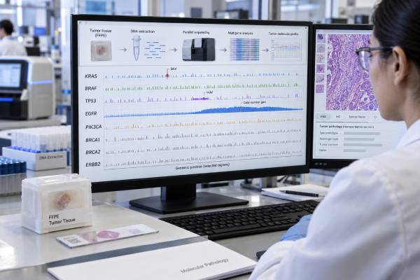
A cancer gene mutation panel analyzes many cancer-related genes at the same time to identify molecular changes that may help classify a tumor, estimate prognosis, reveal an inherited cancer risk, or guide targeted treatment. Most panels use next-generation sequencing, but the exact genes, specimen types, and variant classes covered differ greatly from one laboratory to another. A result is not simply “positive” or “negative.” Reports may include single-nucleotide variants, small insertions or deletions, copy-number gains and losses, gene fusions, splice alterations, microsatellite instability, or tumor mutational burden, depending on the assay. The most useful interpretation asks whether a finding is diagnostic, prognostic, predictive of treatment response or resistance, potentially hereditary, or currently of uncertain significance. Tumor-only testing and germline testing answer different questions, so a tumor mutation does not automatically mean a person inherited it. Molecular profiling is most valuable when its technical limits and clinical context are understood.
- A cancer gene panel tests multiple genes at once and may detect mutations, copy-number changes, and fusions depending on how the assay is designed.
- An actionable result means the finding has a recognized clinical use for diagnosis, prognosis, treatment, resistance, or trial eligibility; it does not necessarily mean an approved drug exists.
- A negative panel does not mean the tumor has no genetic changes: it means no reportable alteration was detected within that assay’s genes, variant types, and sensitivity limits.
- Tumor-only testing is not the same as hereditary testing: some findings can suggest a germline mutation and should be confirmed in blood, saliva, or another normal-tissue source.
- Tissue quality matters: low tumor content, old or damaged specimens, and low circulating tumor DNA can cause false-negative or incomplete results.
Table of Contents
- What a cancer gene panel measures
- Why molecular profiling is ordered
- How testing is performed
- How to read a panel report
- Actionable variants and treatment meaning
- Tumor versus germline findings
- Limitations and next steps
What a cancer gene panel measures
A cancer gene mutation panel is a molecular test that evaluates a selected set of genes known or suspected to play roles in cancer. Unlike a single-gene test, it can examine dozens to hundreds of genes in one assay. The term “panel” does not describe one standard test. Two panels with similar names can cover different genes, different regions of those genes, and different alteration types.
Most modern panels use next-generation sequencing, or NGS. This technology reads millions of DNA fragments in parallel and can identify many variants in a relatively small specimen. Some assays also sequence RNA, which can improve detection of gene fusions and abnormal splice events.
Depending on the panel, the report may include:
- single-nucleotide variants, such as an EGFR L858R substitution;
- small insertions and deletions, such as an EGFR exon 19 deletion;
- copy-number changes, such as ERBB2, MDM2, or CDK4 amplification;
- gene fusions, such as ALK, ROS1, RET, NTRK, or DDIT3 rearrangements;
- genomic signatures such as microsatellite instability or tumor mutational burden; and
- resistance alterations that arise after treatment.
A molecular panel is not the same as a blood tumor-marker panel. It measures DNA or RNA alterations rather than proteins such as CEA, CA-125, PSA, or AFP.
The clinical value depends on cancer type. A lung adenocarcinoma panel may prioritize EGFR, ALK, ROS1, BRAF, MET, RET, KRAS, ERBB2, and NTRK alterations. A myeloid panel focuses on a very different gene set. A broad “pan-cancer” panel may cover both, but larger is not always better if important genes or variant classes are poorly covered.
Why molecular profiling is ordered
Molecular profiling is ordered when genetic information could change diagnosis, prognosis, treatment, or trial selection. The purpose should be clear before testing because the same molecular result can carry different weight in different tumors.
Diagnosis and classification. Some cancers are defined or strongly supported by characteristic molecular abnormalities. Examples include BCR::ABL1 in chronic myeloid leukemia, FUS::DDIT3 in myxoid liposarcoma, and MDM2 amplification in well-differentiated or dedifferentiated liposarcoma. A panel can be useful when morphology is ambiguous or when several diagnoses are being considered.
Treatment selection. In advanced cancers, panels can identify predictive biomarkers that match approved targeted therapies. The relevance is cancer-specific. An alteration that is actionable in one tumor type may not have the same evidence in another.
Resistance analysis. Repeat profiling at progression can reveal acquired resistance mutations. Examples include EGFR T790M after earlier-generation EGFR inhibitors or BTK mutations after BTK-inhibitor treatment.
Prognosis. Some mutations are associated with relapse risk, survival, or transformation. In AML, for example, combinations of molecular and cytogenetic findings contribute to risk classification. In myelofibrosis, several mutations are incorporated into prognostic models.
Tumor-agnostic biomarkers. Some therapies are approved based on a molecular feature rather than the organ where the cancer began. Examples include certain NTRK fusions and high microsatellite instability. Whether a panel reliably measures such features depends on assay design.
Guidelines increasingly support NGS in selected advanced cancers where multiple actionable alterations are relevant. ESMO’s 2024 recommendations expanded routine or context-dependent use of tumor NGS across several common and rare cancers. Testing is most valuable when matched therapies or appropriate clinical trials are accessible.
How testing is performed
Most solid-tumor panels use formalin-fixed, paraffin-embedded tissue from a biopsy or surgery. A pathologist reviews the specimen to confirm tumor type and estimate tumor percentage. Areas rich in tumor may be selected for DNA or RNA extraction.
Low tumor content can reduce sensitivity. If only 5% of cells in a sample are malignant, a heterozygous mutation may be present in only a small fraction of total DNA molecules. Laboratories therefore set minimum tumor-content and quality requirements.
Blood-based circulating tumor DNA, often called liquid biopsy, is another option. It can be helpful when tissue is unavailable, unsafe to obtain, or too old. It can also capture DNA released from multiple metastatic sites. However, some tumors shed little DNA into blood, so a negative liquid biopsy may need tissue confirmation.
For blood cancers, the specimen may be peripheral blood or bone marrow rather than solid tissue.
Testing usually does not require fasting. Turnaround time varies widely, often from about one to several weeks depending on assay complexity and whether confirmatory testing is needed.
Before ordering, useful questions include:
- Which genes are covered?
- Does the assay detect fusions and copy-number changes as well as sequence variants?
- What is the minimum variant allele fraction it can reliably detect?
- Is RNA sequencing included when fusion detection matters?
- Does the panel report tumor mutational burden or microsatellite instability?
- Does the laboratory flag variants that may be germline?
These details can materially change what “negative” means.
How to read a panel report
A molecular report should be read in layers. Start with the variant itself, then the evidence level, then the clinical context.
| Report element | What it tells you | Key caution |
|---|---|---|
| Gene and variant | The exact molecular change, such as KRAS p.G12C | Different variants in the same gene can have different effects |
| Variant allele frequency | Fraction of sequencing reads carrying a DNA variant | Not the same as the percentage of tumor cells |
| Tier or evidence level | How strongly the finding is linked to diagnosis, prognosis, or therapy | Tier systems vary among laboratories |
| Copy number | Evidence of amplification or deletion | Thresholds and purity adjustments differ by assay |
| Fusion | Abnormal joining of two genes | DNA-only assays may miss some fusions better detected by RNA |
| VUS | A change whose clinical significance is not established | Should not be treated as an actionable driver without supporting evidence |
Somatic variants are often categorized by clinical significance rather than by the germline five-class system alone. Professional groups have developed standards for classifying both oncogenicity and clinical actionability. A variant can be clearly oncogenic yet not have an approved matched drug. Conversely, a biomarker can be treatment-relevant only in a specific cancer type or line of therapy.
One of the most common mistakes is assuming that every listed mutation caused the cancer. Tumors accumulate passenger alterations as well as drivers. Another is assuming that every driver is targetable. Molecular reports frequently list biologically important findings for which no standard targeted therapy exists.
Actionable variants and treatment meaning
“Actionable” means that a finding can lead to a clinical action. That action may be an approved drug, a diagnostic reclassification, a prognosis discussion, a resistance interpretation, germline referral, or trial eligibility.
The strongest treatment findings usually have evidence from regulatory approvals or major guidelines in the same cancer type. Lower-level findings may be supported only by small studies, another tumor type, preclinical data, or early clinical trials.
A molecular tumor board can help when a report contains multiple alterations, conflicting biomarkers, rare variants, or off-label treatment possibilities. These boards combine expertise from oncology, pathology, molecular genetics, pharmacy, and often clinical genetics. ESMO published recommendations in 2025 emphasizing structured interpretation and documentation of genomic-informed recommendations.
Treatment should not be chosen from a mutation in isolation. Important modifiers include:
- cancer type and stage;
- prior therapies;
- whether the alteration is a true driver;
- variant clonality;
- coexisting resistance mechanisms;
- organ function and performance status;
- drug approval and access; and
- strength of evidence in that specific clinical setting.
For example, an EGFR mutation test in lung cancer can identify variants with well-established treatment implications, while a BRAF mutation result must be interpreted differently across melanoma, colorectal, thyroid, and lung cancers.
Tumor versus germline findings
Tumor profiling examines DNA from cancer cells. Most findings are acquired, but some can represent an inherited pathogenic variant that is present in every cell of the body.
Genes that may raise this concern include BRCA1, BRCA2, PALB2, TP53, APC, mismatch-repair genes, and others. The probability that a tumor finding is germline depends on the gene, cancer type, variant allele fraction, age, family history, and whether paired normal DNA was tested.
A tumor-only report cannot reliably prove that a variant is inherited. If a potentially germline finding is clinically important, confirmatory testing should use blood, saliva, cultured skin fibroblasts, or another appropriate normal-tissue source. In hematologic malignancies, blood may not be suitable for germline confirmation because the blood itself contains the cancer clone.
The reverse is also true: a negative tumor panel does not exclude hereditary cancer susceptibility. Tumor assays may not cover all germline-relevant genes or variant types. People who meet hereditary testing criteria should receive dedicated germline evaluation regardless of whether tumor profiling is negative.
A broad germline panel is a separate clinical decision. ASCO’s 2024 guideline on germline panel selection emphasizes choosing genes based on personal and family history and the testing indication rather than assuming that tumor profiling answers the hereditary question.
Limitations and next steps
Every panel has blind spots. Some tests do not reliably detect large rearrangements, repeat expansions, epigenetic alterations, low-level subclones, or complex structural variants. Formalin can damage nucleic acids. Decalcified bone specimens may perform poorly. RNA degrades more easily than DNA. Low tumor purity reduces sensitivity. Liquid biopsy can be negative when a tumor is not shedding DNA.
A “no actionable alterations” report therefore should not be translated into “no mutations.” It means no finding met the laboratory’s reporting and actionability criteria within the technical boundaries of that assay.
Other common limitations include:
- outdated interpretation as new drugs and evidence emerge;
- differences in variant classification among laboratories;
- incidental findings with possible hereditary implications;
- uncertain significance of rare variants;
- intratumor heterogeneity; and
- evolution of the tumor after treatment.
Repeat testing can be reasonable when the original specimen was inadequate, when a new biopsy is obtained after major treatment pressure, or when the treatment landscape has changed enough that newly relevant biomarkers should be assessed. It is not automatically useful at every visit.
When reviewing a result, focus on five questions: What alteration was found? Is it technically reliable? What does it mean in this cancer type? Does it change care now? Does it suggest hereditary testing? Those questions turn a long genomic report into a clinically useful plan.
Panel size, coverage, and technical quality
A larger cancer panel is not automatically a better panel. The useful test is one that reliably covers the alteration types that matter for the cancer being evaluated. Some genes are mainly assessed for single-nucleotide variants and small insertions or deletions; others require copy-number analysis, structural-variant detection, or RNA-based fusion testing. A DNA-only assay can be excellent for many mutations yet still be less sensitive for certain fusions. Likewise, a panel that lists hundreds of genes may provide little added clinical value if key exons, introns, copy-number events, or RNA targets are not adequately covered.
Quality indicators on the report help establish whether a negative result is trustworthy. Depending on the method, these can include tumor percentage, sequencing depth, unique molecular coverage, assay limit of detection, specimen age, nucleic-acid quality, and whether internal quality controls passed. Low tumor content can dilute a true variant below the assay threshold. Decalcified bone specimens and heavily treated tissue may also produce degraded nucleic acid. In those situations, “no alteration detected” should be interpreted as a technically qualified negative result rather than proof that the tumor has no relevant genomic abnormality.
Results can change meaning over time
Cancer genomics is a moving field. A variant classified as having no established clinical significance today may become relevant when new trials, drug approvals, or disease-specific evidence appear. Conversely, an early association may become less persuasive as larger studies are completed. Molecular reports therefore have a date and a clinical context. Reinterpretation can be valuable when a patient develops advanced disease, exhausts standard options, or returns years after the original test.
This does not mean every tumor should be sequenced repeatedly. Repeat profiling is most informative when the tumor biology may have changed, when resistance mechanisms are clinically actionable, when the original assay was limited, or when a new specimen can answer a new treatment question. The goal is not to collect the largest possible list of variants. It is to identify findings that are analytically sound and clinically relevant to the patient’s current cancer and treatment setting.
A molecular tumor board can help with complex reports
Some reports contain several potentially actionable alterations, rare variants with limited evidence, or findings that point toward an off-label drug or clinical trial rather than a standard therapy. A multidisciplinary molecular tumor board can bring together oncology, pathology, genetics, pharmacy, and bioinformatics expertise to rank the evidence and decide which findings are most relevant. The purpose is not to turn every alteration into a treatment. It is to distinguish validated biomarkers from hypotheses, account for the cancer type and treatment history, and identify when a trial or confirmatory test is more appropriate than immediate therapy.
References
- Recommendations for the use of next-generation sequencing (NGS) for patients with advanced cancer in 2024: a report from the ESMO Precision Medicine Working Group 2024 (Guideline)
- Clinical practice recommendations for the use of next-generation sequencing in patients with solid cancer: a joint report from KSMO and KSP 2024 (Guideline)
- Standards for the classification of pathogenicity of somatic variants in cancer (oncogenicity): Joint recommendations of Clinical Genome Resource (ClinGen), Cancer Genomics Consortium (CGC), and Variant Interpretation for Cancer Consortium (VICC). 2022 (Guideline)
- Recommendations for reporting tissue and circulating tumour (ct)DNA next-generation sequencing results in non-small cell lung cancer 2024 (Guideline)
- ESMO Precision Oncology Working Group recommendations on the structure and quality indicators for molecular tumour boards in clinical practice 2025 (Guideline)
- Selection of Germline Genetic Testing Panels in Patients With Cancer: ASCO Guideline 2024 (Guideline)
Disclaimer
Cancer gene panel results should be interpreted by clinicians who know the tumor type, treatment history, specimen quality, and assay limitations. A molecular finding may be diagnostic, prognostic, predictive, hereditary, or uncertain, and those meanings are not interchangeable. This article is educational and does not replace individualized oncology or genetics advice.





