
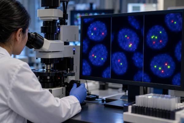
A CLL FISH panel looks for selected chromosome changes inside chronic lymphocytic leukemia cells. The most familiar targets include deletion 17p, deletion 11q, trisomy 12, and deletion 13q, although the exact panel varies by laboratory. These findings help define disease biology and can contribute to prognosis and treatment planning. The most important result for current therapy is often del(17p), because the deleted region contains TP53 and strongly overlaps with high-risk TP53-disrupted CLL. FISH cannot detect every TP53 mutation, so TP53 sequencing is usually needed as a separate test. Del(11q), trisomy 12, and isolated del(13q) also have recognized associations, but their prognostic impact has changed in the era of BTK and BCL2 inhibitors. A “positive” FISH result does not mean the disease suddenly became active or that treatment must start. Results are interpreted with TP53 mutation status, IGHV mutation status, clinical stage, symptoms, blood counts, prior therapy, and the specific treatment being considered.
- Del(17p) is the highest-priority CLL FISH abnormality for treatment planning. It suggests loss of the TP53 region and should be assessed together with TP53 mutation testing.
- Del(11q) historically carried an adverse prognosis, especially with bulky nodal disease. Its effect is less straightforward with modern targeted therapies.
- Trisomy 12 is generally considered an intermediate-risk FISH finding in older prognostic models. It has distinctive biological associations but is not a treatment indication by itself.
- Isolated del(13q) has traditionally been associated with a more favorable course. The size of the clone and deletion pattern can add nuance.
- CLL FISH can change over time. High-risk clones may emerge, so testing is commonly repeated before a new line of treatment, especially to reassess TP53 status.
Table of Contents
- What a CLL FISH Panel Tests
- How CLL FISH Testing Is Done
- del(17p) and TP53
- del(11q), Trisomy 12, and del(13q)
- How FISH Findings Affect Prognosis Today
- How FISH Guides Treatment Decisions
- Limitations, Repeat Testing, and Next Steps
What a CLL FISH Panel Tests
A CLL FISH panel uses fluorescent DNA probes to look for specific chromosome gains, losses, or rearrangements in the leukemia cells. FISH stands for fluorescence in situ hybridization. Unlike a conventional chromosome analysis, it does not require cells to divide in culture and can detect targeted abnormalities directly in interphase nuclei.
A typical CLL panel may evaluate:
- del(17p13): deletion involving the TP53 region;
- del(11q22-23): deletion involving the ATM region;
- trisomy 12: an extra copy of chromosome 12;
- del(13q14): deletion of a common CLL region on chromosome 13; and
- additional probes chosen by the laboratory or clinical question.
The exact panel matters. FISH only answers questions for the probes that were used. A “normal CLL FISH panel” means none of the targeted abnormalities were detected above the laboratory’s validated threshold; it does not mean the CLL genome is normal.
A negative result can also reflect the sample. If only a small fraction of cells in the tube are CLL cells, an abnormal clone may be harder to detect. Laboratories may use enrichment or other strategies when appropriate, but every assay has a detection limit. A later sample can also become positive because CLL evolves over time. Therefore, “not detected” should be read as “not detected in this specimen with these probes at this assay’s sensitivity,” not as a lifetime guarantee that the abnormality will never appear during the future course of CLL in that individual patient.
Historically, these abnormalities formed a widely used prognostic hierarchy. That framework was developed in the chemoimmunotherapy era and remains useful for understanding disease biology, but modern targeted therapy has changed the weight of several findings.
FISH is also only one part of CLL biomarker testing. The CLL IGHV mutation test measures a stable feature of the B-cell receptor gene and provides independent prognostic information. TP53 sequencing is required because a patient can have a clinically important TP53 mutation without a detectable 17p deletion.
How CLL FISH Testing Is Done
CLL FISH is usually performed on peripheral blood when enough circulating CLL cells are present, although bone marrow or other involved specimens can also be used. The laboratory fixes cells on a slide, applies fluorescent probes that bind selected chromosome regions, and counts signal patterns under a specialized microscope or imaging system.
For a deletion probe, cells with the expected two signals are generally normal for that target, while loss of one signal in an abnormal clone supports a deletion. For trisomy 12, three chromosome 12 signals support an extra copy. Laboratories examine a validated number of cells and apply their own cutoff to distinguish a true abnormal clone from background technical variation.
Reports often include both the abnormality and the percentage of analyzed nuclei showing it. For example, a report may state that del(17p) was detected in a certain fraction of cells. The exact percentage can matter, but it should not be interpreted without the laboratory’s threshold and the clinical setting. Small subclones can be biologically important, especially when they affect TP53, but assay sensitivity and sample composition limit what can be detected.
No fasting is needed for FISH. If peripheral blood is used, the sample is collected like a standard blood test. Bone marrow sampling is not required solely because a person has CLL, but marrow may be obtained for other reasons such as unexplained cytopenias or response assessment.
Cell enrichment can sometimes improve analysis when the malignant B-cell fraction is low. This is a laboratory decision and can affect sensitivity. When comparing serial reports, check whether the specimen type and method were similar.
How FISH differs from karyotyping and sequencing
FISH is fast and focused. Conventional chromosome analysis, often called karyotyping, surveys the chromosomes more broadly but usually requires dividing cells. In CLL, stimulated cultures can improve karyotype yield and may reveal a complex karyotype that a limited FISH panel cannot see. Next-generation sequencing answers a different question again: it can identify sequence-level variants in genes such as TP53, NOTCH1, SF3B1, and others, depending on the panel.
These methods are complementary. FISH can detect a 17p deletion, sequencing can find a TP53 point mutation, and karyotyping can show whether the leukemia carries several chromosome abnormalities at once. No single method captures the entire CLL genome. When a report says “cytogenetics negative” or “molecular testing normal,” it is worth checking which technology and targets were actually included.
del(17p) and TP53
Del(17p) is a high-risk CLL finding because it removes one copy of the chromosome region containing TP53, a critical tumor-suppressor gene. TP53 helps cells respond to DNA damage by arresting the cell cycle or triggering cell death. When TP53 function is disrupted, CLL can be more resistant to treatments that depend heavily on intact DNA-damage responses.
The key concept is that del(17p) and TP53 mutation are related but not interchangeable. FISH can show physical loss of the 17p region, but it does not sequence the remaining TP53 gene. Some patients have a TP53 mutation without del(17p), while others with del(17p) may have an additional mutation in the remaining TP53 copy.
For this reason, modern assessment generally includes both FISH for del(17p) and molecular testing for TP53 mutation before treatment. The dedicated TP53 test for CLL explains why both types of abnormality are grouped clinically as TP53 disruption.
TP53-abnormal clones can also evolve. A patient who lacked del(17p) at diagnosis may acquire it after years of disease or under treatment pressure. That is why an old negative FISH result should not automatically be used for a new treatment decision many years later.
A positive del(17p) result does not mean immediate treatment is necessary if the patient has no standard indication for therapy. It means that when treatment is needed, therapy should be selected with the high-risk biology in mind. Modern targeted agents have improved outcomes substantially compared with older chemoimmunotherapy, although TP53-disrupted CLL remains a group requiring careful treatment planning and follow-up.
del(11q), Trisomy 12, and del(13q)
Del(11q), trisomy 12, and del(13q) each describe different CLL biology, but none should be read from an old prognostic hierarchy without considering modern therapy. Their effects are also modified by other abnormalities within the same clone.
del(11q)
Deletion 11q often includes the ATM gene region. Historically, del(11q) was associated with more extensive lymph-node disease, a shorter time to progression, and less favorable outcomes with some older regimens. Targeted therapies have reduced the magnitude of that disadvantage, so del(11q) is no longer interpreted as a simple fixed prognosis label.
Trisomy 12
Trisomy 12 means the CLL clone carries an extra chromosome 12. In classic FISH risk groupings, it fell into an intermediate category. Trisomy 12 CLL can have atypical cell morphology or immunophenotypic features and is enriched for certain molecular changes, including NOTCH1 alterations in some cohorts. These associations may help explain biology but do not make every trisomy 12 case behave the same way.
del(13q)
Deletion 13q14 is the most common FISH abnormality in CLL. When it is the sole detected FISH abnormality, it has traditionally been associated with a more favorable course. That statement is an average, not a guarantee. Large deletion size, biallelic involvement, and the percentage of the clone carrying the abnormality have been studied as sources of additional heterogeneity.
| FISH finding | Major biological association | Modern interpretation |
|---|---|---|
| del(17p) | TP53 region loss | High-priority adverse marker; pair with TP53 sequencing |
| del(11q) | ATM region loss | Historically adverse; impact modified by targeted therapy |
| Trisomy 12 | Extra chromosome 12 | Intermediate historical group with distinct biology |
| Isolated del(13q) | Loss of 13q14 region | Traditionally favorable on average |
| No panel abnormality detected | No targeted FISH lesion found | Does not exclude other genomic risk factors |
How FISH Findings Affect Prognosis Today
CLL prognosis is now estimated from multiple clinical and molecular features rather than from FISH alone. The old FISH hierarchy remains a useful foundation, but targeted agents have changed outcomes enough that historical survival estimates can be misleading.
Several factors now deserve to be considered together:
- TP53 disruption by del(17p) and/or mutation;
- IGHV mutation status;
- clinical stage and disease activity;
- age and medical fitness;
- beta-2 microglobulin and other validated prognostic variables;
- complex karyotype in selected settings;
- prior treatment and resistance mutations in relapsed disease; and
- depth and duration of response to therapy.
The CLL International Prognostic Index combines several of these features for broad risk estimation, but even validated scores do not predict exactly what will happen to one individual. They are most useful for discussing relative risk, counseling, and clinical-trial design.
Del(17p)/TP53 disruption remains especially important in the targeted-therapy era. IGHV status also continues to provide meaningful outcome information, although its predictive effect differs depending on whether therapy uses chemoimmunotherapy, a BTK inhibitor, a BCL2 inhibitor, or a combination.
By contrast, del(11q) and trisomy 12 may carry less independent prognostic weight with current treatments than they did historically. A reader should be cautious when online sources quote survival or time-to-treatment numbers from studies conducted before modern targeted therapy was available.
A FISH percentage is not a direct “cancer burden” measurement either. A higher fraction of nuclei carrying an abnormality means that abnormality is present in a larger sampled clone, but it is not equivalent to total body disease volume, lymph-node size, or symptom severity.
Why a small high-risk clone can still matter
FISH reports often include the percentage of nuclei carrying del(17p) or another lesion. Higher clone fractions can correlate with different outcomes in some studies, but there is no universal percentage below which a TP53-related abnormality can simply be ignored. A small resistant subclone may expand under treatment pressure, particularly when therapy strongly favors cells with impaired TP53 function.
The practical response is not to treat the percentage as a binary danger scale. Instead, clinicians confirm the result, pair it with sensitive TP53 sequencing, review the prior treatment history, and choose therapy according to current evidence for TP53-disrupted disease. This approach is more reliable than assuming a low-percentage del(17p) finding behaves exactly like either a completely negative sample or a dominant clone.
How FISH Guides Treatment Decisions
The most actionable use of a CLL FISH panel is to identify biology that should influence treatment selection once therapy is actually indicated. Treatment is not started simply because FISH is abnormal.
Standard reasons to treat CLL include progressive marrow failure causing significant anemia or thrombocytopenia, symptomatic or progressive lymph-node or spleen enlargement, rapid disease progression under defined criteria, autoimmune complications that do not respond adequately to standard therapy, or significant disease-related symptoms. Many people with newly diagnosed CLL can be observed for years even when prognostic biomarkers suggest higher risk.
When treatment is needed, del(17p) or a TP53 mutation makes conventional chemoimmunotherapy an unattractive strategy because responses are typically less durable. Targeted approaches using BTK or BCL2 pathway inhibition have substantially improved outcomes and are selected according to current guidelines, comorbidities, drug interactions, treatment-duration preferences, and prior therapy.
Other FISH abnormalities can contribute to the overall risk discussion but usually do not dictate a single drug by themselves. The most appropriate regimen depends on the entire molecular and clinical profile.
FISH also helps explain why two patients with the same Rai or Binet stage can have different expected disease courses. Stage describes the current clinical extent of disease; FISH describes part of the leukemia’s underlying biology. Both are useful, but they answer different questions.
Limitations, Repeat Testing, and Next Steps
FISH is targeted, so its biggest limitation is that it cannot detect abnormalities outside the probe set. A normal panel therefore cannot rule out clinically relevant gene mutations, copy-number changes, structural variants, or complex chromosome patterns.
Other limitations include:
- laboratory-specific positivity thresholds;
- small abnormal clones below assay sensitivity;
- differences in specimen type or tumor-cell fraction;
- clonal evolution over time;
- inability to identify most point mutations; and
- incomplete information about whether several abnormalities are present in the same cell unless the probe design demonstrates it.
Before first treatment, patients commonly need current FISH and TP53 sequencing rather than relying only on tests performed years earlier. Reassessment is also important before later treatment lines because TP53-abnormal clones can emerge. IGHV status, by contrast, is generally stable and does not need repeated testing once accurately established.
If a FISH report is positive, useful questions include: Which abnormality was found? What fraction of nuclei carried it? Was TP53 sequencing performed? Was IGHV status tested? Is the result current enough for the treatment decision? Were any abnormalities found that suggest another B-cell neoplasm should be excluded?
No FISH abnormality is an emergency by itself. Urgency depends on symptoms and complications such as severe anemia, bleeding, infection, rapidly enlarging nodes affecting organs, or transformation to an aggressive lymphoma. A hematologist can place the chromosome result into the current disease stage and treatment context.
References
- Chronic Lymphocytic Leukemia: 2025 Update on the Epidemiology, Pathogenesis, Diagnosis, and Therapy 2025 (Review)
- Prognostic factors in chronic lymphocytic leukaemia – the old, the new and the future 2025 (Review)
- The Heterogeneity of 13q Deletions in Chronic Lymphocytic Leukemia: Diagnostic Challenges and Clinical Implications. 2025 (Review)
- Recent Advances in the Molecular Biology of Chronic Lymphocytic Leukemia: How to Define Prognosis and Guide Treatment 2024 (Review)
- Chronic Lymphocytic Leukemia: Prognostic Factors in the Era of Novel Drugs. 2024 (Review)
Disclaimer
This article is for general education and does not diagnose CLL or select treatment. FISH findings must be interpreted with TP53 mutation testing, IGHV status, clinical stage, symptoms, blood counts, treatment history, and current specialist guidance. Seek prompt medical care for severe bleeding, high fever, breathing difficulty, rapidly worsening weakness, or other acute symptoms.





