
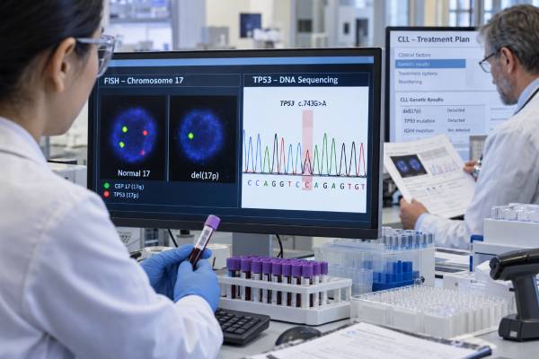
TP53 testing is one of the most important genetic evaluations in chronic lymphocytic leukemia (CLL) because TP53 disruption can change which treatments are expected to work best. In CLL, the TP53 pathway can be damaged by a mutation in the TP53 gene, deletion of chromosome 17p where TP53 is located, or both. These abnormalities are associated with more treatment-resistant disease and historically poor outcomes with chemoimmunotherapy. Modern targeted therapies have substantially improved results, but TP53-aberrant CLL remains a higher-risk group. Testing usually combines TP53 mutation sequencing with fluorescence in situ hybridization (FISH) for del(17p); one test cannot reliably replace the other. The result should be checked before starting a new line of therapy because TP53-abnormal clones can emerge or expand over time. A positive TP53 result does not mean treatment must start immediately, however. People with asymptomatic CLL are still generally observed until standard clinical criteria for active disease are met. The marker mainly guides risk assessment and treatment selection when therapy is actually needed.
- TP53 mutation and del(17p) are related but different abnormalities, so CLL workup commonly includes both sequencing and FISH.
- Either del(17p) or a pathogenic TP53 mutation can identify high-risk CLL, even when the other test is negative.
- TP53 status should be reassessed before each new treatment line, because resistant subclones can evolve during the disease course.
- A positive TP53 test does not by itself trigger treatment; therapy starts when CLL meets accepted criteria for active or symptomatic disease.
- Targeted therapies are preferred over traditional chemoimmunotherapy in TP53-aberrant CLL, with regimen choice based on efficacy, comorbidities, prior therapy, safety, and patient preference.
Table of Contents
- What TP53 Testing in CLL Measures
- del(17p) vs TP53 Mutation
- How to Read the Test Result
- Why TP53 Status Affects Treatment
- Prognosis in Modern CLL Treatment
- When TP53 Testing Should Be Repeated
- Next Steps After a Positive Result
What TP53 Testing in CLL Measures
TP53 encodes p53, a tumor-suppressor protein that responds to DNA damage and other cellular stress. Normal p53 can stop a damaged cell from dividing or trigger cell death. When CLL cells lose normal TP53 function, they can survive treatments that depend on intact DNA-damage responses.
There are two main ways the pathway is assessed in routine CLL practice.
TP53 sequencing looks for pathogenic variants within the gene. Modern laboratories usually use next-generation sequencing (NGS), which can detect smaller mutant subclones than older Sanger sequencing. Reports may include the exact variant and its variant allele frequency (VAF), which estimates how much of the sequenced DNA carries the mutation.
FISH for del(17p) looks for deletion of chromosome 17p, the region containing TP53. This test is typically included in a CLL FISH panel along with other recurrent chromosome abnormalities.
These tests answer different questions. Sequencing finds changes in the TP53 DNA sequence. FISH detects physical loss of the chromosome region. A patient can be positive by either method alone, so both are needed for a complete assessment of TP53 aberration.
Testing is usually performed on peripheral blood when enough CLL cells are circulating. In some situations, another involved sample may be required. The laboratory method and sensitivity matter, particularly for small TP53-mutated subclones.
del(17p) vs TP53 Mutation
The terms del(17p) and TP53 mutation are often grouped together because both can disrupt the p53 pathway, but they are not interchangeable.
A 17p deletion usually removes one copy of TP53 along with nearby chromosome material. The remaining TP53 copy may still be normal or may also carry a mutation. When deletion and mutation occur together, the CLL clone can lose essentially all normal TP53 function.
A TP53 mutation changes the gene sequence. It can occur without a detectable 17p deletion. This is clinically important because a normal FISH result does not rule out high-risk TP53-mutated CLL.
| FISH del(17p) | TP53 sequencing | Practical interpretation |
|---|---|---|
| Negative | Negative | No detected TP53 aberration by these assays; other risk markers still matter |
| Positive | Negative | 17p loss is present and remains a clinically important high-risk feature |
| Negative | Positive | TP53-mutated CLL is present despite no detectable 17p deletion |
| Positive | Positive | Both abnormalities are present; often reflects marked TP53 pathway disruption |
Because the tests can disagree, using “del17p test” as shorthand for all TP53 risk can miss mutation-only cases. The reverse is also true: sequencing alone does not provide the same chromosome information as FISH.
The rest of the molecular profile remains relevant. CLL IGHV mutation status provides separate prognostic information and is generally tested before first treatment. Unlike TP53 status, IGHV status is usually stable over time and does not need repeated testing before every treatment line.
How to Read the Test Result
A TP53 sequencing report should distinguish pathogenic or likely pathogenic mutations from variants of uncertain significance. A VUS does not have the same established clinical meaning as a confirmed disease-associated mutation and should not automatically place a patient into the TP53-aberrant treatment group.
VAF is another important detail. A VAF of 5%, for example, means about 5% of sequencing reads at that position contain the mutation. It does not mean exactly 5% of the CLL cells are mutated because sample composition, copy number, and other factors affect the relationship.
Historically, some laboratories used VAF thresholds partly because older methods could not detect very small clones reliably. Updated European Research Initiative on CLL recommendations no longer endorse a single universal VAF cutoff for reporting TP53 mutations. Instead, laboratories should validate their methods carefully and report clinically credible low-burden variants within the assay’s reliable range.
That change matters because small TP53-mutated subclones can expand under treatment pressure. Their exact prognostic impact can vary, but they should not be automatically ignored simply because the VAF is low.
FISH reports usually provide the percentage of analyzed cells showing del(17p) and the laboratory’s positivity threshold. That percentage should not be directly compared with sequencing VAF because the methods measure different things.
When reviewing a result, useful questions include:
- Was both del(17p) FISH and TP53 sequencing performed?
- Is the TP53 variant pathogenic or a VUS?
- What was the assay’s validated sensitivity?
- What proportion of cells carried del(17p)?
- Is this result new compared with earlier testing?
- Are other CLL markers, including IGHV, known?
A “negative” result means no reportable abnormality was detected at that time and within the assay limits. It does not guarantee that a very small resistant subclone is absent or that TP53 aberration cannot emerge later.
Why TP53 Status Affects Treatment
TP53 status became a critical predictive marker because CLL with TP53 disruption responds poorly to traditional chemotherapy-based approaches. Drugs that cause DNA damage depend partly on an intact p53 response to push damaged CLL cells toward death. When that pathway is lost, resistant cells can survive and quickly repopulate.
Targeted therapies act through different mechanisms and have transformed outcomes. Current treatment strategies generally favor agents such as covalent Bruton tyrosine kinase inhibitors (BTK inhibitors) and BCL2 inhibitor-based regimens rather than chemoimmunotherapy for TP53-aberrant disease.
Second-generation covalent BTK inhibitors such as acalabrutinib or zanubrutinib are commonly favored options for patients with del(17p) and/or TP53 mutation. Venetoclax-based fixed-duration therapy can also be considered in many patients, although experts may favor a BTK inhibitor for some TP53-aberrant cases because long-term disease control data and treatment design differ.
Treatment selection is not based on TP53 alone. Clinicians also consider:
- cardiovascular history and arrhythmia risk;
- bleeding risk and anticoagulant use;
- kidney function and tumor lysis syndrome risk;
- disease bulk and lymphocyte count;
- prior BTK or BCL2 inhibitor exposure;
- drug interactions;
- preference for continuous versus time-limited treatment; and
- access and ability to complete monitoring.
A BTK mutation test addresses a different question—acquired resistance after exposure to certain BTK inhibitors—and should not be confused with baseline TP53 testing.
Most importantly, TP53 positivity does not override the usual criteria for starting treatment. An asymptomatic patient without active-disease criteria can still be managed with watchful waiting. Starting therapy early solely because TP53 is abnormal has not become the routine standard.
Prognosis in Modern CLL Treatment
TP53-aberrant CLL remains a high-risk biological subgroup, but older survival statistics based on chemotherapy should not be applied directly to patients treated in the current targeted-therapy era.
Historically, del(17p) and TP53 mutation predicted short remissions and poor overall survival with fludarabine-, cyclophosphamide-, and antibody-based chemoimmunotherapy. BTK inhibitors and venetoclax-based regimens have substantially improved disease control, and real-world studies show better outcomes after targeted therapies became widely adopted.
The marker still matters. Even with modern drugs, patients with TP53 disruption can have shorter progression-free intervals than lower-risk groups in some studies, and relapse remains a concern. The adverse effect can be especially relevant after multiple lines of therapy or when other high-risk features coexist.
Prognosis is therefore better understood as a combination of factors rather than a single number. Age, comorbidities, disease stage, beta-2 microglobulin, IGHV status, treatment chosen, depth and duration of response, and acquired resistance mutations all affect the individual course.
TP53 result also does not predict that targeted treatment will fail. Many patients have durable responses. The practical purpose of testing is to avoid less effective treatment strategies and choose therapies that can work despite impaired p53 signaling.
For patients who receive time-limited combinations, measurable residual disease may add response information in selected settings, but routine treatment decisions should follow the regimen and guideline context. A molecular high-risk marker and an MRD result answer different clinical questions.
When TP53 Testing Should Be Repeated
TP53 status can change as CLL evolves. A small mutant clone that was absent or below the detection limit at diagnosis may expand after years of disease or after treatment. This clonal evolution is why current expert recommendations call for reassessment before each new line of therapy.
Testing at diagnosis can still be useful for prognostic counseling and baseline characterization, but the most actionable time is before treatment selection. If a patient was tested several years earlier and now meets criteria for therapy, relying on the old result can miss a newly dominant TP53-aberrant clone.
A practical testing timeline is:
- At initial workup: establish major prognostic features as appropriate, including FISH and often TP53 sequencing.
- Before first treatment: ensure current del(17p) and TP53 mutation status is available.
- Before each subsequent treatment line: repeat TP53 mutation testing and relevant FISH because the clone may have evolved.
- At suspected resistance: additional molecular tests may be ordered based on prior therapy, such as BTK or PLCG2-related resistance testing after BTK inhibitor exposure in selected situations.
The same sample type and test method do not always have to be used, but the assay should be validated for the specimen and have adequate sensitivity. A very low CLL cell fraction can make interpretation harder, and laboratories may enrich cells or select another specimen when needed.
This repeated-testing approach is different from IGHV mutation status, which generally reflects the original CLL cell of origin and remains stable.
Next Steps After a Positive Result
If TP53 mutation or del(17p) is detected, the first step is not automatically to start therapy. The clinician should determine whether the patient currently meets accepted criteria for active CLL, such as progressive marrow failure, symptomatic or progressive bulky disease, rapid lymphocyte progression, autoimmune complications that are difficult to control, or significant disease-related symptoms.
If treatment is not yet indicated, active surveillance continues. Follow-up typically includes history, physical examination, and blood counts at intervals based on disease behavior. The TP53 result remains important because it will influence treatment choice later.
If treatment is needed, targeted therapy is generally preferred. A detailed discussion should cover the tradeoffs of continuous BTK inhibition versus fixed-duration combination approaches, including cardiovascular risks, bleeding, tumor lysis monitoring, infusion requirements, duration, convenience, and expected disease control.
Patients with relapsed or multiply treated TP53-aberrant CLL may have additional options depending on previous exposure, including noncovalent BTK inhibitors, cellular therapies, or clinical trials. Sequencing of therapies is individualized and changes as new evidence and approvals emerge.
The most useful questions for the treating hematologist are straightforward: Do I have a TP53 mutation, del(17p), or both? Was the test repeated recently enough to guide this treatment decision? Does my CLL actually need treatment now? Which targeted options fit my cardiac, bleeding, kidney, and medication profile? Those questions connect the genetic result directly to safe clinical decision-making.
The percentage of cells with del(17p) can change substantially over time. A small del(17p) subclone at one point may become dominant after selective pressure from treatment, while a previously dominant clone can shrink under effective targeted therapy. This is another reason an old FISH report should not be assumed to represent the biology years later.
TP53-aberrant CLL can still be observed safely when it is not active. During watchful waiting, clinicians focus on the same clinical triggers used for other CLL: progressive marrow failure, symptomatic or rapidly enlarging nodes or spleen, constitutional symptoms, problematic autoimmune cytopenias, or sufficiently rapid lymphocyte progression. A high-risk genetic result increases the need for thoughtful planning, but it does not turn an otherwise stable CLL into an emergency.
When targeted treatment is required, the choice between continuous and time-limited therapy can be individualized. Continuous second-generation BTK inhibition avoids the tumor lysis ramp-up required with venetoclax, but it carries ongoing risks such as bleeding, hypertension, atrial arrhythmias, drug interactions, and treatment burden. Venetoclax-based fixed-duration therapy offers a planned stopping point but requires careful tumor lysis risk assessment and often an anti-CD20 antibody. In TP53-aberrant disease, expert groups differ somewhat in how strongly they favor one approach, so patient-specific tradeoffs matter.
At relapse, the next treatment depends heavily on what was used before and why it stopped. Progression on a covalent BTK inhibitor is different from stopping it for intolerance; similarly, relapse years after a fixed-duration venetoclax regimen differs from progression while actively taking venetoclax. Updated TP53/FISH testing is combined with this treatment history, and additional resistance testing may be useful in selected cases.
A practical issue is tumor-cell content. If the blood contains very few CLL cells, a mutation can be harder to detect, especially at low VAF. Laboratories may use sensitive NGS, enrich B cells, or test another involved specimen when needed. A negative result from a low-tumor-content sample should be interpreted with the assay’s stated limitations.
Patients should also avoid comparing FISH percentages from different laboratories as if they were exact measures of tumor burden. Probe sets, counting rules, positivity thresholds, and specimen composition differ. The clinically important point is whether a validated TP53 aberration is present and how it affects the treatment decision.
References
- ERIC recommendations for TP53 mutation analysis in chronic lymphocytic leukemia-2024 update 2024 (Recommendations)
- Chronic Lymphocytic Leukemia/Small Lymphocytic Lymphoma, Version 2.2024, NCCN Clinical Practice Guidelines in Oncology 2024 (Guideline)
- Chronic Lymphocytic Leukemia: 2025 Update on the Epidemiology, Pathogenesis, Diagnosis, and Therapy 2025 (Review)
- Consensus recommendations from the 2024 Lymphoma Research Foundation workshop on treatment selection and sequencing in CLL or SLL 2025 (Consensus)
- Guidelines for the diagnosis and treatment of chronic lymphocytic leukemia and small lymphocytic lymphoma from Chronic Lymphocytic Leukemia Spanish Group (GELLC) 2025 (Guideline)
- First-line treatment for CLL in the era of targeted therapy 2026 (Review)
Disclaimer
TP53 and del(17p) results guide CLL risk assessment and treatment selection but do not determine when therapy must begin. Testing methods, variant interpretation, prior treatment, comorbidities, and current disease activity all affect the clinical decision. Review the result with a hematologist before changing or starting treatment.





