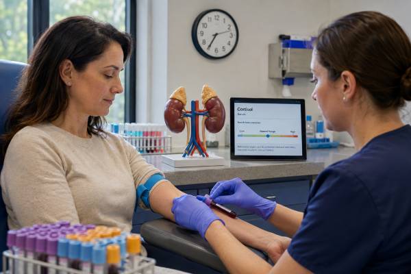
A cortisol blood test measures the adrenal glucocorticoid that supports blood pressure, metabolism, immune regulation, and the response to illness. The number is meaningful only with its collection time and clinical purpose. Cortisol normally peaks around waking, falls through the day, and reaches a low point near midnight. A morning value cannot be compared with an evening range.
Doctors use an 8–9 a.m. cortisol to screen for adrenal insufficiency, often with ACTH. Very low results raise concern, clearly adequate results reduce it, and intermediate results usually require an ACTH stimulation test. A random or morning cortisol is not a reliable screen for Cushing syndrome because healthy and affected values overlap. Suspected cortisol excess is tested with late-night salivary cortisol, 24-hour urine free cortisol, or dexamethasone suppression. Total blood cortisol is also affected by binding proteins: oral estrogen and pregnancy can raise it without raising active free cortisol, while low albumin or cortisol-binding globulin can lower it. Steroid medicines can suppress the axis or interfere with measurement, so every route and dose must be reported.
- For suspected adrenal insufficiency, collect serum cortisol between 8 and 9 a.m. and interpret it with an assay-specific cutoff.
- Low morning cortisol with high ACTH suggests primary adrenal failure; low cortisol with low or normal ACTH suggests central suppression.
- A random high cortisol can reflect pain, illness, exercise, poor sleep, or anxiety and does not diagnose Cushing syndrome.
- Oral estrogen and pregnancy raise total cortisol-binding protein; low albumin can lower total cortisol without the same change in free cortisol.
- Repeated vomiting, dehydration, confusion, fainting, or shock may indicate adrenal crisis and needs immediate treatment.
Table of Contents
- What Cortisol Does
- Why Blood Cortisol Is Tested
- Preparation and Timing
- Normal Range and Interpretation
- High Cortisol Results
- Low Cortisol Results
- Follow-Up Tests and Next Steps
What Cortisol Does
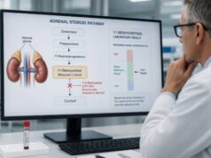
Cortisol is made in the adrenal cortex’s zona fasciculata. The hypothalamus releases corticotropin-releasing hormone, the pituitary releases adrenocorticotropic hormone (ACTH), and ACTH tells the adrenal glands to synthesize cortisol. Cortisol then feeds back to reduce further hypothalamic and pituitary signaling.
Its effects include:
- maintaining vascular responsiveness and blood pressure;
- increasing glucose availability during fasting or illness;
- mobilizing amino acids and fats for energy;
- moderating inflammation and immune activity;
- influencing memory, mood, and wakefulness; and
- helping the body respond to surgery, infection, injury, and other stress.
Secretion is pulsatile and circadian. For a person who sleeps at night, cortisol rises in the hours before waking, peaks around the early morning, and declines toward a midnight nadir. Shift work, jet lag, fragmented sleep, and critical illness can shift or flatten this rhythm.
About 80–90% of blood cortisol binds to cortisol-binding globulin, and much of the rest binds albumin. Only a small fraction is free and biologically available. Most routine blood tests measure total cortisol. Changes in binding proteins can therefore move the reported number without an equivalent change in tissue exposure.
Serum and plasma cortisol are usually reported in mcg/dL or nmol/L. A common conversion is 1 mcg/dL equals about 27.6 nmol/L. The assay may be an immunoassay or LC-MS/MS. More specific modern methods can read lower than older assays, which changes dynamic-test cutoffs.
Cortisol is chemically identical to hydrocortisone. If hydrocortisone was taken before the draw, the test measures the medicine as cortisol. Prednisolone can cross-react with some immunoassays, while dexamethasone generally has little direct cross-reactivity but suppresses ACTH.
The word “stress” covers different physiologic situations. A fever, operation, low blood sugar, major injury, or severe pain creates an immediate cortisol demand. Chronic psychological stress can alter sleep, behavior, autonomic tone, and HPA signaling, but it does not produce one diagnostic blood-cortisol signature. A single value cannot distinguish healthy adaptation from harmful chronic strain.
Cortisol also works with catecholamines and aldosterone to support circulation. During severe deficiency, blood vessels respond poorly to catecholamines and volume may fall, contributing to shock. This is why cortisol becomes lifesaving during acute illness and why emergency replacement should not wait for a perfect laboratory workup.
Why Blood Cortisol Is Tested
The test is most useful when ordered around a specific question. An early-morning sample can assess possible adrenal insufficiency in someone with fatigue plus weight loss, nausea, low blood pressure, low sodium, high potassium, low glucose, salt craving, or hyperpigmentation. Risk factors include pituitary or adrenal disease, adrenal or pituitary surgery, cranial radiation, autoimmune disease, bilateral adrenal hemorrhage, or prolonged glucocorticoid exposure.
Cortisol may be measured before and after cosyntropin to test adrenal reserve. It is also measured after dexamethasone to see whether feedback suppresses cortisol production. A midnight blood sample is occasionally used in specialized inpatient Cushing evaluation, but the stress of hospitalization and venipuncture must be controlled.
Random cortisol may be obtained in emergency or critical illness. A very low value can support adrenal insufficiency, but ordinary outpatient ranges do not define the adequate stress response. In suspected adrenal crisis, treatment must not wait for a final result.
Blood cortisol can monitor selected postoperative states, such as after pituitary surgery for Cushing disease or after adrenal surgery. Interpretation depends on the procedure, time since surgery, and steroid coverage.
It is not a general “stress level” test. Cortisol responds to physical and psychological stress, but one draw cannot quantify chronic stress, burnout, resilience, or mental health. The concept of “adrenal fatigue” has no validated cortisol pattern.
Repeated cortisol measurement is justified only when the samples form a recognized protocol. An 8 a.m. and 4 p.m. pair can demonstrate a decline, but it rarely diagnoses a disorder by itself. Serial samples after cosyntropin or dexamethasone answer a specific physiologic question. Multiple random draws can simply capture normal pulses and create apparent inconsistency.
Saliva and urine are not “better” versions of the same test; they address different aspects. Late-night saliva measures free cortisol at the expected nadir. A 24-hour urine collection integrates filtered free cortisol across a day. Blood total cortisol is best suited to morning deficiency screening and dynamic protocols. Choosing specimen by clinical question prevents unnecessary testing.
After unilateral adrenal surgery, the remaining HPA axis may be suppressed if the removed tumor had produced cortisol. Temporary replacement and repeat morning or stimulated testing may be needed. After pituitary surgery for Cushing disease, low cortisol can be an expected sign of remission but still requires clinical replacement and follow-up.
A normal morning cortisol also does not rule out Cushing syndrome. People with Cushing syndrome often retain daytime values that overlap with healthy people. Validated Cushing syndrome screening examines nighttime rhythm, daily free cortisol excretion, or dexamethasone suppression.
Preparation and Timing
For suspected adrenal insufficiency, current NICE guidance recommends an 8–9 a.m. serum cortisol for people aged 1 year and older. Record the exact draw time and wake time. Babies require pediatric endocrine interpretation rather than an adult clock-time threshold.
Fasting is not always required for cortisol alone, but follow the laboratory’s instructions. Avoid strenuous exercise immediately before a planned baseline sample and arrive early enough to rest. Do not postpone urgent testing or care to create ideal conditions.
Report all steroid exposure:
- hydrocortisone, prednisone, prednisolone, methylprednisolone, dexamethasone, or deflazacort;
- joint, spinal, intramuscular, or skin injections;
- inhalers and nebulized steroids;
- nasal sprays, eye or ear drops, creams, and rectal preparations;
- “adrenal support,” bodybuilding, and unregulated supplements; and
- the exact time and dose of the last exposure.
Do not stop a prescribed glucocorticoid without a clinician’s plan. Abrupt withdrawal after sustained exposure can cause adrenal crisis. A guideline-directed assessment during tapering may pause short-acting steroids for a defined interval, but the timing depends on the drug and patient safety.
Also report oral estrogen, hormonal contraception, pregnancy, opioids, antiseizure medicines, biotin, acute illness, pain, surgery, sleep disruption, and liver or kidney disease. Oral estrogen raises cortisol-binding globulin and total cortisol. NICE advises a planned six-week pause before some adrenal-insufficiency assessments when safe, with alternative contraception where needed; this decision must be individualized.
Pregnancy increases cortisol-binding globulin, total cortisol, and free cortisol as gestation progresses. Standard nonpregnant morning and stimulated cutoffs may misclassify the result. Persistent vomiting, weight loss, salt craving, or low pressure should still prompt assessment, especially with autoimmune disease or steroid exposure. Obstetric and endocrine teams should coordinate interpretation.
Shift workers need a plan based on their sleep-wake schedule and the available validated ranges. A person drawing blood at 8 a.m. after working all night is not in the same biologic state as someone who just woke. For urgent safety questions, clinicians may use dynamic testing or temporary coverage rather than forcing a conventional clock-time interpretation.
High-dose biotin can interfere with some immunoassay designs. Ask the laboratory how long to withhold nonessential biotin supplements. Prescription biotin for a medical condition should not be interrupted without advice. Ordinary dietary biotin is not usually the issue.
For dexamethasone suppression, take the exact dose at the instructed time and draw cortisol the next morning within the stated window. Medicines that accelerate dexamethasone metabolism can cause false nonsuppression; those that slow it can increase exposure. Measuring dexamethasone with cortisol can confirm adequate drug concentration in selected cases.
Normal Range and Interpretation
Normal ranges depend on clock time, age, assay, and laboratory. One LC-MS/MS laboratory uses an adult interval of 5–25 mcg/dL in the morning and 2–14 mcg/dL in the afternoon. Another immunoassay may use different limits. A reference interval describes a population; an adrenal-insufficiency decision cutoff answers a diagnostic question and may not match the lower reference limit.
For 8–9 a.m. adrenal-insufficiency screening, NICE 2024 guidance for modern immunoassays uses broad decision bands. A value below 150 nmol/L, about 5.4 mcg/dL, raises concern and warrants endocrine evaluation. Higher values may need repeat or stimulation depending on the band and context. Local guidance and assay calibration take priority.
Other reviews use approximate guideposts below 3 mcg/dL as strongly suggestive and above 15 mcg/dL as usually reassuring in stable outpatients. These are not interchangeable with NICE bands or with stimulated cortisol cutoffs.
| Test context | What the result addresses | Main limitation |
|---|---|---|
| 8–9 a.m. baseline | Initial adrenal-insufficiency probability | Gray zone requires dynamic testing |
| 30/60 minutes after cosyntropin | Adrenal cortisol reserve | Assay-specific peak cutoff; may miss recent central disease |
| Morning after dexamethasone | Suppressibility for cortisol-excess screening | Drug absorption and interactions matter |
| Random during severe illness | Possible inadequate stress response | Outpatient reference ranges do not apply |
An isolated afternoon value has little value for excluding adrenal insufficiency because normal cortisol has already declined. A paired morning ACTH result helps classify a low cortisol as primary or central.
Reference ranges do not account for the expected cortisol response to major illness. A result of 12 mcg/dL could be normal on a quiet morning yet inadequate during refractory septic shock. Critical illness changes albumin, cortisol-binding globulin, metabolism, and tissue sensitivity, so outpatient cutoffs should not be imported into intensive care.
Conversely, a value slightly above the morning interval after a difficult blood draw does not prove disease. The upper reference limit is not a Cushing threshold. Clinical pretest probability and a validated suppression, rhythm, or urine test determine whether further workup is warranted.
Free serum cortisol can help when binding proteins are abnormal. One laboratory uses distinct baseline, 30-minute, and 60-minute free-cortisol cutoffs after cosyntropin. Those values cannot be substituted for total cortisol or used with another method. Salivary cortisone may also be useful in selected adrenal-insufficiency protocols, but availability varies.
High Cortisol Results
A high random or morning result is commonly caused by normal physiologic activation. Acute infection, pain, surgery, low glucose, intense exercise, sleep loss, anxiety around venipuncture, depression, alcohol withdrawal, and uncontrolled diabetes can increase cortisol. Pregnancy and oral estrogen can raise total cortisol through binding protein without the same free-cortisol increase.
Endogenous Cushing syndrome is a less common cause. Features that increase suspicion include wide purple striae, easy bruising, facial plethora, progressive proximal muscle weakness, unexplained osteoporosis, growth slowing with weight gain in a child, and difficult hypertension or diabetes. Common weight gain, fatigue, or anxiety alone has low specificity.
The most common cause of Cushing syndrome overall is external glucocorticoid exposure. A person can have Cushingoid effects while blood cortisol and ACTH are low because the medicine suppresses endogenous production. Medication history is more informative than a random high-cortisol search.
To assess endogenous excess, clinicians use two late-night salivary samples, at least two 24-hour urine free cortisol collections, or a 1-mg overnight dexamethasone suppression test. A post-dexamethasone serum cortisol above the test cutoff, often 1.8 mcg/dL for sensitive screening, suggests inadequate suppression but needs confirmation and context.
After hypercortisolism is established, ACTH identifies ACTH-dependent versus adrenal causes. Imaging before biochemical confirmation can mislead because incidental pituitary and adrenal nodules are common.
Non-neoplastic hypercortisolism can occur with severe depression, alcohol use disorder, poorly controlled diabetes, eating disorders, or substantial physiologic stress. The biochemical pattern can overlap with mild Cushing syndrome. Treating or stabilizing the underlying condition and repeating independent tests can clarify the diagnosis.
Cyclic Cushing syndrome creates periods of abnormal and normal cortisol production. A normal test during a quiet phase does not exclude it when progressive discriminatory features are strong. A dated symptom record can help time repeated late-night saliva or urine testing. Random morning cortisol still has poor value for this purpose.
High cortisol from a prescribed glucocorticoid may not be visible as “high cortisol” on the assay. Synthetic steroids have different cross-reactivity, and endogenous ACTH and cortisol may be suppressed. The external exposure history, physical features, and sometimes a synthetic glucocorticoid screen are more useful.
Low Cortisol Results
Low morning cortisol can result from primary adrenal failure, pituitary or hypothalamic disease, or glucocorticoid suppression. The paired ACTH separates them:
- low cortisol with high ACTH supports primary adrenal insufficiency;
- low cortisol with low or inappropriately normal ACTH supports central adrenal insufficiency; and
- low endogenous cortisol with known external steroid exposure supports HPA-axis suppression.
Primary adrenal insufficiency may also cause high renin, low aldosterone, low sodium, high potassium, volume depletion, salt craving, and hyperpigmentation. Causes include autoimmune Addison disease, bilateral adrenal damage, infection, hemorrhage, surgery, and congenital enzyme disorders.
Central disease often preserves aldosterone and potassium. Pituitary surgery, tumor, radiation, trauma, apoplexy, and long-term glucocorticoids are common contexts. Other pituitary hormones may be abnormal.
A low total cortisol can be misleading when cortisol-binding globulin or albumin is low. Liver disease, nephrotic syndrome, and critical illness can reduce binding. Free serum cortisol may help in selected cases, using validated protocol-specific cutoffs.
Etomidate, ketoconazole, metyrapone, osilodrostat, and other steroidogenesis inhibitors can lower cortisol intentionally or as an adverse effect. Immune checkpoint inhibitors can cause primary adrenalitis or pituitary inflammation. Opioids can suppress hypothalamic-pituitary signaling. Medication indication and timing help distinguish disease from a pharmacologic effect.
New pituitary injury deserves caution. A standard 250-mcg cosyntropin test may remain normal for a period because the adrenal glands have not yet atrophied. If cortisol is low and the history is recent, the endocrinologist may provide temporary replacement, retest later, or use an integrated HPA-axis test.
Children with adrenal insufficiency may present with poor weight gain, recurrent low glucose, vomiting, or reduced stamina. Primary congenital disorders can include genital or pubertal changes. Pediatric thresholds, weight-based treatment, and the urgency of dehydration differ from adult practice.
The ACTH stimulation test is used when baseline results are uncertain. Modern assays often use peak thresholds around 14–15 mcg/dL rather than the historical 18–20 mcg/dL. A recent central deficiency can still pass because the adrenal glands have not yet atrophied.
Follow-Up Tests and Next Steps
For possible adrenal insufficiency, the next step depends on the morning value, symptoms, and risk. A very low result with compatible features may prompt immediate replacement and cause testing. An intermediate value leads to cosyntropin stimulation. High ACTH may lead to 21-hydroxylase antibodies, renin, aldosterone, electrolytes, and adrenal cause evaluation. Low ACTH may lead to other pituitary hormones and MRI.
For possible cortisol excess, repeat a recommended first-line test rather than repeating random cortisol. Agreement between different validated tests is more convincing. Treat non-neoplastic hypercortisolism drivers such as alcohol misuse, severe depression, uncontrolled diabetes, or sleep apnea and reassess when appropriate.
Keep the complete report with collection time, units, assay, cortisol range, ACTH, and medication record. A portal flag without time or context can be meaningless.
If results are repeated, use the same laboratory when practical and preserve comparable medication timing. A change from immunoassay to LC-MS/MS may lower the number without a physiologic decline. The report should identify both the analytical method and any dynamic-test decision limit.
Long-term glucocorticoid tapering is a clinical process, not a race to a normal laboratory value. Symptoms can reflect adrenal insufficiency, withdrawal syndrome, or recurrence of the treated disease. Morning cortisol is generally assessed only after the dose approaches a physiologic range and under a safe pause specified for the drug.
When replacement is confirmed, the aim is physiologic coverage rather than a high serum cortisol throughout the day. Random on-treatment cortisol values are difficult to interpret because they depend on dose timing and absorption. Symptoms, safety, growth in children, blood pressure, weight, bone health, and structured testing guide care.
Ask the clinician:
- Was the sample drawn at the right time for the question?
- Is the result total or free cortisol, and which assay was used?
- Could oral estrogen, pregnancy, low albumin, or a steroid medicine change it?
- Was ACTH drawn at the same time and handled correctly?
- Do I need cosyntropin testing, late-night salivary cortisol, urine free cortisol, or dexamethasone suppression?
- Which symptoms require immediate stress dosing or emergency care?
Adrenal crisis can cause repeated vomiting, severe weakness, abdominal pain, dehydration, confusion, low blood pressure, low glucose, and shock. Clinicians may draw cortisol and ACTH immediately if it does not delay treatment, then give hydrocortisone and fluids. A pending or “normal-range” portal result should never postpone emergency care in an unstable patient.
One cortisol value is a timed snapshot, not a complete verdict on stress or adrenal health. Matching the specimen to the diagnostic question, accounting for binding and medicines, and using dynamic testing when needed makes the result clinically useful.
References
- Cortisol, Serum 2026
- Adrenal insufficiency: identification and management 2024 (Guideline)
- Hypercortisolism (Cushing Syndrome) 2025 (Review)
- Physiology, Cortisol 2023 (Review)
- European Society of Endocrinology and Endocrine Society Joint Clinical Guideline: Diagnosis and therapy of glucocorticoid-induced adrenal insufficiency 2024 (Guideline)
Disclaimer
This article is educational and cannot diagnose cortisol excess or deficiency. Ranges and decision thresholds vary by time, assay, binding proteins, medicines, and clinical state. Suspected adrenal crisis requires immediate medical treatment.