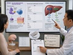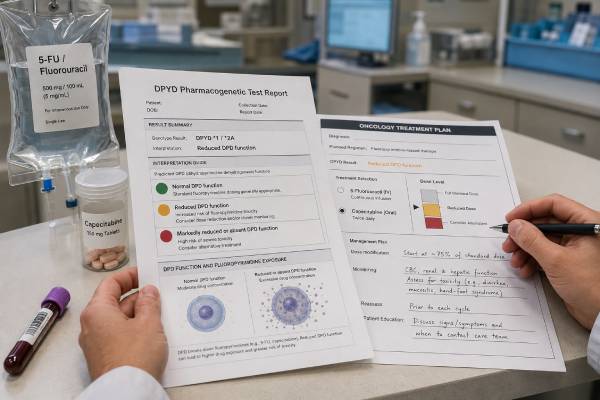
A DPYD genetic test identifies inherited variants that can reduce dihydropyrimidine dehydrogenase, or DPD, the main enzyme that breaks down fluorouracil. Reduced DPD activity can expose the body to much more 5-fluorouracil from intravenous 5-FU or oral capecitabine, causing early and sometimes life-threatening toxicity. Testing before treatment can identify many patients who need a lower starting dose or a different medicine. It cannot detect every person at risk, because targeted panels cover only selected variants and toxicity also depends on the regimen, organ function, age, other medicines, and illness. Results are translated into a DPYD activity score and predicted metabolizer status. In February 2026, updated U.S. labeling advised DPYD testing before capecitabine or 5-FU unless treatment must begin immediately. The oncology team should interpret the genotype before the first dose and continue close clinical and laboratory monitoring throughout treatment.
- A DPYD activity score of 2 usually indicates normal predicted DPD function and standard starting-dose eligibility.
- Activity scores of 1 or 1.5 indicate partial DPD deficiency and commonly support a substantially reduced starting dose.
- Activity scores of 0 or 0.5 suggest severe deficiency; fluoropyrimidines are generally avoided when an alternative exists.
- A negative targeted test lowers risk but does not rule out rare DPYD variants or non-genetic causes of severe toxicity.
- No fasting or medication pause is usually needed for a blood, saliva, or cheek-swab DNA test.
- Severe diarrhea, mouth sores, fever, confusion, weakness, or vomiting during the first treatment days requires immediate oncology contact.
Table of Contents
- How DPYD Controls Fluoropyrimidine Exposure
- When Testing Should Occur
- Variants, Activity Scores, and Phenotypes
- How Results Change Treatment
- What a Negative or Uncertain Result Means
- Testing Methods and Complementary Assays
- Recognizing and Responding to Toxicity
- Questions to Resolve Before the First Dose
How DPYD Controls Fluoropyrimidine Exposure
DPYD is the gene that provides instructions for making DPD. The enzyme begins the normal breakdown of uracil and thymine, two naturally occurring pyrimidines. It also clears most of an administered fluorouracil dose. When DPD activity is low, fluorouracil can persist at high concentrations and damage rapidly dividing normal cells as well as cancer cells.
Fluoropyrimidines include intravenous 5-FU and capecitabine, a tablet converted through several steps into 5-FU in the body. They are used in colorectal, breast, stomach, pancreatic, head and neck, and other cancers. They may be given alone, with radiation, or in combinations such as FOLFOX, FOLFIRI, CAPOX, or modified regimens. The DPYD result applies to the fluoropyrimidine component, not automatically to oxaliplatin, irinotecan, leucovorin, or other drugs in the regimen.
A person inherits one DPYD copy from each biological parent. Some variants have normal function, some reduce function, and some produce little or no functional enzyme. The combined effect of both copies is expressed as an activity score. This score estimates inherited DPD capacity before the patient receives chemotherapy.
DPD deficiency exists on a spectrum. Complete or near-complete deficiency is rare but creates extreme danger from conventional doses. Partial deficiency is more common and can still lead to severe diarrhea, mucositis, neutropenia, infection, neurotoxicity, dehydration, and death. Many carriers have no symptoms in everyday life because the problem becomes apparent only when the fluoropyrimidine pathway is heavily challenged.
The test is a safety tool, not a cancer test. It does not describe tumor mutations or predict whether a cancer will shrink. A separate tumor molecular profile may guide targeted treatment, while DPYD testing examines inherited DNA to help select a safer fluoropyrimidine dose.
When Testing Should Occur
Testing is most useful before the first exposure to systemic 5-FU or capecitabine. The result can then shape the starting dose rather than explain a crisis after it occurs. For a planned course, the oncology team should order the test early enough that the laboratory report is available before treatment authorization, pharmacy preparation, and patient education are complete.
Current U.S. labeling advises testing for DPYD variants before starting capecitabine or 5-FU unless immediate treatment is necessary. European regulators have recommended pre-treatment testing for several years. Local practice still varies, and a clinician may combine genotyping with a DPD phenotype test depending on national policy and laboratory access.
Testing is also appropriate after unusually early or severe fluoropyrimidine toxicity, even if treatment has already stopped. A result may help explain the event, guide future treatment, and alert relatives that an inherited variant could be present. However, clinicians must treat the toxicity immediately rather than wait for genetic confirmation.
A previous history of tolerating 5-FU or capecitabine does not always make testing unnecessary. Tolerance depends on the dose, schedule, combination therapy, organ function, and duration. A person who tolerated a short radiosensitizing course may react differently to full-dose systemic therapy. Conversely, a variant carrier may have tolerated a reduced or interrupted regimen by chance.
The urgency exception needs careful handling. Some cancers require treatment before a result can return. The team may choose a non-fluoropyrimidine option, use an expedited test, begin with an individualized conservative plan, or proceed under close monitoring based on the clinical stakes. The decision should be documented rather than treating an absent result as normal.
Testing is usually germline, meaning the result is intended to represent inherited DNA. Blood is acceptable for most patients. After an allogeneic stem-cell transplant, blood can contain donor DNA; the laboratory may need a cheek sample, cultured skin cells, or a pretransplant specimen to determine the recipient’s genotype.
Variants, Activity Scores, and Phenotypes
Clinical panels often include four well-established variants: c.1905+1G>A, also called DPYD2A; c.1679T>G, associated with DPYD13; c.2846A>T; and the HapB3-associated c.1236G>A or linked intronic c.1129-5923C>G variant. Laboratories may also include c.557A>G and other variants important in populations that were underrepresented in early studies.
Each allele receives a function value. A normal-function allele contributes 1. A decreased-function allele commonly contributes 0.5, while a no-function allele contributes 0. Adding the two alleles produces the activity score.
| Activity score | Predicted phenotype | General treatment implication |
|---|---|---|
| 2 | Normal metabolizer | Use the usual starting dose if otherwise appropriate, with standard toxicity monitoring. |
| 1.5 | Intermediate metabolizer | Begin with a major dose reduction and titrate according to tolerance and response. |
| 1 | Intermediate metabolizer | Begin with a major dose reduction; close follow-up is essential. |
| 0.5 | Poor metabolizer | Avoid fluoropyrimidines when possible; any use requires specialist dosing and early monitoring. |
| 0 | Poor metabolizer | Avoid 5-FU and capecitabine because no conventional dose has been proven safe. |
A heterozygous result means one altered allele and one other allele, but the consequence depends on the altered allele’s function. One no-function allele usually produces an activity score of 1. One decreased-function allele usually produces 1.5. Two altered alleles can produce 0, 0.5, or 1 depending on their combination and whether they are on opposite chromosomes.
Phasing can matter. Two variants detected in the same person may be located on one chromosome together or on separate chromosomes. The clinical effect can differ. HapB3 also illustrates why exact variant nomenclature matters: some assays test a tagging variant, while others test the linked causal candidate. The report should explain how the haplotype was assigned.
“Wild type” or “no clinically significant variant detected” does not mean zero toxicity risk. It means the assay did not identify one of the variants it was designed to detect. The tested-variant list is therefore an essential part of the result.
How Results Change Treatment
A DPYD result guides the starting exposure, followed by dose titration. It should not lock a patient permanently at the initial reduced dose if treatment is tolerated and the oncology team judges that cautious escalation is appropriate.
For an activity score of 2, genotype-based guidance generally supports the standard starting dose. The patient still needs routine blood counts, organ-function tests, symptom review, and regimen-specific monitoring because most fluoropyrimidine toxicity occurs in people without a tested high-risk variant.
For activity scores of 1 or 1.5, CPIC-based guidance supports reducing the starting dose by about 50%, then adjusting based on toxicity and effectiveness. The exact calculation depends on the regimen and variant. Some centers use therapeutic drug monitoring for continuous-infusion 5-FU to refine exposure. Capecitabine does not have the same routine concentration-monitoring approach, so clinical assessment is especially important.
A 50% start does not mean the treatment is expected to be half as effective. Variant carriers can experience exposure similar to a normal metabolizer at a full dose because they clear the drug more slowly. If the reduced dose is tolerated, stepwise increases may help avoid chronic underexposure. Escalation should occur only under the oncology protocol; severe early toxicity argues against it.
For an activity score of 0, fluoropyrimidines are generally avoided. For 0.5, avoidance is also preferred because a safe dose is difficult to establish. When no effective alternative exists, a highly reduced dose with very early pharmacokinetic monitoring may be considered by specialists, but this is not a routine patient-directed calculation.
The treatment intent affects risk tolerance. In curative therapy, maintaining anticancer intensity matters, but preventing fatal toxicity is equally important. In palliative therapy, quality of life and alternatives may lead to a different choice. The genotype informs the discussion without determining it alone.
Schedule changes do not erase genetic risk. Bolus 5-FU tends to produce more bone-marrow suppression, while continuous infusion more often produces mucositis and hand-foot effects, but a deficient patient can develop severe toxicity with either approach. Capecitabine creates prolonged oral exposure and depends on correct daily dosing, kidney function, and rapid reporting of symptoms. Switching among these formulations without accounting for DPYD status is not a reliable safety strategy.
Dose intensity should be reviewed over the entire course rather than judged by the first cycle alone. A reduced first dose may be increased in small steps after blood counts, gastrointestinal symptoms, skin effects, and overall recovery are evaluated. If therapeutic drug monitoring is available for infused 5-FU, measured exposure can support escalation or further reduction. Every change should be recorded so a future clinician can distinguish the genotype-based starting dose from later tolerance-based adjustments.
A result can also affect future care. The same DPYD status should be considered whenever systemic fluorouracil or capecitabine is proposed, even for a different cancer years later. Topical fluorouracil has much lower systemic exposure, but the prescriber should still know about severe DPD deficiency, extensive application, damaged skin, or prior reactions.
What a Negative or Uncertain Result Means
Targeted genotyping has high specificity for the variants it detects but incomplete sensitivity for DPD deficiency. DPYD is a large and variable gene. Rare harmful variants, regulatory changes, and combinations not included on a standard panel can reduce function. Variant frequencies also differ across ancestry groups, so a panel optimized for one population may miss relevant variants in another.
A normal genotype therefore cannot be used to dismiss severe symptoms. Early-onset, unusual, or rapidly progressive toxicity should be managed as a clinical emergency regardless of the report. Further testing may include broader DPYD sequencing, deletion and duplication analysis, plasma uracil measurement, or a specialized DPD activity assay.
A variant of uncertain significance is not automatically a reason to avoid treatment, and it is not equivalent to a normal result. The laboratory may lack enough functional or clinical evidence to assign an activity value. The oncology team can consult a pharmacogenomics specialist, review phenotype testing, and choose a cautious plan that reflects treatment urgency.
Some sequencing reports identify a rare variant predicted to be damaging by computer models. Prediction alone does not establish how much DPD activity is lost. Functional evidence, population data, variant location, and the patient’s phenotype all matter. Large dose changes should not rely on a prediction score without expert interpretation.
A negative result also says nothing about non-genetic risk factors. Kidney impairment can increase capecitabine and metabolite exposure. Liver dysfunction, low performance status, older age, low body reserves, combination chemotherapy, radiation, infection, and drug interactions can amplify toxicity. Dosing must account for the complete regimen and baseline health.
Testing Methods and Complementary Assays
DPYD genotyping can be performed from blood, saliva, or a cheek swab. Because the assay reads DNA, fasting is not required and chemotherapy medicines do not need to be held for collection. A poor-quality cheek sample can fail, so patients should follow instructions about eating, drinking, smoking, and mouth care before swabbing.
Targeted genotyping is fast and focuses on well-validated variants. It is practical for pre-treatment programs but leaves rare variants undetected. Sequencing examines more of the gene and may improve detection across diverse populations, although it also produces more uncertain findings. Some laboratories combine sequencing with copy-number analysis.
Phenotyping estimates actual DPD function. Plasma uracil concentration is used in some countries because DPD deficiency can cause uracil to accumulate. The dihydrouracil-to-uracil ratio and peripheral blood mononuclear-cell enzyme assays are other approaches. Preanalytic handling, illness, kidney function, and laboratory method can affect phenotype results, so thresholds are not perfectly interchangeable.
Genotype and phenotype answer related but different questions. Genotype is stable and specific for recognized inherited variants. Phenotyping may capture reduced activity from rare genetic or non-genetic causes, but it can vary with current physiology and sample handling. Combining them can increase detection, particularly when local policy supports both.
For continuous-infusion 5-FU, therapeutic drug monitoring measures drug exposure after treatment starts. A blood sample is used to estimate area under the concentration-time curve, and later doses can be adjusted toward a target exposure. This does not replace pre-treatment DPYD screening because a severely deficient patient can become critically ill before routine adjustment.
Laboratory turnaround should fit the oncology schedule. The report needs the exact variants tested, genotype, activity score, phenotype, dosing interpretation, limitations, and date or version of the guidance. A chemotherapy pharmacogenetic panel may include DPYD with UGT1A1, TPMT, or NUDT15, but only the gene relevant to each drug should direct that drug’s dosing.
Recognizing and Responding to Toxicity
DPD-related toxicity often appears unusually early, severe, or prolonged. Warning signs include frequent watery diarrhea, inability to drink, repeated vomiting, painful mouth ulcers, difficulty swallowing, fever, marked weakness, confusion, unsteady walking, severe hand-foot symptoms, chest pain, and rapidly worsening fatigue. Neutropenia may not be visible, which is why fever during chemotherapy requires urgent assessment.
Patients should receive written instructions and a 24-hour oncology contact before the first dose. “Wait until the next appointment” is unsafe for severe symptoms. Capecitabine should be held according to the treatment team’s instructions when significant toxicity begins; patients should not restart it on their own after symptoms improve.
Acute early toxicity or overdose may be treated with uridine triacetate, an antidote that is most effective when started as soon as possible and generally within 96 hours of the end of fluorouracil or capecitabine exposure. Emergency clinicians need the drug name, dose, timing, and treatment center contact. Genetic results should never delay antidote assessment.
Hospital care may include intravenous fluids, electrolyte correction, infection treatment, blood counts, cardiac and neurologic evaluation, nutrition support, and intensive monitoring. Severe toxicity can continue to worsen after the drug has been stopped because cellular injury has already occurred.
After recovery, the oncology team should reassess the indication, genotype coverage, possible phenotype testing, organ function, and alternatives. Rechallenge is not automatic. When treatment resumes, it may use a lower dose, a different schedule, therapeutic drug monitoring, or a non-fluoropyrimidine regimen.
Questions to Resolve Before the First Dose
The patient and oncology team should confirm that the result belongs to the correct person, that the assay covered variants appropriate to the patient’s ancestry, and that the activity score matches the listed alleles. A bare “positive” result is not enough for dosing.
The treatment order should state how the genotype changed the dose, whether later escalation is planned, and what monitoring will be used. For combination regimens, the patient should know which component was reduced. This prevents the mistaken belief that every chemotherapy drug was lowered.
Baseline review should include blood counts, kidney and liver function, nutrition, bowel symptoms, infection, prior fluoropyrimidine exposure, and all medicines. The team should discuss treatment intent and reasonable alternatives if the result indicates severe deficiency.
Patients should know the first-day contact plan, after-hours number, symptoms that trigger an immediate call, and whether to stop oral capecitabine while awaiting instructions. They should not use anti-diarrheal medicines to conceal rapidly worsening symptoms without contacting the oncology service.
Store the genotype and activity score in the permanent record, not only in a scanned report. Pharmacists, oncologists, radiation oncologists, and future cancer teams may need it. Family members should not assume they carry the same variant, but they can discuss targeted testing with a qualified clinician if a pathogenic allele is found.
DPYD testing reduces preventable risk when it is completed before exposure, translated into an individualized starting plan, and paired with rapid response to symptoms. It cannot make fluoropyrimidine therapy risk free, but it can prevent a predictable inherited vulnerability from being treated as an unexpected complication.
References
- Safety labeling update for capecitabine and fluorouracil (5-FU) on risks associated with dihydropyrimidine dehydrogenase (DPD) deficiency 2026 (Official Safety Update)
- A Guide for Implementing DPYD Genotyping for Systemic Fluoropyrimidines into Clinical Practice 2025 (Implementation Guide)
- DPYD Genotyping Recommendations: A Joint Consensus Recommendation of the Association for Molecular Pathology, American College of Medical Genetics and Genomics, Clinical Pharmacogenetics Implementation Consortium, College of American Pathologists, Dutch Pharmacogenetics Working Group of the Royal Dutch Pharmacists Association, European Society for Pharmacogenomics and Personalized Therapy, and Pharmacogenomics Knowledgebase 2024 (Consensus Statement)
- Dihydropyrimidine dehydrogenase gene variants for predicting grade 4-5 fluoropyrimidine-induced toxicity: FUSAFE individual patient data meta-analysis 2024 (Meta-Analysis)
- Pharmacogenetic-guided dosing for fluoropyrimidine and irinotecan chemotherapy: A prospective implementation study 2024 (Prospective Study)
- Clinical Pharmacogenetics Implementation Consortium (CPIC) Guideline for Dihydropyrimidine Dehydrogenase Genotype and Fluoropyrimidine Dosing: 2017 Update 2018 (Guideline)
Disclaimer
DPYD results and fluoropyrimidine doses require interpretation by an oncology team familiar with the full treatment regimen. Do not begin, stop, restart, or alter 5-FU or capecitabine without direct instructions. Early severe diarrhea, mouth sores, fever, vomiting, confusion, chest pain, or profound weakness during treatment requires urgent medical assessment.