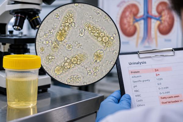
Fatty casts are urinary casts that contain lipid droplets, lipid-filled cells, or both. They form inside kidney tubules and are classically associated with heavy protein leakage and nephrotic syndrome. In nephrotic states, the damaged glomerular filter allows large amounts of protein and lipoprotein material to enter the urine. Tubular cells reabsorb some of that lipid, become packed with fat, and may later shed into the urine as oval fat bodies. When lipid droplets or lipid-filled cells become trapped in a cast matrix, a fatty cast forms. Under polarized light, cholesterol-containing droplets can produce a characteristic Maltese-cross appearance. Fatty casts are highly useful clues, but they do not identify the exact kidney disease and are not present in every patient with nephrotic syndrome. Their significance depends on urine protein, serum albumin, edema, blood lipids, kidney function, and the rest of the sediment.
- Fatty casts usually point to lipiduria: lipid in urine is most strongly associated with heavy glomerular protein loss and nephrotic syndrome.
- Oval fat bodies are closely related: they are lipid-filled tubular cells or macrophage-like cells that may accompany free fat droplets and fatty casts.
- A Maltese cross is a microscopy clue: cholesterol esters can show a bright cross pattern under polarized light, but starch and other contaminants can mimic it.
- Fatty casts do not name the kidney disease: minimal change disease, FSGS, membranous nephropathy, diabetic kidney disease, amyloidosis, and other proteinuric disorders can produce lipiduria.
- Follow protein and kidney function: urine protein, serum albumin, edema, creatinine, and the underlying cause matter more for management than the cast count alone.
Table of Contents
- What Fatty Casts Are
- Why Fat Appears in Urine
- What Fatty Casts Look Like Under the Microscope
- Fatty Casts and Nephrotic Syndrome
- Other Causes and Important Mimics
- How a Fatty-Cast Result Is Interpreted
- Fatty Casts vs Oval Fat Bodies and Cholesterol Crystals
- Common Result Patterns and What They Suggest
- Follow-Up, Treatment, and Monitoring
- When the Finding Needs Urgent Evaluation
What Fatty Casts Are
A cast is a cylindrical structure formed inside a renal tubule, usually on a matrix of uromodulin. Cast contents provide clues about what is happening inside the nephron. Red blood cell casts point toward glomerular bleeding, white blood cell casts toward interstitial inflammation or kidney infection, and epithelial or granular casts toward tubular injury. Fatty casts indicate that lipid-rich material has entered the tubular system.
The “fat” may appear as free droplets, as lipid inside cells, or as lipid incorporated directly into a cast. A lipid-filled renal tubular epithelial cell is often called an oval fat body. These cells can be round or oval and packed with refractile droplets.
Fatty casts and oval fat bodies belong to a broader finding called lipiduria. Lipiduria is most familiar in nephrotic syndrome, where glomerular permeability is markedly increased. A detailed urine sediment test can show the combination of lipid droplets, oval fat bodies, fatty casts, and sometimes cholesterol crystals.
There is no standard “normal range” for fatty casts. They are considered pathologic when confidently identified, but laboratories may report them as present/absent, rare/few/many, or per low-power field. The clinical importance depends on the degree of proteinuria and the underlying disease.
Why Fat Appears in Urine
The normal glomerular filtration barrier restricts large proteins and lipoprotein particles. When that barrier becomes highly permeable, albumin and other proteins spill into the filtrate. Lipoprotein-associated lipids can enter as well.
Proximal tubular epithelial cells try to reclaim filtered proteins and lipids. With heavy protein leakage, these cells can accumulate lipid droplets in lysosomes and other intracellular compartments. Some lipid-loaded cells then detach into the tubule. Once in the urine sediment, they appear as oval fat bodies.
If these cells or free lipid droplets become embedded in a uromodulin matrix, they create fatty casts. The mechanism therefore connects glomerular leakage to downstream tubular handling: the initial problem is often at the kidney filter, while the visible cast forms in the tubule.
This explains why fatty casts often coexist with severe proteinuria rather than occurring as an isolated sediment finding. It also explains why lipiduria can decrease when proteinuria improves, even if blood cholesterol does not normalize immediately.
Nephrotic syndrome also commonly causes hyperlipidemia because the liver increases lipoprotein production in response to low oncotic pressure and altered protein metabolism. However, high blood cholesterol by itself does not usually create fatty casts if the glomerular barrier is intact.
What Fatty Casts Look Like Under the Microscope
Under bright-field or phase-contrast microscopy, fatty casts contain round, highly refractile lipid droplets within a cylindrical cast outline. Some casts include intact oval fat bodies; others show only dispersed droplets.
Polarized light adds an important clue. Cholesterol and cholesterol esters can show Maltese crosses—bright, symmetric cross-like patterns within droplets. This appearance is memorable but not completely specific. Starch granules from glove powder or external contamination can also polarize in a cross pattern, often with a different shape or asymmetry.
Sudan stains can highlight lipid. Sudan III or IV stains neutral fat and cholesterol-containing material, while other stains can help distinguish lipid composition. In routine clinical practice, phase-contrast plus polarized microscopy is often enough to recognize lipiduria.
The main related findings are:
- free lipid droplets: isolated or clustered round droplets;
- oval fat bodies: lipid-filled cells, often renal tubular in origin;
- fatty casts: lipid droplets or lipid-filled cells trapped in a cast matrix;
- cholesterol crystals: large flat plates with notched corners, which are distinct from droplets.
Manual microscopy has an advantage here because automated analyzers may not classify lipids and pathological casts reliably. A 2023 study comparing urinary sediment with kidney biopsy emphasized that manual microscopy can detect lipids and cast composition that automated systems may miss.
Fatty Casts and Nephrotic Syndrome
Nephrotic syndrome is the classic clinical setting for fatty casts. In adults, it is generally characterized by heavy proteinuria, low serum albumin, edema, and often hyperlipidemia. A commonly used threshold for nephrotic-range proteinuria is at least 3.5 g of protein per 24 hours, although spot urine protein-to-creatinine measurements are frequently used instead of a full 24-hour collection.
The underlying causes vary. Important examples include minimal change disease, focal segmental glomerulosclerosis, membranous nephropathy, diabetic kidney disease, amyloidosis, and several immune or systemic disorders. Fatty casts cannot distinguish among them.
A person with nephrotic syndrome may have:
- foamy urine from heavy protein;
- swelling of the legs, eyelids, or abdomen;
- rapid weight gain from fluid retention;
- low serum albumin;
- elevated cholesterol or triglycerides;
- increased risk of blood clots and infection in some settings;
- lipiduria with oval fat bodies and fatty casts.
Not every patient has visible fatty casts. In one modern review, lipiduria was present in only a portion of nephrotic cases examined with phase-contrast microscopy. So the absence of fatty casts does not rule out nephrotic syndrome.
Likewise, the presence of fatty casts does not tell you whether proteinuria is transient or persistent. A quantitative urine protein or albumin measurement is still needed. A nephrotic syndrome urine test panel combines protein measurements with sediment and other kidney findings, while the fatty cast remains a microscopy clue rather than a quantitative measure of protein loss.
Other Causes and Important Mimics
Although nephrotic syndrome is the strongest association, lipiduria can occur in other settings. Reported examples include Fabry disease, autosomal dominant polycystic kidney disease, chyluria, severe proteinuric states that do not meet every formal criterion for nephrotic syndrome, and occasional contamination from oils or lipid-containing products.
Fabry disease can produce characteristic lipid-containing urinary cells and particles because glycosphingolipids accumulate in kidney cells. This is a different mechanism from filtered lipoprotein in ordinary nephrotic syndrome, and diagnosis relies on the broader clinical and genetic context.
Chyluria occurs when lymphatic fluid enters the urinary tract and can produce milky urine with lipid. It is uncommon in many regions and has a different diagnostic pathway.
The biggest practical problem is misidentification. Yeast, RBCs, air bubbles, starch, and some crystals can resemble fat droplets under ordinary light. Polarization, cell morphology, and a true cast outline help distinguish them.
Another related but separate finding is the vacuolar cast. Recent work describes vacuolar casts in advanced proteinuric glomerular disease. These contain clear nonpolarizable vesicles and should not be automatically labeled as fatty casts. Their recognition reinforces how detailed urine microscopy can reveal different consequences of severe proteinuric kidney disease.
How a Fatty-Cast Result Is Interpreted
A useful interpretation asks whether the rest of the data support heavy glomerular protein loss.
First, clinicians quantify urine protein. A spot urine protein-to-creatinine ratio or albumin-to-creatinine ratio is usually convenient. A 24-hour urine collection may be used in selected situations. Dipstick protein alone is not precise enough to quantify heavy proteinuria.
Second, serum albumin, creatinine, estimated GFR, and lipids are reviewed. Low albumin plus edema and heavy proteinuria creates the classic nephrotic picture. Kidney function may be normal, reduced, or acutely worsened depending on the cause.
Third, the rest of the sediment is examined. A mostly bland sediment with fatty casts and oval fat bodies is compatible with a noninflammatory nephrotic pattern. Dysmorphic RBCs or RBC casts suggest a more nephritic or mixed glomerular process. Granular or epithelial casts may indicate superimposed tubular injury.
A broader urine microscopy evaluation for kidney disease is valuable because nephrotic and nephritic features can overlap. Some glomerular diseases cause both heavy proteinuria and active hematuria.
The cast count itself is not used to stage nephrotic syndrome. Protein amount, albumin level, edema, complications, kidney function, and the specific diagnosis drive management.
Fatty Casts vs Oval Fat Bodies and Cholesterol Crystals
Several lipid findings can appear together, but they are not identical. Distinguishing them helps explain what the laboratory actually saw.
A fatty cast has a recognizable cylindrical cast matrix. Lipid droplets, lipid-filled cells, or both sit within that matrix. Because casts form in renal tubules, a true fatty cast confirms that lipid-rich material was present within the nephron.
An oval fat body is a single lipid-laden cell rather than a cast. It often represents a tubular epithelial cell that reabsorbed filtered lipoprotein and then sloughed into the urine. Under polarized light, cholesterol-rich droplets within the cell may show multiple Maltese crosses. Oval fat bodies can be numerous even when fatty casts are sparse.
Free lipid droplets are round refractile particles not enclosed in a cell or cast. They may cluster, and their small size can make them easy to confuse with RBCs, yeast, or artifacts. Polarization and phase contrast improve identification.
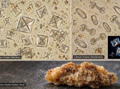
Cholesterol crystals are structurally different. They are flat, rectangular plates with one or more notched corners. They may appear in lipid-rich urine, but they do not have the rounded droplet appearance of neutral fat and do not by themselves make a fatty cast.
The phrase Maltese cross also needs context. It is a pattern seen when some lipid droplets are viewed under polarized light; it is not a diagnosis of nephrotic syndrome. Starch granules can produce a cross-like birefringent pattern too. A trained observer looks at the symmetry, particle outline, location within a cell or cast, and the rest of the sediment.
These distinctions are clinically useful because a combined pattern of heavy proteinuria, oval fat bodies, and fatty casts strongly supports glomerular protein leakage. An isolated cross-shaped particle in a specimen with no protein may instead be contamination or another particle type. Manual review is especially helpful when the automated report says only “lipids” or “unclassified particles.”
Common Result Patterns and What They Suggest
Fatty casts become easier to interpret when they are paired with urine protein and other sediment findings.
Fatty casts + heavy proteinuria + low serum albumin is the classic nephrotic pattern. If edema and hyperlipidemia are also present, the laboratory picture strongly supports nephrotic syndrome, although the specific cause still requires disease-focused evaluation.
Oval fat bodies without obvious fatty casts can still indicate lipiduria. Casts may be sparse or missed in a small sample, while lipid-loaded cells are easier to find. The absence of a visible cast does not negate significant protein leakage.
Fatty casts + dysmorphic RBCs or RBC casts suggests a mixed nephrotic-nephritic picture. Some glomerular diseases cause both heavy protein loss and active inflammation. In that setting, clinicians pay close attention to creatinine, blood pressure, complement levels, serologies, and the possibility of biopsy.
Fatty casts + granular or epithelial casts may indicate superimposed tubular injury. Heavy protein traffic itself can stress tubules, and severe glomerular disease can coexist with ischemia, medication toxicity, or other AKI triggers.
Maltese crosses with little or no proteinuria should prompt caution. Starch contamination, other birefringent particles, or a non-nephrotic source of lipid may be more plausible than classic nephrotic lipiduria. Confirming that the crosses are inside oval fat bodies or a true cast can prevent overinterpretation.
These patterns are why quantitative protein testing is more useful than trying to grade nephrotic severity by the number of fatty casts. A urine protein-creatinine ratio or another appropriate quantitative protein measure gives the clinician a reproducible way to follow disease activity over time.
Follow-Up, Treatment, and Monitoring
Treatment targets the underlying cause of protein leakage. That may involve supportive therapy alone, disease-specific immunosuppression, treatment of diabetes or systemic disease, or targeted therapy for a particular glomerulopathy.
General management often includes control of blood pressure, reduction of proteinuria, management of edema, and attention to blood lipids and thrombosis risk. The exact plan depends on kidney function, blood pressure, age, cause, and comorbidities. Diuretics may relieve edema but require careful monitoring because excessive fluid removal can reduce kidney perfusion.
Follow-up focuses on measurable disease markers. Urine protein should decline if therapy is effective. Serum albumin may rise, edema may improve, and lipiduria may lessen. Serum creatinine is monitored for kidney function, while potassium and other electrolytes may be important depending on medications.
A kidney biopsy is often needed in adults with unexplained nephrotic syndrome unless the clinical context or a disease-specific biomarker makes the diagnosis sufficiently clear. Microscopy helps define the urine pattern, but a fatty cast does not replace tissue diagnosis when the underlying disease remains uncertain.
Because nephrotic syndrome can increase clotting risk, sudden leg swelling, chest pain, shortness of breath, or neurologic symptoms require urgent evaluation. Infection risk can also be increased, especially in severe disease or during immunosuppressive treatment.
When the Finding Needs Urgent Evaluation
A fatty cast on an otherwise stable outpatient urinalysis is usually not an emergency, but it should not be ignored if proteinuria is substantial or new. Timely follow-up is appropriate for new edema, rapidly increasing weight from fluid retention, very foamy urine with heavy protein, or a low serum albumin.
Urgent medical assessment is warranted for shortness of breath, chest pain, one-sided leg swelling, markedly reduced urine output, severe weakness, confusion, very high blood pressure, or a rapid rise in creatinine. These symptoms can signal complications of nephrotic syndrome, acute kidney injury, thrombosis, or severe fluid overload.
The key message is that fatty casts are a visual marker of lipiduria, usually reflecting heavy glomerular protein leakage. They are most useful when they prompt quantitative protein testing and a focused search for the kidney disease causing that leakage.
References
- Diagnostic Utility of Urine Microscopy in Kidney Diseases 2024 (Review)
- Are oval fat bodies useful diagnostic clues in kidney disease? 2025
- An Updated Comprehensive Review on Diseases Associated with Nephrotic Syndromes 2024 (Review)
- Urinary Vacuolar Casts Are a Unique Type of Casts in Advanced Proteinuric Glomerulopathies 2024
- Urinary Sediment Microscopy and Correlations with Kidney Biopsy: Red Flags Not To Be Missed 2023
- Urine Microscopy: The Enduring Value of a Classic Test. 2025
Disclaimer
Fatty casts and lipiduria can support a diagnosis of heavy glomerular protein loss, but they do not identify the exact kidney disease or determine severity by themselves. Interpretation should include quantitative urine protein, serum albumin, kidney function, edema, blood pressure, and the overall urine sediment. Seek urgent care for severe shortness of breath, chest pain, one-sided leg swelling, markedly reduced urine, or other signs of serious illness.