
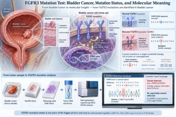
An FGFR3 mutation test looks for activating changes in the FGFR3 gene, an important molecular driver in many urothelial bladder cancers. FGFR3 alterations are especially common in low-grade, non-muscle-invasive bladder tumors, but a clinically important subset of locally advanced and metastatic urothelial cancers also carries actionable FGFR3 mutations or fusions. In advanced disease, identifying a susceptible FGFR3 alteration can affect treatment selection because FGFR-targeted therapy is available for appropriately selected patients. The test is usually performed on tumor tissue using next-generation sequencing or another validated molecular method, although circulating tumor DNA from blood can sometimes provide complementary information. Results are not reported as a concentration with a normal range. Instead, the report identifies whether a pathogenic or actionable FGFR3 alteration was detected, which variant is present, and whether it matches a treatment-relevant category. A positive result does not by itself predict how fast the cancer will grow, and a negative result does not exclude bladder cancer. Its meaning depends on disease stage, specimen quality, alteration type, previous treatments, and the purpose of testing.
- A positive FGFR3 result means the tumor carries an FGFR3 alteration; in advanced urothelial cancer, some alterations are predictive biomarkers for FGFR-targeted therapy.
- Common activating FGFR3 mutations include S249C, Y373C, R248C, and G370C, while some tumors harbor FGFR3 fusions such as FGFR3::TACC3.
- FGFR3 mutations are frequent in non-muscle-invasive bladder cancer but are not limited to low-stage disease.
- Tumor tissue is the main testing source; plasma ctDNA may help when tissue is unavailable or when updated molecular information is needed.
- A negative test means no covered alteration was detected in that sample, not that bladder cancer is absent or that every FGFR3 abnormality has been excluded.
Table of Contents
- What FGFR3 Testing Measures
- FGFR3 in Bladder and Urothelial Cancer
- How FGFR3 Testing Is Performed
- How to Read an FGFR3 Result
- Treatment Meaning in Advanced Disease
- Prognosis, Recurrence, and Test Limitations
- What to Discuss After Testing
What FGFR3 Testing Measures
FGFR3 stands for fibroblast growth factor receptor 3. The gene encodes a cell-surface receptor tyrosine kinase. When normal signaling molecules bind the receptor, FGFR3 can activate pathways that regulate cell growth, survival, differentiation, and tissue development.
In cancer, certain FGFR3 alterations switch this signaling on abnormally. The tumor may become partly dependent on the FGFR pathway, creating both a biological marker and, in some settings, a treatment target.
The main alteration types include:
- missense mutations such as S249C, Y373C, R248C, and G370C;
- gene fusions or rearrangements, including FGFR3::TACC3;
- less commonly, other sequence or structural alterations whose significance may vary.
An FGFR3 mutation test is not the same as an FGFR3 protein stain. It directly evaluates DNA or, for some fusion assays, RNA. It is also not a urine cytology test and does not provide a numeric tumor-marker level.
There is no universal normal range. The clinically useful categories are usually pathogenic/actionable alteration detected, no relevant alteration detected, variant of uncertain significance, or test unsuccessful/insufficient.
A single-gene assay may be enough when the treatment question is narrowly focused on FGFR3. Broader profiling may be preferred when the oncology team also needs information about other actionable tumor genes, tumor mutational burden, microsatellite instability, or resistance mechanisms. A solid tumor NGS panel can combine many of those questions in one analysis.
FGFR3 in Bladder and Urothelial Cancer
FGFR3 is one of the best characterized genomic drivers in urothelial carcinoma. Its frequency varies strongly by tumor grade and stage.
In low-grade non-muscle-invasive bladder cancer, FGFR3 mutations are very common and are part of a molecular pathway associated with papillary tumor development. These tumors often differ biologically from the TP53/RB1-dominated pathway more typical of many high-grade muscle-invasive cancers.
However, FGFR3 is not only a marker of low-grade disease. A meaningful subset of muscle-invasive and metastatic urothelial carcinomas also carries FGFR3 alterations. Those cases are particularly important because the alteration may be therapeutically actionable.
Urothelial carcinoma can arise in the bladder, renal pelvis, ureter, or urethra. FGFR3 testing may therefore be relevant beyond the bladder when the diagnosis is advanced urothelial cancer.
The test is not used to determine whether a bladder mass is malignant by itself. Diagnosis still relies on cystoscopy, imaging, tissue sampling, and histopathology. In non-muscle-invasive disease, grade, stage, carcinoma in situ, tumor size, multiplicity, and recurrence history remain central to risk assessment.
Urine-based molecular assays that include FGFR3 mutations are being studied and used in some diagnostic or surveillance contexts, but they are not interchangeable with tissue companion-diagnostic testing for targeted therapy. A urine assay may answer whether tumor-associated DNA is detectable in urine; a treatment-selection assay must establish the specific genomic alteration required for a drug indication.
How FGFR3 Testing Is Performed
Tumor-tissue testing
Tumor tissue is the main source for FGFR3 molecular testing. DNA is extracted from a transurethral resection specimen, cystectomy specimen, metastatic biopsy, or another adequate tumor sample. Depending on the platform, RNA may also be analyzed for fusion detection.
Validated methods include:
- targeted PCR assays for selected FGFR3 alterations;
- DNA-based next-generation sequencing;
- combined DNA/RNA NGS panels; and
- other laboratory-developed assays with defined analytical performance.
The pathology laboratory estimates tumor content before testing. If the specimen contains too few tumor cells, a low-frequency mutation may be missed. Old blocks can still be usable, but nucleic-acid quality may deteriorate over time or after certain processing steps.
Blood-based ctDNA testing
Circulating tumor DNA can provide a less invasive way to profile advanced urothelial cancer. A blood sample may be particularly useful when tissue is unavailable, unsafe to obtain, or too old to represent the current disease.
The limitation is that plasma sensitivity depends on how much tumor DNA is circulating. A positive ctDNA result can be highly informative when the alteration is well validated. A negative plasma result is less definitive when the ctDNA fraction is low.
Recent expert guidance supports early molecular profiling in locally advanced or metastatic urothelial cancer so that an FGFR3 result is available before a treatment decision becomes urgent. Waiting until every previous therapy has failed can create avoidable delays.
No fasting is usually needed for the molecular test itself. The practical issues are specimen adequacy, assay choice, and turnaround time. Reports may take several days to a few weeks depending on the laboratory.
How to Read an FGFR3 Result
The exact variant matters more than the broad label “FGFR3 positive.”
| Result | Likely meaning | Next question |
|---|---|---|
| Susceptible/actionable FGFR3 alteration | The tumor has a validated FGFR3 driver that may qualify for an FGFR-directed treatment in the appropriate disease setting. | Does the exact alteration and treatment history match the current drug indication? |
| Other pathogenic FGFR3 alteration | The alteration is biologically important but may not have the same level of treatment evidence. | Is it included in an approved label, guideline, or clinical trial? |
| Variant of uncertain significance | A sequence change was detected but its cancer-driving role is unclear. | Should it be considered nonactionable unless stronger evidence emerges? |
| No alteration detected | No covered FGFR3 alteration was identified in the tested specimen. | Was tumor content adequate, and were both mutations and fusions assessed if needed? |
The report may include a variant allele fraction. VAF reflects the proportion of sequencing reads carrying a mutation, but it is not a simple measure of tumor burden. Copy-number changes, normal-cell contamination, and tumor heterogeneity can alter the number.
A fusion result needs special attention because not every DNA panel detects rearrangements equally well. RNA-based methods can be particularly sensitive for expressed fusion transcripts. If the clinical question includes both point mutations and rearrangements, the laboratory’s panel design should cover both.
Some reports classify alterations by tiers of clinical actionability. This can help distinguish a variant with established treatment relevance from one that is mainly of research interest.
Treatment Meaning in Advanced Disease
The clearest treatment implication of FGFR3 testing is in locally advanced or metastatic urothelial carcinoma.
Erdafitinib is an oral pan-FGFR inhibitor with an FDA indication for adults with locally advanced or metastatic urothelial carcinoma carrying susceptible FGFR3 genetic alterations under defined prior-treatment conditions. In January 2024, the FDA converted and revised the indication based on phase 3 evidence and an FDA-approved companion diagnostic.
The THOR phase 3 trial provided key evidence. In patients with FGFR-altered advanced urothelial cancer whose disease had progressed after prior systemic therapy including PD-1 or PD-L1 treatment, erdafitinib improved overall survival compared with chemotherapy. This established FGFR3 as a genuinely predictive biomarker rather than only a biological curiosity.
A positive FGFR3 result still does not mean erdafitinib is automatically the next treatment. Current sequencing of therapy in metastatic urothelial cancer is influenced by previous platinum therapy, immune checkpoint inhibitors, antibody-drug conjugates, renal function, performance status, comorbidities, disease tempo, and other factors. The approved indication and guidelines should be checked at the time of treatment because the urothelial-cancer landscape changes rapidly.
FGFR inhibitors also have distinctive toxicities. Erdafitinib can cause hyperphosphatemia, nail and skin changes, mouth symptoms, and important ocular toxicity including central serous retinopathy or retinal pigment epithelial detachment. Treatment therefore requires laboratory and ophthalmologic monitoring. A biomarker result should be connected not only to potential benefit but also to whether the patient can be monitored safely.
The presence of FGFR3 may also affect clinical-trial eligibility as next-generation FGFR inhibitors and combination strategies are studied.
Prognosis, Recurrence, and Test Limitations
FGFR3 has a complicated relationship with prognosis because its frequency differs across bladder-cancer subtypes.
In non-muscle-invasive disease, FGFR3 mutations are common in low-grade papillary tumors, which often have a lower risk of progression than high-grade muscle-invasive tumors but can recur repeatedly. That association does not mean an FGFR3 mutation guarantees a favorable course. Stage, grade, carcinoma in situ, tumor size, multiplicity, previous recurrence rate, and response to intravesical therapy remain more directly useful for individual risk decisions.
In advanced disease, FGFR3 is better viewed as a molecular subtype and treatment biomarker than as a simple survival marker. The availability of effective targeted therapy further complicates older prognostic associations because outcome now depends on what treatments a patient receives.
Important limitations include:
- Assay coverage: some tests detect common mutations but not fusions, or vice versa.
- Tissue age and tumor content: poor-quality specimens can produce false negatives.
- Tumor evolution: a metastatic lesion may not have the same profile as the original bladder tumor.
- Plasma shedding: ctDNA testing can be falsely negative when little tumor DNA enters the blood.
- Variant interpretation: not every FGFR3 change is a validated drug-sensitive alteration.
- Surveillance confusion: a tissue FGFR3 mutation test is not the same as a urine-based recurrence assay.
A negative result may prompt evaluation of other actionable biomarkers rather than ending molecular testing. A broader cancer gene mutation panel can identify other genomic alterations that may matter for trials or treatment planning.
What to Discuss After Testing
After an FGFR3 report is available, useful questions include:
- What exact mutation or fusion was detected?
- Is this alteration considered susceptible to an approved FGFR inhibitor?
- Was testing performed on a recent tumor sample or an older archival specimen?
- Did the assay evaluate both FGFR3 mutations and fusions?
- If the tissue result was negative, was tumor content sufficient?
- Would plasma ctDNA add useful information if tissue is limited?
- How does my treatment history affect whether an FGFR-directed drug is appropriate now?
- What monitoring would be required if an FGFR inhibitor is used?
- Does the result affect a clinical-trial option?
- In non-muscle-invasive disease, does this molecular finding change surveillance or is standard risk stratification still the main guide?
The central point is that FGFR3 testing has two very different roles depending on context. In earlier bladder cancer, it helps describe tumor biology and may contribute to molecular surveillance approaches. In advanced urothelial cancer, a susceptible FGFR3 alteration can be a direct treatment-selection biomarker. The report is most valuable when the exact variant, disease stage, specimen quality, and treatment history are considered together.
The distinction between mutation, fusion, and expression is particularly important for FGFR3. A sequencing panel may find an activating point mutation such as S249C, while another tumor may carry an FGFR3::TACC3 fusion. Both can activate the pathway, but companion-diagnostic eligibility depends on the exact alteration and the current drug label. High FGFR3 messenger RNA or protein expression alone is not automatically equivalent to a susceptible genomic alteration.
Specimen timing can also matter in metastatic disease. Urothelial cancer often spreads to lymph nodes, liver, lung, bone, and other sites, and treatment can change the relative size of different tumor clones. An archival transurethral-resection sample may still provide a valid actionable result, but a newer metastatic sample or plasma assay may better reflect the current genomic landscape when the old material is inadequate or the disease has evolved substantially.
For non-muscle-invasive bladder cancer, readers should be cautious about transferring the advanced-disease treatment meaning of FGFR3 to earlier-stage disease. The presence of an FGFR3 mutation in a low-grade papillary tumor does not mean systemic erdafitinib is indicated. Management of localized disease is driven by cystoscopic findings, pathology, recurrence and progression risk, intravesical-treatment history, and surgical considerations. Molecular urine tests that include FGFR3 may become useful adjuncts in selected settings, but they do not replace cystoscopy or standard pathologic staging when those are required.
Another subtle point is that an FGFR3-positive tumor can still contain other clinically important alterations. The oncologist may need to prioritize among biomarkers, therapies, and clinical trials rather than assuming the FGFR pathway should always be targeted first. In advanced urothelial cancer, the sequence of antibody-drug conjugates, immunotherapy, chemotherapy, and FGFR-directed treatment continues to evolve. A molecular report is most useful when it is available early enough to support that sequencing discussion rather than after options have narrowed.
One additional detail can prevent a major treatment error: not every FGFR3 sequence change is a susceptible alteration. Molecular reports can contain pathogenic drivers, likely pathogenic variants, and variants of uncertain significance. The current U.S. erdafitinib indication is tied to susceptible FGFR3 genetic alterations detected with an appropriate companion diagnostic, so a rare or uncertain change should be checked against the current label and the laboratory’s interpretation before it is treated as actionable. The same caution applies when a report uses broad wording such as “FGFR pathway alteration.” An FGFR1 amplification, an FGFR2 change, and an FGFR3 activating mutation are not interchangeable biomarkers. Keeping the exact gene, variant, specimen, and test method in the record helps prevent an older shorthand label from being used incorrectly when treatment is reconsidered later.
For the same reason, a molecular result should be reviewed again when therapy is being chosen months or years later. Drug labels, companion-diagnostic requirements, and the relative place of FGFR-directed treatment can change even though the underlying variant name stays the same.
References
- FGFR3 Testing in Urothelial Carcinoma: An Expert Commentary on Best Practices From Patient Identification to Result Reporting 2026 (Review)
- FGFR Testing in Metastatic Urothelial Carcinoma-Who, When, and How to Test 2026 (Review)
- FGFR Inhibition in Urothelial Carcinoma 2025 (Review)
- Erdafitinib or Chemotherapy in Advanced or Metastatic Urothelial Carcinoma 2023 (RCT)
- FDA approves erdafitinib for locally advanced or metastatic urothelial carcinoma 2024
Disclaimer
FGFR3 testing should be interpreted with the bladder or urothelial cancer stage, pathology, specimen quality, prior therapies, and the exact detected alteration. A positive result does not by itself determine prognosis or treatment sequence, and a negative result does not exclude bladder cancer or every FGFR3 abnormality. Targeted-therapy indications and companion-diagnostic requirements can change, so treatment decisions should use current oncology guidance and prescribing information.





