A gynecomastia hormone test panel helps investigate true glandular breast enlargement in a male, especially when it begins suddenly, is painful, occurs before or after the usual pubertal period, or is accompanied by testicular, sexual, or systemic symptoms. Typical tests include total testosterone, estradiol, LH, hCG, and prolactin, with additional liver, kidney, thyroid, or adrenal testing selected from the history and examination. The panel does not diagnose gynecomastia by itself. A clinician first distinguishes firm glandular tissue beneath the nipple from fatty chest tissue, a breast mass, infection, or another condition. Results are interpreted as patterns: low testosterone with high LH suggests testicular failure, low testosterone with low or normal LH suggests central suppression, and detectable hCG may point to a tumor or another important source. Many cases are related to puberty, medicines, substance use, obesity, aging, or chronic disease. Prompt assessment is important for a hard one-sided mass, nipple discharge, a testicular lump, rapid progression, or systemic illness.
- Gynecomastia is glandular breast tissue, not simply increased chest fat; examination usually comes before hormone testing.
- Testosterone, estradiol, LH, hCG, and prolactin are interpreted together rather than as isolated “male hormone” numbers.
- Detectable or rising hCG in a male requires timely medical evaluation and confirmation.
- Medicines, anabolic steroids, cannabis, alcohol-related liver disease, thyroid disease, and testicular disorders can all contribute.
- A hard fixed breast mass, skin dimpling, bloody discharge, enlarged lymph nodes, or a testicular lump needs prompt assessment.
Table of Contents
- What the panel investigates
- Who needs gynecomastia testing
- Tests in the hormone panel
- How result patterns are interpreted
- Common causes of gynecomastia
- Examination, imaging, and other tests
- Treatment and follow-up
- Urgent findings and next steps
What the panel investigates
Gynecomastia develops when estrogenic stimulation of male breast tissue outweighs androgen action at the breast. That imbalance can result from increased estrogen production, reduced testosterone production, altered hormone binding, conversion of androgens to estrogens in fat tissue, medication effects, or reduced hormone clearance. The absolute estradiol value may be normal; the clinical problem can arise from the relationship between estrogenic and androgenic signals rather than a single markedly abnormal number.
A hormone panel looks for endocrine patterns that change management. It may identify primary testicular failure, pituitary or hypothalamic suppression, hyperprolactinemia, an hCG-secreting tumor, hyperthyroidism, or an adrenal or testicular source of excess estrogen or androgen precursors. It can also show that the major reproductive hormones are unremarkable, shifting attention toward medicines, physiologic puberty, obesity, or chronic disease.
The panel cannot determine by itself whether breast enlargement is true gynecomastia. Palpable glandular tissue typically feels like a rubbery or firm disc centered beneath the nipple and areola. Pseudogynecomastia is mainly fat without a discrete glandular disc. A separate breast lesion may feel eccentric, hard, fixed, irregular, or associated with skin or nipple changes. These distinctions require examination and sometimes imaging.
Testing is most useful when the findings are unexplained or clinically concerning. A broad panel is not always necessary for a stable pubertal case with a typical examination and no warning signs. Conversely, normal hormone results do not justify ignoring a suspicious breast or testicular mass.
Who needs gynecomastia testing
Laboratory evaluation is commonly considered when gynecomastia is new in an adult, rapidly enlarging, painful, persistent, markedly asymmetric, or not explained by an obvious medicine or life stage. Testing is also appropriate when there are signs of low testosterone, infertility, delayed or abnormal puberty, hyperthyroidism, liver or kidney disease, adrenal disease, or a testicular disorder.
The history should establish when the change began, whether one or both sides are affected, how quickly it progressed, and whether there is tenderness or nipple discharge. Sexual desire, erections, fertility, testicular size, body-hair changes, weight change, headaches, visual symptoms, tremor, heat intolerance, and symptoms of chronic illness provide useful context.
A complete medication and substance history is essential. Possible contributors include spironolactone, antiandrogens, 5-alpha-reductase inhibitors, some antipsychotics, certain antidepressants, metoclopramide, cimetidine, ketoconazole, antiretroviral drugs, chemotherapy, and some cardiovascular medicines. Exogenous testosterone and anabolic steroids may cause gynecomastia during use through aromatization or after withdrawal because endogenous testosterone production is suppressed. Supplements may contain undeclared hormones or prohormones.
Alcohol use, cannabis, opioids, and other substances should be discussed without judgment because they can affect hormones, liver function, or medication choices. A clinician also asks about family history, prior cancer treatment, testicular injury, mumps orchitis, and occupational or topical estrogen exposure.
Adolescent gynecomastia is common and often resolves, but testing may be needed when it begins before typical puberty, is severe, persists, or occurs with small testes, rapid virilization, under-virilization, a mass, or other endocrine features. In older men, a new breast change deserves careful evaluation rather than being attributed automatically to age.
Tests in the hormone panel
The exact panel should be tailored, but several tests address the most important endocrine pathways.
Total testosterone
Total testosterone is usually collected in the morning and repeated if low. A low value can increase the estrogen-to-androgen effect at breast tissue even when estradiol is not elevated. Because SHBG can distort total testosterone, free testosterone or SHBG may be added when the total result is borderline or inconsistent with symptoms. A total testosterone test must be interpreted using the laboratory method, collection time, health status, and repeat results.
Estradiol
Estradiol is the principal circulating estrogen measured in men. A sensitive method may be preferable because male concentrations are much lower than typical premenopausal female concentrations. Elevated estradiol can occur with obesity, liver disease, estrogen exposure, aromatase excess, or estrogen-producing testicular or adrenal tumors. A normal result does not exclude gynecomastia because local tissue conversion and relative hormone balance also matter. The estradiol test in men should not be judged against a reference interval from another assay.
LH
LH signals Leydig cells in the testes to produce testosterone. Low testosterone with high LH supports primary testicular dysfunction. Low testosterone with low or inappropriately normal LH suggests hypothalamic or pituitary suppression. High testosterone with high LH is unusual and may indicate assay interference, androgen resistance, or medication effects. LH is a pattern-defining test, not a direct measure of breast stimulation.
hCG
Human chorionic gonadotropin can stimulate the same receptor as LH. In males, hCG is generally expected to be undetectable or within a very low laboratory reference range. A confirmed elevation can be associated with testicular germ-cell tumors and some tumors outside the testes. Assay interference and exogenous hCG use are possible, so an unexpected result is repeated and evaluated urgently rather than interpreted online. Testicular examination and ultrasound are often important.
Prolactin
Prolactin does not commonly cause breast growth directly, but a persistent elevation can suppress gonadotropins and testosterone, contributing to gynecomastia, low libido, erectile dysfunction, or infertility. Stress, sleep, exercise, medicines, hypothyroidism, kidney disease, and pituitary tumors can raise it. A mildly high prolactin result often needs repeat testing under calmer conditions and review for macroprolactin or medication effects.
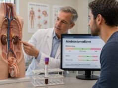
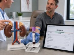
Thyroid-stimulating hormone, free thyroxine, liver tests, kidney function, and sometimes DHEA-S or androstenedione are added when indicated. No single fixed panel fits every patient.
How result patterns are interpreted
The relationship among results is more informative than whether one value is marked high or low.
| Laboratory pattern | Possible explanation | Typical follow-up |
|---|---|---|
| Low testosterone, high LH | Primary testicular failure or damage | Repeat testosterone; FSH, testicular examination, fertility assessment, and cause-specific testing |
| Low testosterone, low or normal LH | Pituitary/hypothalamic suppression, obesity, illness, medicine effect, or high prolactin | Prolactin, medication review, pituitary assessment when indicated |
| High estradiol with suppressed LH and testosterone | Exogenous estrogen or an estrogen-producing source | Confirm by sensitive assay; testicular/adrenal evaluation and imaging as directed |
| Elevated hCG | Germ-cell tumor, another hCG-producing tumor, exogenous hCG, or assay interference | Prompt repeat/confirmatory testing, tumor markers, testicular ultrasound, specialist referral |
| High prolactin with low testosterone | Medication effect, hypothyroidism, pituitary lesion, kidney disease, or stress-related elevation | Repeat prolactin, TSH, medicine review, macroprolactin and pituitary workup as appropriate |
| Normal panel | Physiologic, medication-related, obesity-associated, idiopathic, or resolved endocrine trigger | Review history and examination; observe or treat symptoms; image suspicious findings |
Timing and assay quality matter. Testosterone should generally be confirmed on another morning before diagnosing deficiency. Estradiol results near the low male range may be inaccurate with less sensitive immunoassays. hCG assays can be affected by heterophile antibodies, and prolactin may be temporarily elevated by venipuncture stress. Clinicians verify surprising results rather than building a diagnosis on one discordant number.
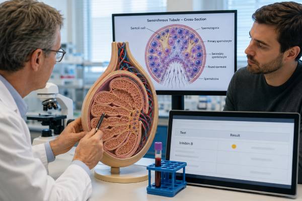
The direction of LH often provides the clearest first branch. For example, low testosterone plus high LH points toward the testes, while low testosterone plus low LH points toward central regulation. FSH adds information about seminiferous tubule and sperm-producing function. SHBG helps when total testosterone may be misleading.
Reference intervals vary by laboratory. The “ratio” of testosterone to estradiol is sometimes discussed, but there is no universally accepted diagnostic cutoff for gynecomastia. Converting values incorrectly or comparing assays can create a meaningless ratio. Clinical interpretation should use the original units, validated methods, and the complete pattern.
Common causes of gynecomastia
Physiologic gynecomastia occurs at life stages when hormone balance changes. Newborn breast tissue reflects maternal and placental hormones and usually resolves. Pubertal gynecomastia is common, often tender, and frequently improves over months to a few years. In older men, falling androgen production, increased body fat, medicines, and chronic disease can contribute.
Medicines are among the most actionable causes. Spironolactone has antiandrogenic and progestational effects; antiandrogens block testosterone action; 5-alpha-reductase inhibitors change androgen metabolism; and dopamine-blocking drugs may raise prolactin. The strength of evidence differs by drug, and not every breast symptom that occurs during treatment is caused by the medicine. Do not stop an essential prescription abruptly; the prescriber can assess alternatives.
Obesity can enlarge the chest through fat deposition and may increase aromatase activity, converting more androgen to estrogen. It can also lower SHBG and total testosterone. True glandular tissue and excess fat can coexist. Weight reduction may improve chest contour and metabolic hormone suppression, but established fibrotic glandular tissue may remain.
Testicular conditions include primary failure, orchitis, trauma, torsion, Klinefelter syndrome, and hormone-producing tumors. A testicular germ-cell tumor may raise hCG even before a mass is obvious. Some Leydig- or Sertoli-cell tumors produce estrogen or alter steroid production.
Liver disease can reduce hormone clearance and change SHBG, while kidney failure can disrupt the gonadal axis. Hyperthyroidism raises SHBG and changes estrogen-androgen balance. Malnutrition and recovery from severe illness can also alter gonadotropins. Adrenal tumors and congenital adrenal hyperplasia are uncommon but considered when adrenal androgens or estrogen precursors are abnormal.
Anabolic steroid cycles deserve specific attention. During use, aromatization may increase estrogenic stimulation. After discontinuation, suppressed LH and testosterone can leave estrogen action relatively unopposed. Products sold as post-cycle therapy can add further risk and confound laboratory results. Full disclosure allows safer testing and treatment.
When no cause is identified after appropriate evaluation, the condition may be labeled idiopathic. That does not mean symptoms are imaginary; it means no specific driver was found with available evidence.
Examination, imaging, and other tests
The physical examination covers both breasts, nipples, regional lymph nodes, testes, body-hair pattern, muscle mass, thyroid signs, liver signs, and features of chronic illness. The clinician documents whether tissue is centered under the areola, whether it is tender, and whether a separate mass is present. Testicular volume and consistency help interpret LH, FSH, and testosterone.
Breast imaging is not automatically required for classic, symmetric gynecomastia with a reassuring examination. Ultrasound or mammography may be used when the finding is indeterminate or suspicious. Imaging does not replace biopsy when cancer remains a concern. Male breast cancer is uncommon, but delay is possible if every male breast lump is assumed to be hormonal.
Testicular ultrasound is appropriate when a lump, asymmetry, pain, abnormal hCG, elevated estradiol, or otherwise unexplained gynecomastia raises concern for a testicular lesion. Some guidelines support ultrasound during specialist assessment because small tumors may not be palpable. Tumor markers such as alpha-fetoprotein and lactate dehydrogenase may be added to hCG when germ-cell cancer is suspected.
Liver enzymes, bilirubin, creatinine, thyroid tests, and a full blood count are selected based on history and examination. DHEA-S, androstenedione, 17-hydroxyprogesterone, or adrenal imaging are not routine for every patient, but may be useful when adrenal steroid excess is plausible. Genetic testing or karyotyping may be appropriate for small firm testes, infertility, tall stature, or suspected Klinefelter syndrome.
A semen analysis is more important than hormone testing alone when fertility is a concern. The FSH test in men helps assess testicular signaling, but normal FSH cannot guarantee normal sperm concentration or function.
Treatment and follow-up
Treatment depends on duration, pain, cause, severity, and patient preference. Recent tender gynecomastia is more likely to respond to medical treatment than long-standing fibrotic tissue. Many pubertal cases can be observed with periodic review when the examination is typical and distress is manageable.
The first step is to treat an identified cause where possible. A contributing medicine may be changed by the prescriber, hyperthyroidism treated, prolactin excess managed, liver or kidney disease addressed, and a tumor referred urgently. Weight management can reduce fatty chest enlargement and improve obesity-related hormone suppression. Stopping non-prescribed anabolic steroids is important, but recovery should be medically supported because mood symptoms, sexual dysfunction, and prolonged gonadal suppression can follow.
Selective estrogen receptor modulators such as tamoxifen may reduce pain and gland size in selected recent cases. They are prescription medicines with potential adverse effects and should not be self-administered. Aromatase inhibitors have not shown consistent benefit for ordinary gynecomastia and are not a universal solution to an estradiol value.
Surgery provides the most definitive contour correction for persistent, fibrotic, severe, or highly distressing gynecomastia. Techniques may combine gland excision and liposuction. The endocrine cause should be addressed first when feasible to reduce recurrence. Expectations, scars, nipple sensation, asymmetry, and surgical risks should be discussed.
Follow-up may track pain, size, symmetry, medication changes, and repeat hormone values when an abnormality was found. Repeated broad panels are unnecessary when the cause and clinical course are clear. A new change after stability warrants re-examination.
Urgent findings and next steps
Arrange prompt medical assessment for a hard or fixed breast mass, a lump away from the nipple, skin dimpling, nipple retraction, bloody discharge, enlarged underarm nodes, or rapid one-sided enlargement. These features do not prove cancer, but they require direct examination and appropriate imaging rather than hormone testing alone.
A testicular lump, new testicular asymmetry, significant pain, or confirmed hCG elevation also needs urgent evaluation. Severe headache, loss of peripheral vision, double vision, or neurological symptoms with high prolactin may indicate a pituitary mass. Acute shortness of breath or chest pain is an emergency regardless of hormone results.
For a nonurgent presentation, bring a complete list of medicines, injections, supplements, and substance use; note when symptoms started; and identify any fertility plans. Do not begin testosterone merely because gynecomastia is present. Testosterone can help confirmed hypogonadism but may worsen breast symptoms through aromatization and can suppress fertility.
A carefully selected panel answers whether an endocrine disorder is driving the breast change. The most reliable pathway combines examination, targeted testing, confirmation of unexpected results, and cause-specific follow-up. This approach finds serious conditions without turning a common symptom into indiscriminate testing.
Duration changes the likely response to treatment
Early gynecomastia is often tender because glandular tissue is actively proliferating. Over time, inflammation and cellular growth give way to more fibrotic tissue. A recent painful case is therefore more likely to shrink with removal of the cause or selected medical therapy than a stable enlargement present for several years. The laboratory panel may identify the driver, but it cannot determine the exact tissue age or predict cosmetic response.
Observation should still include a plan. The clinician may document size, tenderness, symmetry, testicular findings, medicines, and weight, then reassess after a defined interval. New growth, skin change, discharge, or a testicular symptom should bring the review forward. Repeating the entire hormone panel at every visit is unnecessary when the initial findings were normal and the examination remains stable.
Interpreting results during testosterone or anabolic steroid use
Exogenous androgens complicate every component of the panel. LH and FSH are usually suppressed. Testosterone may be high, normal, or low depending on the product and sampling time. Estradiol may rise through aromatization, while hCG use can stimulate testicular testosterone and estradiol despite a low measured LH. Prolactin can be unrelated or affected by other medicines.
A man may develop breast tenderness during a high-dose cycle, during withdrawal when endogenous testosterone is low, or while using multiple drugs intended to block estrogen. The safest interpretation requires the actual compounds, doses, dates, and injection schedule. Nonprescribed aromatase inhibitors or selective estrogen receptor modulators can cause thrombotic, bone, lipid, visual, mood, or sexual adverse effects and may obscure an underlying tumor or endocrine disorder.
For prescribed testosterone, breast symptoms do not automatically mean the dose must be stopped, but the regimen and indication should be reassessed. A physiological dose, appropriate sampling, weight management, and treatment of liver or thyroid disease may be more useful than reflexively adding another hormone drug. Persistent unilateral or suspicious findings still require breast evaluation even when the timing suggests a medication effect.
Pain severity and emotional distress should be assessed directly. Even benign gynecomastia can affect clothing, exercise, intimacy, and mental health. Reassurance is useful only after the examination is sound and the patient’s concerns are heard. Adolescents may need privacy, protection from bullying, and a clear plan for review rather than a casual statement that the condition is common.
When surgery is considered, photographs, stable weight, duration, gland versus fat distribution, smoking status, and expectations help planning. Hormone testing does not select the surgical technique, but untreated endocrine disease or ongoing androgen use can increase recurrence risk.
A stable normal panel can be reassuring, but it does not override examination. Conversely, a mildly abnormal hormone result does not prove the breast change is hormonal. The clinician should connect the timing and magnitude of each result to tissue findings before recommending observation, medicine, imaging, or surgery.
References
- EAA clinical practice guidelines-gynecomastia evaluation and management 2019 (Clinical Guideline)
- Gynecomastia: Etiology, Diagnosis, and Treatment 2023 (Clinical Review)
- Gynecomastia 2023 (Clinical Review)
- Features of true gynecomastia in adult males 2024 (Observational Study)
- Sexual and Reproductive Health 2026 (Clinical Guideline)
Disclaimer
This article is educational and is not a diagnosis or treatment plan. A new breast or testicular mass, nipple discharge, rapid growth, or abnormal hCG needs direct medical assessment. Do not stop prescription medicine or use hormones, antiestrogens, or bodybuilding products without a qualified clinician.