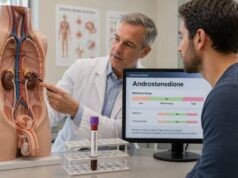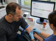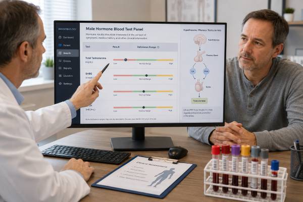
An androgen deficiency blood test panel looks beyond a single testosterone number. It combines total testosterone with sex hormone-binding globulin (SHBG), free or calculated free testosterone, luteinizing hormone (LH), follicle-stimulating hormone (FSH), and sometimes prolactin to determine whether low androgen activity is likely, whether the result is reliable, and where the problem may begin. Symptoms still matter: low sexual desire, fewer morning erections, erectile difficulty, reduced body hair, hot flashes, infertility, loss of muscle, and unexplained anemia are more suggestive than fatigue alone. Testing also needs the right timing because testosterone changes through the day and may fall temporarily during illness, calorie restriction, sleep loss, or medication use. A carefully collected and repeated panel can separate testicular failure from pituitary or hypothalamic suppression, reveal misleading SHBG effects, and guide the next step without treating a laboratory value in isolation.
- Diagnosis usually requires symptoms plus consistently low morning testosterone, not one unexpected result.
- Total testosterone should generally be checked on two separate mornings, preferably when fasting and medically stable.
- SHBG helps explain a misleading total testosterone result and supports calculation of free testosterone when binding is abnormal.
- High LH and FSH with low testosterone suggest primary testicular failure; low or normal gonadotropins suggest secondary suppression.
- Very low testosterone, high prolactin, headaches, or visual symptoms may require prompt pituitary evaluation.
Table of Contents
- What the androgen deficiency panel measures
- Who should be tested
- How to prepare and time the blood draw
- Understanding testosterone, SHBG, and free testosterone
- Using LH and FSH to locate the cause
- Common result patterns and causes
- Follow-up tests and clinical next steps
- Common mistakes and signs that need faster care
What the androgen deficiency panel measures
Androgens are hormones that support sexual development, libido, erectile function, sperm production, muscle and bone maintenance, red blood cell production, and several aspects of mood and energy. Testosterone is the main circulating androgen in adult men. The testes make most of it under stimulation from LH, while FSH acts mainly on Sertoli cells to support sperm production.
A useful panel answers three separate questions:
- Is circulating testosterone repeatedly low? Total testosterone is the usual first-line measurement.
- Is the available fraction also low? SHBG and free testosterone help when binding changes make total testosterone hard to interpret.
- Is the signal from the pituitary appropriate? LH and FSH help distinguish a testicular problem from inadequate stimulation by the hypothalamus or pituitary.
The exact panel varies. A focused evaluation commonly includes:
- Total testosterone
- SHBG
- Albumin, if calculated free testosterone will be reported
- Free testosterone by a validated method or a calculation based on total testosterone, SHBG, and albumin
- LH
- FSH
- Prolactin when secondary hypogonadism, low libido, infertility, or pituitary disease is possible
Estradiol may be added when breast enlargement, breast tenderness, obesity-related aromatization, unexplained fluid retention, or use of testosterone or anabolic steroids is relevant. A broader male hormone panel may also include thyroid tests, complete blood count, iron studies, or metabolic markers when symptoms are nonspecific.
The word “deficiency” should not be assigned from the panel alone. Testosterone levels overlap between healthy men and men with symptoms, laboratory methods differ, and reversible illness can suppress the reproductive axis. Clinical diagnosis combines symptoms, repeated measurements, examination findings, health history, and the pattern across the panel.
Who should be tested
Testing is most useful when symptoms or medical conditions make androgen deficiency plausible. Sexual and reproductive symptoms have greater diagnostic value than vague symptoms by themselves.
More specific reasons to test include:
- Reduced sexual desire that persists
- Fewer spontaneous or morning erections
- Erectile dysfunction, especially with low libido
- Infertility or abnormal semen results
- Hot flashes or sweats without another explanation
- Reduced beard growth, loss of body hair, or small testes
- Gynecomastia or breast tenderness
- Osteoporosis, a low-trauma fracture, or unexpectedly low bone density
- Unexplained anemia
- Delayed puberty or incomplete sexual development
Less specific complaints such as tiredness, low motivation, depressed mood, reduced concentration, weight gain, or poor exercise recovery can occur with low testosterone, but they are also common with sleep apnea, depression, thyroid disease, medication effects, chronic pain, overtraining, nutritional deficiency, and many other conditions. Testing may still be reasonable when several symptoms cluster or when a risk factor is present.
Risk factors include pituitary disease, testicular injury, chemotherapy or radiation, certain genetic conditions, chronic opioid use, past anabolic steroid use, severe obesity, type 2 diabetes, HIV, chronic kidney or liver disease, hemochromatosis, and prolonged glucocorticoid treatment. Men with a history of undescended testes, testicular torsion, orchitis, or testicular cancer treatment may have primary gonadal impairment.
Symptoms also change with the age at which androgen deficiency begins. A boy with congenital or early-onset deficiency may have delayed puberty, limited facial and body hair, a high-pitched voice, reduced muscle development, long limbs relative to the trunk, or small testes. An adult who loses testicular or pituitary function after normal puberty is more likely to notice reduced libido, fewer erections, falling shaving frequency, infertility, hot flashes, loss of strength, or declining bone density. Slow functional suppression associated with obesity or chronic illness may cause subtler changes that overlap with the underlying condition.
Physical findings can strengthen or weaken the case for testing. Testicular size and consistency, body-hair pattern, breast tissue, waist circumference, blood pressure, and signs of pituitary, thyroid, liver, or systemic disease provide context that a blood draw cannot. A clinician may also ask about puberty, prior fertility, head injury, sense of smell, testicular infection, radiation, chemotherapy, snoring, sleep schedule, sexual function, and use of performance-enhancing drugs. Loss of smell with absent or incomplete puberty can point toward a congenital gonadotropin-releasing hormone disorder, while a history of normal puberty followed by symptoms suggests an acquired process.
Symptoms should be separated from expected variation. Sexual desire differs greatly among healthy people and can fall during relationship stress, depression, sleep deprivation, or medication treatment. Erectile dysfunction frequently has vascular, neurologic, psychological, or medication-related causes even when testosterone is normal. Muscle gain depends on training, protein intake, sleep, age, and illness as well as androgens. The panel is therefore most convincing when several compatible findings occur together and improve the probability that low androgen action, rather than a single unrelated problem, is present.
Routine population screening of symptom-free men is generally not recommended. Testing every tired adult can produce borderline values that reflect timing, sleep, illness, or body weight rather than a persistent endocrine disorder. A clinician should first decide whether the result would change evaluation or treatment.
Fertility goals must be discussed before any treatment. Standard testosterone therapy can suppress LH and FSH and sharply reduce sperm production. Men trying to conceive need an evaluation centered on the male fertility hormone panel and semen analysis rather than automatic testosterone replacement.
How to prepare and time the blood draw
Testosterone has a daily rhythm, with the highest concentrations usually occurring after sleep and in the earlier part of the waking day. For men with a conventional schedule, laboratories commonly collect the first sample between about 7:00 and 10:00 or 11:00 a.m. A shift worker should discuss timing based on the main sleep period rather than relying only on clock time.
Fasting is often preferred because food, especially a substantial glucose-containing meal, may lower testosterone for several hours in some men. Follow the laboratory or clinician’s instructions. Water is usually allowed.
For the most interpretable result:
- Sleep as normally as possible before testing.
- Avoid testing during an acute infection, immediately after surgery, or during another major illness unless urgent assessment is needed.
- Avoid unusually hard exercise the evening before if it is not part of the normal routine.
- Report testosterone, anabolic steroids, “test boosters,” DHEA, opioids, glucocorticoids, antipsychotics, anticonvulsants, and supplements.
- Do not stop a prescribed medicine without medical advice.
- Tell the clinician about recent weight loss, very low calorie intake, sleep deprivation, or a major change in training.
A low result should usually be repeated on a different morning under similar conditions. Biological variation is substantial; one measurement can differ meaningfully from the next. Repeating only total testosterone may be enough when SHBG is clearly normal and the result is not borderline. Repeating total testosterone with SHBG and calculated free testosterone is often more informative when the first result is near the laboratory threshold or when a binding abnormality is likely.
Assays matter. Liquid chromatography–tandem mass spectrometry is often favored when accurate steroid measurement is important, especially at low concentrations, although many clinical laboratories use well-standardized immunoassays. Free testosterone by equilibrium dialysis is considered a strong reference method, but it is less available. Direct analogue free-testosterone immunoassays may be less reliable and should not be treated as interchangeable with equilibrium dialysis or a validated calculation.
Understanding testosterone, SHBG, and free testosterone
Most testosterone in blood is bound to proteins. A large portion binds tightly to SHBG, another portion binds more loosely to albumin, and only a small fraction circulates unbound. Total testosterone counts all three fractions. Free testosterone measures or estimates the unbound fraction. “Bioavailable testosterone” generally includes free testosterone plus the albumin-bound portion, which can dissociate readily.
Reference intervals depend on the assay, age distribution, and laboratory. Many guidelines use a total testosterone threshold around 300 ng/dL, while some European guidance uses approximately 12 nmol/L, which is about 346 ng/dL, as a practical diagnostic boundary in symptomatic men. These are not universal cutoffs. A result of 295 ng/dL is not automatically disease, and a result just above a threshold does not rule out a problem when SHBG is markedly abnormal.
To convert total testosterone:
- ng/dL × 0.0347 = nmol/L
- nmol/L × 28.8 = ng/dL
SHBG can change the relationship between total and free testosterone.
How SHBG can change interpretation
| SHBG pattern | Possible effect | Common associations |
|---|---|---|
| High SHBG | Total testosterone may look adequate while free testosterone is low | Aging, hyperthyroidism, liver disease, some anticonvulsants, estrogen exposure, HIV |
| Low SHBG | Total testosterone may look low while free testosterone is less reduced | Obesity, insulin resistance, type 2 diabetes, hypothyroidism, glucocorticoids, nephrotic syndrome |
| Normal SHBG | Total testosterone usually tracks available testosterone more closely | No major binding disturbance identified |
Calculated free testosterone is especially useful when total testosterone is borderline, SHBG is outside range, or symptoms and total testosterone do not agree. The calculation should use reliable total testosterone and SHBG values and an accepted equation. Different equations can produce different numbers, so the laboratory’s method and interval matter.
A SHBG test in men does not diagnose androgen deficiency by itself. It explains distribution and binding. Likewise, free testosterone should not be interpreted without total testosterone, SHBG, symptoms, and the clinical setting.
Using LH and FSH to locate the cause
LH and FSH are gonadotropins made by the pituitary gland. The hypothalamus releases gonadotropin-releasing hormone in pulses, which prompts the pituitary to release LH and FSH. LH stimulates Leydig cells in the testes to make testosterone. FSH, together with high intratesticular testosterone, supports Sertoli cells and sperm development.
When testosterone is low, the pituitary should normally increase LH. The response creates a useful diagnostic pattern:
- Low testosterone with high LH suggests primary hypogonadism, meaning the testes are not responding adequately.
- Low testosterone with low or inappropriately normal LH suggests secondary hypogonadism, meaning hypothalamic or pituitary stimulation is reduced.
- Low testosterone with high FSH strengthens concern for impaired seminiferous-tubule function and reduced sperm production.
- Normal testosterone with elevated LH may represent compensated testicular dysfunction, assay variation, medication effects, or an early response to declining testicular reserve.
FSH often reflects sperm-producing tissue more than testosterone production. A man can have normal testosterone and high FSH because Leydig-cell function remains sufficient while seminiferous-tubule function is impaired. Conversely, FSH can be normal in some men with poor sperm production, so a normal result does not replace semen analysis.
The relationship is also altered by external hormones. Testosterone injections, gels, pellets, anabolic steroids, and some selective androgen receptor modulators suppress hypothalamic and pituitary signaling. LH and FSH can become very low, testicular volume may fall, and sperm production can decline or stop even when serum testosterone is high.
A detailed LH test interpretation can therefore reveal whether the low testosterone result is accompanied by an appropriate pituitary response. FSH adds information about fertility, but neither hormone should be read as a stand-alone diagnosis.
Common result patterns and causes
Panel interpretation becomes clearer when values are viewed together rather than labeled one by one.
Common androgen panel patterns
| Testosterone pattern | LH and FSH | Likely category | Examples |
|---|---|---|---|
| Low total and low free testosterone | High | Primary testicular failure | Klinefelter syndrome, chemotherapy, orchitis, torsion, severe testicular injury |
| Low total and low free testosterone | Low or normal | Secondary hypogonadism | Pituitary tumor, hyperprolactinemia, opioids, obesity, energy deficit, systemic illness |
| Low total testosterone, free testosterone near range | Variable | Low SHBG effect | Obesity, insulin resistance, hypothyroidism |
| Total testosterone in range, low free testosterone | Variable | High SHBG effect | Aging, hyperthyroidism, liver disease, selected medicines |
| High testosterone | Suppressed | Exogenous androgen exposure | Prescribed testosterone, anabolic steroids, contaminated supplements |
Primary hypogonadism may affect testosterone, fertility, or both. In Klinefelter syndrome, for example, FSH is often high, testes are usually small and firm, and testosterone may be low or low-normal. Acquired causes include testicular trauma, infection, radiation, chemotherapy, vascular injury, or surgical loss of testicular tissue.
Secondary hypogonadism includes structural pituitary disease but is often functional and potentially reversible. Obesity, untreated sleep apnea, severe chronic illness, opioid use, glucocorticoids, undernutrition, excessive endurance training, and major stress can reduce hypothalamic-pituitary signaling. The term “functional” does not mean imaginary; it describes suppression without a fixed destructive lesion.
Prolactin is especially relevant when LH is low or normal despite low testosterone. A persistently high prolactin can inhibit gonadotropin-releasing hormone and reduce LH, FSH, testosterone, libido, and fertility. Common medication causes include antipsychotics, some antidepressants, metoclopramide, and other dopamine-blocking drugs. Hypothyroidism can raise prolactin through increased thyrotropin-releasing hormone.
A mixed pattern is possible. A man with testicular damage may also take opioids, have obesity, or use testosterone. The panel may not fit a single textbook box, and repeated testing plus history often resolves the apparent contradiction.
Follow-up tests and clinical next steps
The next step depends on the pattern, severity, symptoms, fertility plans, and examination. A clinician may order:
- Repeat morning total testosterone
- SHBG, albumin, and calculated free testosterone
- Prolactin, repeated under calm conditions if mildly high
- Thyroid-stimulating hormone and free T4
- Complete blood count to assess anemia or a high hematocrit
- Iron saturation and ferritin when hemochromatosis is possible
- Comprehensive metabolic panel for liver and kidney clues
- Hemoglobin A1c or fasting glucose for metabolic disease
- Estradiol when gynecomastia or an estrogen-related issue is present
- Semen analysis when fertility is relevant
- Karyotype or Y-chromosome testing in selected severe infertility patterns
- Pituitary MRI when biochemical and clinical findings support it
Pituitary imaging is not needed for every borderline low testosterone result. It becomes more important with very low testosterone and low or normal LH, persistently elevated prolactin, multiple pituitary-hormone abnormalities, severe headaches, visual-field changes, or symptoms of a sellar mass.
Treatment targets the cause when possible. Weight reduction can raise testosterone in some men with obesity. Treating sleep apnea, improving nutrition, changing an interfering medicine, reducing opioid exposure under supervision, or allowing recovery after anabolic steroid use may restore the axis. Structural pituitary or testicular disorders need specialist care.
Testosterone therapy is considered only after confirming a compatible diagnosis and discussing benefits, limits, contraindications, monitoring, and fertility. It can improve sexual symptoms, anemia, bone density, and body composition in appropriately selected men, but it is not a general treatment for fatigue or aging. It also requires monitoring of testosterone concentration, hematocrit, symptoms, adverse effects, and prostate-related factors according to age and risk.
Men who want children should avoid assuming that higher serum testosterone means better fertility. Exogenous testosterone can lower intratesticular testosterone and suppress sperm. Depending on the cause, fertility-preserving treatment may involve gonadotropins, selective estrogen receptor modulators, an aromatase inhibitor, management of hyperprolactinemia, surgery for selected conditions, or assisted reproduction under specialist guidance.
Common mistakes and signs that need faster care
Several errors can turn a useful panel into a misleading one.
Testing at the wrong time. An afternoon result may be lower than a morning value, especially in younger men. Shift work and poor sleep further complicate timing.
Diagnosing from one sample. Transient suppression and ordinary biological variation are common. Most men need confirmation on a separate day.
Ignoring SHBG. Low SHBG can make total testosterone look more deficient than the free fraction; high SHBG can conceal low free testosterone.
Treating symptoms as proof. Fatigue, low mood, and weight gain are not specific. Sleep, thyroid status, medication use, mental health, anemia, and chronic disease still need attention.
Starting testosterone before discussing fertility. Suppression of sperm production may be profound and recovery can take months or longer after treatment stops.
Using supplement-driven “optimal” ranges. Commercial targets may be much narrower or higher than validated laboratory intervals and may encourage treatment without a clinical diagnosis.
Overreacting to a mildly high prolactin. Stress, exercise, venipuncture difficulty, sleep, and medicines can raise prolactin. A calm repeat sample and macroprolactin assessment may prevent unnecessary imaging, while a marked or persistent elevation deserves proper evaluation.
Seek prompt medical assessment for a new severe headache, loss of side vision, double vision, fainting, confusion, sudden testicular pain, a hard testicular mass, or rapidly enlarging breast tissue with concerning features such as nipple retraction or bloody discharge. These findings are not typical consequences of uncomplicated age-related testosterone decline.
An androgen panel is most valuable when it narrows the diagnosis. Repeated testosterone establishes persistence, SHBG and free testosterone clarify hormone availability, and LH and FSH reveal whether the pituitary response fits the result. That combination supports safer, more individualized decisions than any single number taken out of context.
References
- Sexual and Reproductive Health 2026 (Guideline)
- The British Society for Sexual Medicine Guidelines on Male Adult Testosterone Deficiency, with Statements for Practice 2023 (Guideline)
- Male hypogonadism: recommendations from the Fifth International Consultation on Sexual Medicine 2025 (Position Statement)
- 2025 Update on Male Hypogonadism, Erectile Dysfunction, Premature Ejaculation, and Penile Curvature: From the EAU Guidelines on Sexual and Reproductive Health 2025 (Guideline)
- Testosterone Therapy in Men With Hypogonadism: An Endocrine Society Clinical Practice Guideline 2018 (Guideline)
Disclaimer
This information is educational and cannot diagnose androgen deficiency or identify its cause. Testosterone and gonadotropin results must be interpreted with symptoms, timing, medications, fertility plans, examination findings, and the laboratory’s own methods and ranges. Do not start, stop, or change hormone treatment without a qualified clinician.