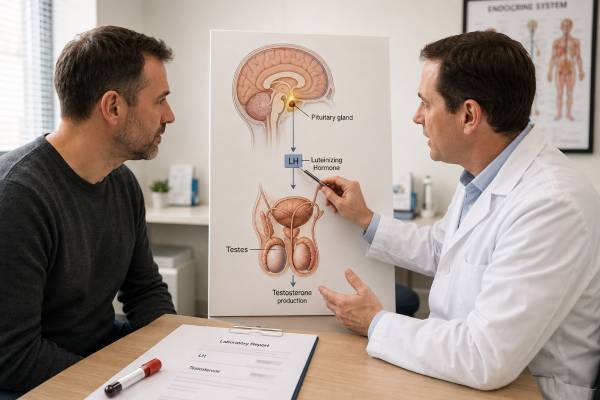
A luteinizing hormone (LH) test in men measures the pituitary signal that tells Leydig cells in the testes to produce testosterone. Its main clinical value is not whether LH is simply high or low, but whether it is appropriate for the testosterone level. Low testosterone with high LH suggests primary testicular failure. Low testosterone with low or inappropriately normal LH suggests a pituitary, hypothalamic, medication-related, or functional cause. LH may also be used in infertility, delayed puberty, anabolic steroid suppression, and monitoring of selected endocrine treatments. Because LH is released in pulses, one result can vary, and it should be interpreted with morning testosterone, FSH, prolactin, symptoms, medication use, and the laboratory range. Acute illness, obesity, severe calorie restriction, opioids, glucocorticoids, and exogenous androgens can suppress the axis. An LH result does not measure sperm count and cannot diagnose pituitary disease by itself, but it often provides the clearest first clue about where a low-testosterone problem begins.
- High LH with repeatedly low testosterone usually points toward primary testicular dysfunction.
- Low or normal LH with low testosterone can indicate central hypogonadism, functional suppression, or medication effects.
- A numerically “normal” LH may still be abnormal if the pituitary should be responding to very low testosterone.
- Testosterone or anabolic steroid use commonly suppresses LH and FSH, even when serum testosterone is high.
- LH should be interpreted with FSH, prolactin, SHBG or free testosterone when needed, and fertility goals.
Table of Contents
- What LH does in men
- When an LH test is ordered
- Preparation, timing, and variation
- Normal range and result patterns
- Causes of high LH
- Causes of low LH
- LH in fertility and puberty
- Follow-up tests and treatment
What LH does in men
LH is produced by gonadotroph cells in the anterior pituitary. The hypothalamus releases gonadotropin-releasing hormone in pulses, and those pulses stimulate the pituitary to release LH and FSH. LH binds receptors on Leydig cells in the testes, promoting testosterone synthesis. Testosterone then provides negative feedback to the hypothalamus and pituitary.
This feedback loop explains why LH is interpreted relative to testosterone. If testosterone falls because the testes cannot respond, the brain and pituitary usually increase LH. If testosterone falls because pituitary or hypothalamic signaling is inadequate, LH remains low or fails to rise enough. The same LH value can therefore mean different things at different testosterone concentrations.
LH also supports the very high testosterone concentration inside the testes that is required for normal spermatogenesis. Serum testosterone can be replaced with medication, but exogenous testosterone suppresses LH and reduces intratesticular testosterone. A man may have a normal blood testosterone level while sperm production is markedly impaired.
FSH is the companion gonadotropin. It acts mainly on Sertoli cells and supports seminiferous tubules and sperm development. LH is more directly connected with Leydig-cell testosterone production, while FSH often reflects sperm-producing tissue. A complete assessment may use both, but they should not be expected to move identically.
LH is not a direct measure of testosterone action in tissues. Normal LH and testosterone do not exclude erectile dysfunction, infertility, androgen-receptor disorders, depression, sleep problems, vascular disease, or medication effects. The test answers a narrow physiological question about pituitary signaling.
When an LH test is ordered
LH is commonly ordered after a low or borderline testosterone result. It helps classify hypogonadism as primary, secondary, or mixed. It may also be included at the initial visit when symptoms are strong, infertility is present, or there is a known risk of pituitary or testicular disease.
Symptoms that can prompt testing include reduced libido, fewer morning erections, erectile difficulty, infertility, hot flushes, loss of body hair, reduced shaving frequency, small testes, gynecomastia, low bone density, unexplained anemia, delayed puberty, or incomplete virilization. Fatigue and low mood are less specific and should be assessed broadly.
Risk factors for primary testicular failure include Klinefelter syndrome, prior torsion, orchitis, chemotherapy, radiation, severe trauma, and advanced testicular disease. Risk factors for central suppression include pituitary tumors, surgery or radiation near the pituitary, traumatic brain injury, iron overload, congenital gonadotropin deficiency, high prolactin, obesity, severe illness, undernutrition, opioids, and glucocorticoids.
LH is important when anabolic steroids or testosterone are used. Suppressed LH is an expected pharmacological effect, not proof that the pituitary is permanently damaged. After discontinuation, recovery can be slow and depends on duration, dose, compounds, age, and baseline function. The exact exposure history is essential.
In infertility, LH helps explain low testosterone and can guide treatment in hypogonadotropic hypogonadism. It does not replace semen analysis. In delayed puberty, pediatric endocrinologists interpret LH using age, pubertal stage, testicular size, and sometimes stimulation testing because adult ranges are not appropriate.
Routine LH screening in an asymptomatic man with normal testosterone is rarely useful. Mild isolated abnormalities can reflect pulsatile secretion, assay variation, or a compensated response and may lead to unnecessary testing if clinical context is ignored.
Preparation, timing, and variation
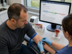
LH is measured from blood and often collected with morning total testosterone. Testosterone has a stronger daily rhythm than LH, but pairing the samples makes the relationship easier to interpret. Fasting may be requested because it standardizes testosterone testing; follow the laboratory’s instructions.
LH secretion is pulsatile. A short burst can make one result higher, while a sample between pulses can be lower. In most adult evaluations, a random morning LH is sufficient when interpreted with testosterone, but an unexpected result may be repeated. Specialized pooled sampling or stimulation tests are reserved for selected endocrine cases.
Avoid diagnosing chronic hypogonadism during acute severe illness if testing can wait. Infection, surgery, major sleep disruption, undernutrition, and physiological stress can suppress gonadotropins and testosterone. A repeat after recovery may look different.
List all medicines and substances. Testosterone, anabolic steroids, hCG, selective estrogen receptor modulators, aromatase inhibitors, opioids, glucocorticoids, dopamine-modifying drugs, and some cancer treatments alter LH. Do not stop a prescribed medicine without medical advice. Biotin and assay interference can affect some laboratory systems.
Women’s fertility products and men’s hormone products can contain hCG or compounds that change feedback. “Test boosters” may include undeclared androgens. An honest record of product names, doses, injection dates, and last use helps prevent a false pituitary diagnosis.
For men receiving hCG, LH may remain low because hCG stimulates the LH receptor while suppressing endogenous pituitary signaling. Some LH assays do not cross-react meaningfully with hCG, so a low measured LH does not mean Leydig cells are unstimulated. Treatment monitoring must account for the drug’s mechanism.
Normal range and result patterns
LH reference intervals differ by assay, age, and laboratory. Results are usually reported in international units per liter. The printed interval is necessary, but the appropriateness of the pituitary response is more important than the flag.
| LH and testosterone pattern | Possible interpretation | Common next step |
|---|---|---|
| High LH, low testosterone | Primary testicular failure | Repeat testosterone; FSH, examination, fertility and cause-specific testing |
| Low or normal LH, low testosterone | Secondary or functional hypogonadism | Prolactin, medication review, pituitary and systemic assessment |
| High LH, normal testosterone | Compensated Leydig-cell dysfunction or transient variation | Repeat and monitor symptoms; investigate risk factors |
| Low LH, normal or high testosterone | Exogenous androgen or hCG effect; less commonly autonomous production | Review all hormones, supplements, timing, and related results |
| Very low LH and FSH with very low testosterone | Marked central suppression or pituitary/hypothalamic disease | Prompt endocrine evaluation and additional pituitary testing |
Low testosterone should generally be confirmed on a second properly timed sample. If SHBG is abnormal, total testosterone may not reflect the free fraction accurately. A free testosterone test or calculated value may clarify the diagnosis.
“Normal LH” is not always reassuring. If testosterone is profoundly low, a healthy pituitary would ordinarily increase LH. A midrange result is therefore inappropriately normal and supports a central pattern. Conversely, a mildly high LH with normal testosterone may show that the pituitary is compensating successfully; it is not identical to overt testicular failure.
LH and FSH can diverge. Seminiferous tubule damage may raise FSH while LH and testosterone remain relatively preserved. Leydig-cell dysfunction may raise LH more prominently. This difference helps localize which testicular compartment is most affected.
Causes of high LH
High LH most often reflects reduced negative feedback from testicular testosterone production. In primary hypogonadism, Leydig cells cannot produce enough testosterone despite increased pituitary stimulation. Causes include Klinefelter syndrome, mumps orchitis, torsion, trauma, chemotherapy, radiation, surgical loss of testicular tissue, and some genetic or autoimmune conditions.
Klinefelter syndrome may present with small firm testes, infertility, gynecomastia, tall stature, reduced body hair, or learning and psychosocial differences. Testosterone can be low or low-normal earlier in life while LH and FSH rise. Karyotype testing confirms the diagnosis when clinical findings support it.
After chemotherapy or radiation, FSH may rise before LH because germinal tissue is often more vulnerable than Leydig cells. Over time, Leydig-cell reserve may also fall. Cancer survivors need interpretation based on treatment type, age at exposure, fertility plans, and previous baseline results.
A high LH with normal testosterone is sometimes called compensated hypogonadism or subclinical Leydig-cell dysfunction. It may be seen with aging, chronic disease, prior testicular injury, or reduced reserve. Evidence does not support automatically treating a man who has normal testosterone and no compatible symptoms. Repeat testing and attention to the cause are more appropriate.
High LH with high testosterone is discordant. Possible explanations include selective estrogen receptor modulator use, aromatase inhibitor use, androgen resistance, assay interference, or unusual hormone-producing conditions. A medication such as clomiphene blocks estrogen feedback and intentionally raises LH and testosterone. The result should be interpreted in light of treatment, not against an untreated range.
Transient mild elevation can occur because LH is pulsatile. A single isolated high result without a matching clinical or testosterone abnormality often warrants confirmation rather than extensive imaging.
Causes of low LH
Low LH with low testosterone indicates inadequate central stimulation, but the cause may be structural, congenital, medication-related, or functional. Pituitary adenomas, surgery, radiation, infiltrative disease, traumatic brain injury, and iron overload can impair gonadotropin secretion. Other pituitary hormones may also be deficient.
High prolactin can suppress hypothalamic gonadotropin-releasing hormone. Causes include prolactinomas, dopamine-blocking medicines, hypothyroidism, kidney disease, chest-wall stimulation, stress, and macroprolactin. A prolactin test in men is often part of the follow-up when LH is low or inappropriately normal.
Functional suppression is common. Severe obesity, insulin resistance, sleep apnea, uncontrolled systemic illness, overtraining, significant calorie deficit, and psychological or physiological stress can lower gonadotropin drive. The axis may recover when the underlying condition improves.
Opioids suppress hypothalamic signaling and can cause low libido, erectile dysfunction, infertility, and low bone density. Glucocorticoids and some cancer therapies can also suppress the axis. Medication adjustment must balance endocrine effects against the condition being treated.
Exogenous testosterone and anabolic steroids predictably suppress LH. The serum testosterone concentration depends on the compound and time since dosing; it may be high during use and low during withdrawal. Suppressed LH alone cannot distinguish prescribed testosterone from undisclosed anabolic use, and it cannot predict recovery time.
Congenital hypogonadotropic hypogonadism may present with absent or incomplete puberty, small testes, infertility, and sometimes reduced sense of smell. Early diagnosis is important for pubertal development, bone health, and fertility options. Genetic and specialized endocrine evaluation may be needed.
LH in fertility and puberty
LH supports fertility through intratesticular testosterone. Serum levels of testosterone are far lower than the concentration inside the testes. When LH is suppressed by exogenous testosterone, intratesticular testosterone falls and sperm production may stop even if sexual symptoms improve.
A fertility evaluation begins with semen analysis, reproductive history, and examination. LH, FSH, testosterone, and prolactin are selected according to sperm concentration, testicular size, and symptoms. A male fertility hormone panel can identify endocrine causes, but a normal panel cannot guarantee normal sperm or conception.
In hypogonadotropic hypogonadism, hCG can act at the LH receptor to stimulate Leydig cells. FSH may be added to stimulate Sertoli cells and spermatogenesis. Treatment can take many months, particularly when testes did not develop normally during puberty. Doses are individualized and monitored by specialists.
Primary testicular failure behaves differently. High LH shows that the pituitary is already sending a strong signal, so adding hCG is less likely to restore Leydig-cell function. Fertility options depend on residual spermatogenesis, genetic diagnosis, and potential surgical sperm retrieval.
During puberty, rising nighttime LH pulses precede and support testosterone production and testicular growth. A single daytime sample early in puberty may be low despite normal development. Pediatric endocrinologists use growth, bone age, testicular volume, family timing, and sometimes stimulation tests to distinguish constitutional delay from permanent gonadotropin deficiency.
Infants have a temporary “mini-puberty” with active gonadotropin secretion. LH values outside this period should not be interpreted with neonatal or adult expectations. Age-specific laboratory ranges are essential.
Follow-up tests and treatment
Follow-up begins by confirming testosterone and reviewing SHBG when needed. FSH adds information about sperm-producing tissue. Prolactin, thyroid tests, iron studies, liver and kidney function, and a complete blood count are selected from the clinical pattern. Semen analysis is required for fertility questions.
Pituitary MRI is considered when testosterone is very low with low gonadotropins, prolactin remains elevated, headaches or visual symptoms are present, or other pituitary hormones are abnormal. MRI is not triggered by every mildly low LH in a man with obesity and transient low testosterone.
Testicular examination and sometimes ultrasound are appropriate with a mass, pain, asymmetry, small testes, high gonadotropins, or infertility. Genetic testing may include karyotype or Y-chromosome microdeletion analysis in severe sperm-production disorders.
Treatment targets the cause. A prolactinoma may respond to a dopamine agonist. Functional suppression may improve with treatment of sleep apnea, weight reduction, recovery from illness, adequate nutrition, or adjustment of a suppressive medicine. Primary testicular failure may require testosterone for symptoms and long-term health when fertility is not an immediate goal.
Men who want fertility should not begin exogenous testosterone without specialist advice. Gonadotropins or other selected treatments may be more appropriate for central hypogonadism. A complete hypogonadism blood test panel should be obtained before treatment changes erase the diagnostic pattern.
Seek urgent evaluation for severe headache with visual change, a new testicular mass, acute testicular pain, or neurological symptoms. Otherwise, the most useful response to an abnormal LH is a repeatable, cause-focused evaluation—not an attempt to raise or lower LH as an isolated target.
Compensated testicular dysfunction and changing patterns
LH may rise before testosterone falls below range. This compensated pattern suggests that the pituitary is working harder to preserve Leydig-cell output. It can appear with aging, prior chemotherapy, testicular injury, chronic disease, or reduced testicular reserve. It is not identical to symptomatic hypogonadism, and testosterone treatment is not automatically indicated while testosterone remains adequate.
Follow-up depends on symptoms and risk. Repeating LH and morning testosterone after a clinically meaningful interval can show whether the pattern is stable, transient, or progressing. FSH, testicular volume, semen analysis, and fertility plans add context. A small isolated increase after one pulsatile sample may disappear, while a persistent rise with declining testosterone deserves closer evaluation.
The opposite transition can occur during recovery from central suppression. After illness, weight change, opioid reduction, or anabolic steroid withdrawal, LH may begin to rise before testosterone fully recovers. One snapshot can therefore look discordant. Serial results should be spaced according to the expected biology rather than repeated every few days.
LH and medicines that alter feedback
Selective estrogen receptor modulators reduce estrogen feedback at the hypothalamus and pituitary, raising LH and often testosterone. Aromatase inhibitors can have a similar feedback effect by lowering estradiol. These drugs may create high LH with normal or high testosterone, a pattern that would be unusual without treatment. They are not interchangeable and can affect bone, vision, mood, lipids, clotting risk, and fertility differently.
Antiandrogens block androgen action and may raise LH and testosterone if the central feedback loop is intact. In contrast, gonadotropin-releasing hormone agonists and antagonists used in prostate cancer suppress LH and testosterone profoundly. The expected pattern depends on treatment phase; some agonists cause an initial flare before suppression.
hCG stimulates the LH receptor but is not the same molecule as LH. A man receiving hCG can have low endogenous LH, rising testosterone, and preserved or improved intratesticular stimulation. The treatment dose and fertility purpose should be documented so the low LH is not misclassified as pituitary failure.
Protecting the diagnosis before starting treatment
LH is most informative before exogenous hormones are started. Once testosterone suppresses the pituitary, the original distinction between primary and secondary hypogonadism becomes harder to recover. Previous untreated laboratory reports, puberty history, testicular size, fertility history, and risk factors become especially important.
When treatment has already begun without a complete evaluation, stopping it solely to recreate the baseline may cause significant symptoms and is not always necessary or safe. An endocrinologist or urologist can decide whether records and the current pattern provide enough information or whether a supervised washout is justified.
The final clinical goal is not to normalize LH itself. In primary failure, high LH may remain despite appropriate testosterone replacement. In central infertility treatment, endogenous LH may remain low while hCG provides receptor stimulation. Management follows testosterone, symptoms, semen goals, safety markers, and the underlying diagnosis.
Reference intervals cannot fully account for pulsatility. When an LH value would change the diagnosis from primary to central hypogonadism, repeating it with testosterone can be worthwhile. The repeated pattern, not the most convenient single value, should direct imaging and treatment.
Men with headache, visual-field loss, double vision, faintness, or other pituitary-hormone symptoms need faster assessment than men with a mild isolated low LH. Clinical urgency comes from the whole presentation.
A concise record of untreated testosterone, LH, FSH, prolactin, symptoms, and exposures is especially valuable before any hormone therapy changes the feedback pattern.
This preserves the evidence needed for future cause-specific care and fertility planning.
A result should always be connected to the testosterone concentration measured at the same stage of evaluation.
References
- Sexual and Reproductive Health 2026 (Clinical Guideline)
- Male hypogonadism: recommendations from the Fifth International Consultation on Sexual Medicine 2025 (Consensus Recommendations)
- The British Society for Sexual Medicine Guidelines on Male Adult Testosterone Deficiency, with Statements for Practice 2023 (Clinical Guideline)
- Diagnosis and Treatment of Infertility in Men: AUA/ASRM Guideline 2024 (Clinical Guideline)
- Male Infertility 2024 (Clinical Review)
Disclaimer
This information is educational and cannot diagnose pituitary or testicular disease. LH must be interpreted with testosterone, FSH, symptoms, medicines, fertility goals, age, and the laboratory method. Do not stop hormones or other prescription medicine or begin fertility treatment without a qualified clinician.