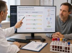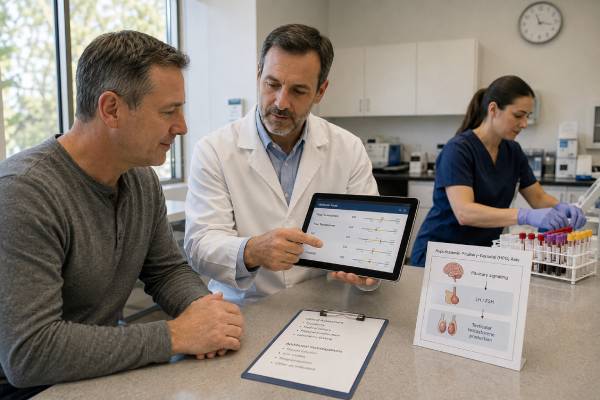
A hypogonadism blood test panel checks whether a man has consistently low testosterone and helps locate the cause in the testes, pituitary, hypothalamus, hormone-binding system, or a reversible health condition. The core tests are total testosterone, free testosterone when indicated, LH, FSH, and prolactin. Diagnosis is not based on one low number. It generally requires compatible symptoms or signs plus repeat low morning testosterone measured with a reliable method. LH and FSH then separate primary testicular failure from secondary or central hypogonadism, while prolactin can reveal medication effects or pituitary disease. SHBG, thyroid tests, iron studies, semen analysis, and pituitary imaging may be added according to the pattern. Obesity, acute illness, sleep loss, severe calorie restriction, opioids, glucocorticoids, anabolic steroids, and several chronic diseases can temporarily or functionally suppress the axis. Testing before treatment is important because exogenous testosterone can hide the original pattern, raise hematocrit, and suppress sperm production.
- Hypogonadism requires symptoms or signs plus consistently low testosterone; one abnormal result is not enough.
- Total testosterone is usually tested on two separate mornings, with free testosterone added when SHBG may make the total value misleading.
- Low testosterone with high LH suggests primary testicular failure; low testosterone with low or normal LH suggests central or functional suppression.
- High prolactin can suppress LH and testosterone and may require medication review, repeat testing, or pituitary evaluation.
- Men who want fertility should discuss alternatives before testosterone therapy because treatment can sharply reduce sperm production.
Table of Contents
- What hypogonadism means
- Who should have the panel
- How to prepare and repeat testing
- Tests in the panel
- Result patterns and causes
- Additional tests and imaging
- Treatment choices and fertility
- Monitoring and safety
What hypogonadism means
Male hypogonadism is a clinical condition in which the testes do not produce enough testosterone, sperm, or both because of a problem in the testes or in the hormonal signals that control them. It is not simply a testosterone value below a laboratory flag. Symptoms, signs, repeat biochemical evidence, age, health status, fertility goals, and the cause all matter.
The hypothalamus releases gonadotropin-releasing hormone in pulses. This stimulates the pituitary to release LH and FSH. LH acts mainly on Leydig cells to produce testosterone. FSH, together with intratesticular testosterone, supports Sertoli cells and sperm production. Testosterone and inhibin B provide feedback to the brain and pituitary. A disturbance at any point can alter the panel.
Primary hypogonadism originates in the testes. Testosterone is low and LH is usually high because the pituitary is trying to stimulate underperforming Leydig cells. FSH may be high when sperm-producing tissue is damaged. Secondary hypogonadism originates in the hypothalamus or pituitary. Testosterone is low, but LH and FSH are low or not appropriately elevated. Combined forms occur, particularly with aging, chronic disease, obesity, or multiple treatments.
Some suppression is potentially reversible and is often described as functional hypogonadism. Severe obesity, uncontrolled diabetes, sleep apnea, systemic illness, undernutrition, excessive endurance exercise, opioids, glucocorticoids, and anabolic steroid withdrawal can reduce signaling without permanent structural damage. The distinction matters because treating the underlying factor may restore the axis.
Late-onset symptoms are not automatically hypogonadism. Fatigue, low mood, reduced strength, and erectile difficulty are common and have many causes. The panel is designed to connect clinically meaningful symptoms to a reproducible hormonal pattern.
Who should have the panel
Testing is appropriate when symptoms or signs raise a reasonable suspicion of androgen deficiency. More specific features include reduced libido, fewer spontaneous or morning erections, infertility, hot flushes, loss of body hair, reduced shaving frequency, small or soft testes, gynecomastia, low-trauma fractures, osteoporosis, unexplained anemia, or delayed puberty. Erectile dysfunction alone can arise from vascular, neurological, medication, psychological, or relationship factors, but testosterone assessment may be part of a broader evaluation.
Less specific symptoms include fatigue, poor concentration, low mood, sleep disturbance, reduced exercise capacity, increased body fat, and loss of muscle. These support testing when several occur together or when risk factors are present, but they should not be treated as proof of low testosterone.
Risk factors include pituitary or hypothalamic disease, testicular injury or torsion, mumps orchitis, chemotherapy, pelvic or testicular radiation, genetic conditions such as Klinefelter syndrome, HIV, iron overload, chronic opioid use, high-dose glucocorticoids, severe obesity, and a history of anabolic steroid use. Men with an incidental pituitary mass, very low bone density, or unexplained infertility may also need evaluation.
Routine population screening of men without symptoms is generally not recommended. Testing solely because of age, bodybuilding goals, online symptom scores, or a desire to optimize an already normal value can lead to false diagnoses and unnecessary treatment.
Pubertal development requires age-specific assessment. Delayed puberty, absent testicular enlargement, or incomplete virilization should be evaluated by a pediatric endocrinologist rather than interpreted with adult cutoffs. Likewise, men already using testosterone require a monitoring strategy tied to their formulation; their results cannot establish the untreated cause unless prior records are available.
How to prepare and repeat testing
For an untreated adult, total testosterone is usually collected in the early morning, often between 7 and 10 a.m., when concentrations are generally highest. The sample may be requested fasting because meals and glucose intake can temporarily lower testosterone. Shift workers should discuss timing relative to their main sleep period rather than assuming a fixed clock time is optimal.
Do not deliberately test during an acute infection, immediately after surgery, after major sleep deprivation, or during severe calorie restriction unless assessment cannot wait. Acute stress and illness can suppress the reproductive axis. A low value in those settings may reflect temporary physiology rather than stable hypogonadism.
Bring a full list of prescribed medicines, nonprescription drugs, supplements, injections, and topical hormones. Opioids, glucocorticoids, antiandrogens, some psychotropic medicines, and anabolic steroids can change the pattern. Biotin can interfere with some immunoassays. Do not stop a medicine without instructions from the prescriber.
A low total testosterone result should usually be confirmed on a different morning under similar conditions. Biological variation, sleep, food, laboratory imprecision, and transient illness can produce a single low result. The total testosterone test is interpreted using the method-specific reference interval and clinical threshold rather than an online universal range.
Free testosterone is added when total testosterone is borderline or SHBG is likely abnormal. Equilibrium dialysis is a reference method; calculated free testosterone can be useful when total testosterone and SHBG are accurately measured. Direct analog assays are less dependable. Repeating the same laboratory method improves comparability.
Men receiving injections, gels, patches, pellets, or oral testosterone need collection timed to the treatment. A sample soon after an injection peak cannot be compared with a trough before the next dose. Gel contamination near the blood-draw site can cause a falsely high result.
Tests in the panel
Total testosterone
Total testosterone measures free and protein-bound hormone. It is the usual first biochemical test. Low values on two properly timed samples support deficiency when compatible symptoms are present. SHBG can make total testosterone look low or high without a proportional change in the active fraction.
Free testosterone
Free testosterone is helpful when SHBG is unusually high or low or when total testosterone and the clinical picture disagree. Obesity and insulin resistance often lower SHBG; aging, hyperthyroidism, and some liver conditions may raise it. A free testosterone test should identify whether the value was measured or calculated and use a method-specific range.
LH
LH shows how strongly the pituitary is signaling the testes. A high LH with low testosterone points toward primary testicular failure. A low or inappropriately normal LH with low testosterone points toward central or functional suppression. The word “normal” can be misleading: an LH in the middle of its range is not an adequate response when testosterone is clearly low.
FSH
FSH provides information about Sertoli-cell and seminiferous tubule function. High FSH can indicate impaired sperm production, although normal FSH does not guarantee normal fertility. FSH is particularly important when infertility, small testes, prior chemotherapy, or testicular damage is part of the presentation.
Prolactin
Persistent prolactin elevation can suppress gonadotropin secretion and lower testosterone. Common explanations include dopamine-blocking medicines, hypothyroidism, kidney disease, stress, and pituitary adenomas. Mild elevations often require repeat testing and sometimes macroprolactin assessment. A very high or persistently high result, especially with headaches or visual symptoms, needs prompt pituitary evaluation.
SHBG and albumin may be required to calculate free testosterone. The panel may be expanded, but ordering every reproductive hormone without a clinical question often creates incidental abnormalities rather than clarity.
Result patterns and causes
The panel is interpreted as a physiological pattern.
| Pattern | Likely category | Examples |
|---|---|---|
| Low testosterone, high LH; FSH may be high | Primary hypogonadism | Klinefelter syndrome, orchitis, torsion, chemotherapy, radiation, severe testicular injury |
| Low testosterone, low or normal LH and FSH | Secondary or functional hypogonadism | Pituitary disease, high prolactin, obesity, illness, opioids, glucocorticoids, undernutrition |
| Low total testosterone, normal free testosterone, low SHBG | Binding-related low total value | Obesity, insulin resistance, hypothyroidism, nephrotic syndrome |
| Normal total testosterone, low free testosterone, high SHBG | Binding may conceal deficiency | Aging, hyperthyroidism, liver disease, some medicines |
| Low testosterone, suppressed LH/FSH after androgen use | Exogenous suppression | Testosterone therapy, anabolic steroids, prohormones |
| Low testosterone, high prolactin | Hyperprolactinemic suppression | Medication effect, hypothyroidism, kidney disease, prolactinoma |
Primary causes can be congenital or acquired. Klinefelter syndrome is a common chromosomal cause and may present with small firm testes, infertility, tall stature, gynecomastia, or learning differences. Acquired damage includes infection, trauma, torsion, cancer treatment, and advanced testicular disease. Testosterone production and sperm production are related but not identical; FSH may be more abnormal than LH when seminiferous tissue is predominantly affected.
Central causes include pituitary tumors, surgery, radiation, infiltrative disease, traumatic brain injury, congenital gonadotropin deficiency, and iron overload. Additional pituitary hormones may be abnormal. Functional central suppression is common with obesity and chronic disease and may improve when the underlying problem is treated.
Anabolic-androgenic steroid withdrawal deserves separate interpretation. During use, LH and FSH are suppressed. After discontinuation, testosterone can remain low while the axis recovers, and semen parameters may take longer to improve. The exact substances and dates are more informative than a generic “supplement” label.
A normal LH or FSH result does not exclude disease when testosterone is low. The pituitary response should be appropriate to the degree of hormone deficiency. Conversely, mildly elevated LH with normal testosterone can represent compensated testicular dysfunction, which is not the same as established hypogonadism and does not automatically require testosterone.
Additional tests and imaging
Further testing should answer a specific question raised by the initial panel. SHBG and albumin help resolve discordant testosterone values. Thyroid-stimulating hormone and free thyroxine identify thyroid disease. Iron studies can detect hemochromatosis, which may affect the pituitary or testes. A complete blood count may reveal anemia before treatment or an elevated hematocrit that changes treatment safety.
Liver and kidney tests help explain altered SHBG, chronic illness, or prolactin elevation. Hemoglobin A1c, lipids, blood pressure, waist measures, and sleep-apnea assessment may identify metabolic contributors. These conditions deserve treatment whether or not testosterone rises.
Pituitary MRI is considered when central hypogonadism is severe or accompanied by persistently high prolactin, headaches, visual-field symptoms, multiple pituitary hormone deficits, or another sign of a sellar lesion. It is not required for every mildly low testosterone result associated with obesity. The prolactin test in men is usually repeated before imaging if the elevation is small and an obvious medicine or stress effect is possible.
Semen analysis is central when fertility is a concern. Hormone results cannot establish sperm count, motility, morphology, obstruction, or ejaculatory function. FSH, inhibin B, genetic tests, and testicular imaging may be selected after semen analysis and examination. A male fertility hormone panel supports this evaluation but does not replace the semen sample.
Bone-density scanning may be indicated with long-standing severe deficiency, fractures, height loss, glucocorticoid exposure, or other osteoporosis risks. Genetic testing or karyotyping is considered with primary failure, very small testes, azoospermia, or suggestive physical features.
Before testosterone therapy, clinicians assess fertility plans, hematocrit, prostate and urinary history, sleep apnea, cardiovascular status, and contraindications. Baseline prostate-specific antigen is considered according to age, risk, and individual discussion rather than as part of every young man’s hormone panel.
Treatment choices and fertility
Treatment should address the cause whenever possible. Weight loss in men with obesity, treatment of sleep apnea, recovery from systemic illness, improved diabetes management, adequate nutrition, and review of suppressive medicines may restore testosterone. A medication change must be coordinated with the prescriber; stopping opioids, glucocorticoids, or psychiatric medicines abruptly can be dangerous.
Pituitary causes require specific treatment. A prolactinoma often responds to a dopamine agonist, which can lower prolactin and allow gonadal function to recover. Iron overload, thyroid disease, and other endocrine disorders have their own therapies. Structural testicular failure is less likely to reverse, although fertility options may still be available.
Testosterone replacement can improve sexual symptoms, body composition, anemia, and bone health in appropriately selected men with confirmed deficiency. Formulations include gels, injections, patches, pellets, and oral products, each with different peaks, transfer risks, convenience, cost, and monitoring needs. Treatment aims for a physiological range and symptom benefit, not a supraphysiologic free testosterone target.
Exogenous testosterone suppresses LH and FSH, lowers intratesticular testosterone, and can greatly reduce or eliminate sperm from the ejaculate. Men who want children now or soon should discuss fertility-preserving options with a reproductive urologist or endocrinologist before starting. Depending on the cause, gonadotropin therapy or selected off-label medicines may stimulate endogenous production, but these are specialist treatments and not safe substitutes bought online.
The decision to treat includes uncertainty. Nonspecific symptoms may not improve even when testosterone rises. A time-limited, monitored therapeutic trial may be reasonable in some confirmed cases, with discontinuation if meaningful benefit does not occur. Treatment should not continue indefinitely merely because stopping would lower a treatment-created level.
Monitoring and safety
Monitoring begins after the formulation has reached a suitable steady state. Testosterone is measured at the time recommended for the product: an injection trough, a midpoint, or a post-application interval may be used depending on the regimen. Symptoms, adverse effects, adherence, and application technique are reviewed alongside the number.
Hematocrit is important because testosterone can stimulate red blood cell production. A substantial rise may require dose adjustment, interruption, assessment for sleep apnea or smoking, or another clinical response. Simply donating blood repeatedly without addressing an excessive dose can hide the problem.
Prostate monitoring is individualized by age and risk. New urinary symptoms, a concerning examination, or a significant PSA change requires evaluation. Testosterone is not a treatment for erectile dysfunction caused primarily by vascular disease when testosterone is normal, and it does not replace cardiovascular risk management.
Other concerns include acne, oily skin, edema, breast tenderness, worsening untreated sleep apnea, mood changes, testicular shrinkage, and infertility. Transference is possible with gels. Injections can produce peaks and troughs. Pellets require a procedure. Each formulation requires counseling specific to its risks.
Seek urgent care for chest pain, severe shortness of breath, symptoms of a stroke, a painful swollen leg, sudden severe headache with visual change, or acute testicular pain. These symptoms should not wait for a routine hormone appointment.
A well-run hypogonadism panel does more than confirm a low testosterone value. It defines whether the signal points to the testes, the brain and pituitary, abnormal binding, medication exposure, or a reversible health condition. That distinction protects fertility, avoids inappropriate therapy, and makes treatment more likely to address the actual cause.
Distinguishing permanent disease from functional suppression
The label “functional” should not be used to dismiss symptoms. It means that a structural lesion or irreversible testicular failure has not been established and that health conditions or exposures may be reducing the axis. Severe obesity, sleep apnea, uncontrolled diabetes, undernutrition, chronic inflammation, opioids, glucocorticoids, and recent illness can produce a central pattern. Treating these contributors may improve testosterone and overall health, although recovery is not guaranteed.
Reassessment should be timed to the condition. A man recovering from acute illness may repeat testing after clinical stability. Weight-related suppression is better followed over months than days. Opioid or glucocorticoid changes require the prescribing clinician and may not be feasible. When testosterone remains very low, symptoms are severe, or other pituitary hormones are abnormal, the evaluation should not be delayed under the assumption that lifestyle alone will correct it.
Why symptoms should be tracked before treatment
Documenting baseline symptoms makes a therapeutic trial more meaningful. Libido, spontaneous erections, hot flushes, anemia, bone density, muscle performance, mood, and fatigue do not respond at the same speed or with the same reliability. Sexual symptoms may improve within weeks to months, while bone effects take longer. Nonspecific symptoms may not change even when the blood level normalizes.
Treatment should be reconsidered when there is no meaningful benefit after an adequate, safely monitored trial. Escalating the dose to supraphysiological levels can raise hematocrit, worsen acne or sleep apnea, cause edema, suppress fertility, and increase breast symptoms without treating the actual cause of fatigue or erectile dysfunction. A clear baseline and follow-up plan protects against indefinite treatment based only on a laboratory target.
Bone and blood findings can reveal clinically important deficiency even when sexual symptoms are not volunteered. Long-standing low testosterone may contribute to osteoporosis or unexplained normocytic anemia. Conversely, testosterone treatment can raise hematocrit excessively. Baseline and follow-up blood counts therefore serve different but equally important purposes.
Cardiovascular risk factors should be treated on their own merits. Testosterone is not a substitute for blood-pressure control, lipid management, smoking cessation, diabetes care, or exercise. An individual discussion should separate established treatment goals from claims that hormone therapy broadly prevents aging or cardiovascular disease.
Men should receive a copy of the untreated baseline results whenever possible. These values become difficult to reconstruct after therapy suppresses LH and FSH. Keeping the assay, units, collection time, and medication status with the record improves future decisions about treatment, fertility, and recovery.
References
- Sexual and Reproductive Health 2026 (Clinical Guideline)
- Male hypogonadism: recommendations from the Fifth International Consultation on Sexual Medicine 2025 (Consensus Recommendations)
- The British Society for Sexual Medicine Guidelines on Male Adult Testosterone Deficiency, with Statements for Practice 2023 (Clinical Guideline)
- Testosterone Deficiency Guideline 2024 (Clinical Guideline)
- Testosterone Therapy in Men With Hypogonadism: An Endocrine Society Clinical Practice Guideline 2018 (Clinical Guideline)
Disclaimer
This material is for education and cannot diagnose hypogonadism or determine whether testosterone is appropriate for an individual. Hormone results require interpretation with symptoms, examination, medications, fertility plans, and method-specific ranges. Do not start, stop, or change testosterone, fertility drugs, or other prescription treatment without a qualified clinician.