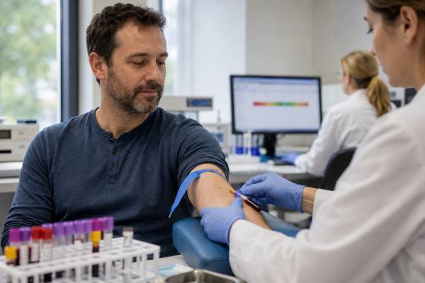
A total testosterone test measures all testosterone circulating in blood, including hormone bound to SHBG and albumin plus the small free fraction. It is the usual first laboratory test when male androgen deficiency is suspected, but one low result does not establish hypogonadism. Diagnosis generally requires compatible symptoms or signs and a second low morning value obtained under suitable conditions. Total testosterone can be misleading when SHBG is unusually low or high, so free testosterone and SHBG may be needed when the result is borderline or does not match the clinical picture. Low levels may reflect primary testicular failure, pituitary or hypothalamic suppression, obesity, illness, sleep disruption, medicines, or anabolic steroid withdrawal. High levels most often result from testosterone treatment, performance-enhancing drugs, collection timing, or laboratory issues; endogenous tumors are much less common. Results must be interpreted using the laboratory method and units, not a universal online range. Treatment decisions also require fertility planning because exogenous testosterone suppresses LH, FSH, intratesticular testosterone, and sperm production.
- Total testosterone is usually checked in the morning on two separate days when diagnosing deficiency.
- A low total result with low SHBG may not mean free testosterone is low; a high SHBG can hide low free testosterone.
- Low testosterone with high LH suggests primary testicular failure, while low testosterone with low or normal LH suggests central or functional suppression.
- High results should be reviewed against testosterone dosing, anabolic steroid use, supplements, sample timing, and possible gel contamination.
- Men seeking fertility should not start testosterone without specialist advice because treatment can greatly reduce sperm production.
Table of Contents
- What total testosterone measures
- When the test is used
- Preparation, timing, and test methods
- Normal range and result interpretation
- Causes of low testosterone
- Causes of high testosterone
- Symptoms and follow-up tests
- Treatment, fertility, and monitoring
What total testosterone measures
Testosterone is produced mainly by Leydig cells in the testes under stimulation from pituitary LH. A smaller amount of androgen precursor comes from the adrenal glands. Testosterone supports sexual desire, erectile physiology, sperm production through high intratesticular concentrations, muscle and bone, body hair, red blood cell production, mood, and energy-related functions.
In blood, most testosterone is attached to proteins. SHBG binds it tightly, albumin binds it more loosely, and usually only a small percentage remains free. Total testosterone adds all three fractions together. This makes it a practical first test, but the number depends on both hormone production and binding-protein concentration.
A low total value can occur because the testes produce less testosterone, because pituitary signaling is reduced, or because SHBG is low and the bound reservoir is smaller. A high total value can occur with excess production or treatment, but also with high SHBG. The result therefore does not show the cause by itself.
Free testosterone is the unbound fraction. Bioavailable testosterone generally includes free plus albumin-bound hormone. These measures may clarify cases near a diagnostic threshold. A free testosterone test is particularly helpful when obesity, thyroid disease, liver disease, aging, or medicine use is likely to alter SHBG.
Total testosterone also cannot measure androgen-receptor sensitivity or explain all sexual symptoms. A normal result does not exclude vascular erectile dysfunction, depression, medication effects, infertility, thyroid disease, sleep apnea, or relationship factors. A low result does not prove that fatigue or low mood is caused by testosterone.
When the test is used
Testing is appropriate when symptoms or signs suggest androgen deficiency. More specific features include reduced libido, fewer spontaneous or morning erections, infertility, hot flushes in severe deficiency, reduced body hair or shaving frequency, small or soft testes, gynecomastia, low-trauma fracture, osteoporosis, delayed puberty, and unexplained anemia.
Less specific symptoms include fatigue, low mood, irritability, reduced concentration, sleep disturbance, loss of strength, increased body fat, and lower exercise performance. These are common in many conditions, so a testosterone result should be part of a broader medical assessment.
The test is also used in evaluation of pituitary or testicular disease, delayed puberty, anabolic steroid suppression, gynecomastia, infertility, and selected bone-health problems. It may monitor testosterone replacement, antiandrogen treatment, or recovery from a suppressive exposure, with collection timing adjusted to the therapy.
Risk factors include Klinefelter syndrome, undescended testes, torsion, orchitis, chemotherapy, radiation, traumatic brain injury, pituitary surgery or tumors, iron overload, chronic opioids, glucocorticoids, severe obesity, diabetes, sleep apnea, undernutrition, and prior anabolic steroid use.
Routine population screening in men without symptoms is not usually recommended. Testing to find a maximized “optimal” level in a healthy man can identify normal biological variation and expose him to unnecessary treatment. Age alone is not a diagnosis.
In infertility, testosterone is one component of the assessment. Serum testosterone does not measure sperm count or intratesticular testosterone directly. A male fertility hormone panel and semen analysis provide the relevant reproductive context.
Preparation, timing, and test methods
For diagnosis in an untreated adult man, blood is generally collected in the early morning, often between 7 and 10 a.m., because testosterone is linked to sleep and usually peaks after waking. The rhythm is stronger in younger men and can be shifted in night workers. A shift worker should discuss testing after the main sleep period rather than relying only on clock time.
Many clinical guidelines favor fasting samples because food and glucose intake can temporarily lower testosterone. Follow the ordering clinician’s instructions. Drinking water is usually acceptable unless the laboratory says otherwise.
Adequate sleep and stable health improve reliability. Acute infection, hospitalization, surgery, severe sleep loss, major endurance exercise, and marked calorie restriction can lower testosterone temporarily. When safe, testing after recovery prevents a transient result from being labeled chronic hypogonadism.
Bring a complete list of medicines, hormones, supplements, and injections. Opioids, glucocorticoids, antiandrogens, some anticonvulsants, and cancer treatments can lower testosterone. Testosterone and anabolic steroids may raise the measured value during use while suppressing natural production. Biotin can interfere with some laboratory tests. Do not stop treatment without advice.
A low result is usually repeated on another morning. Day-to-day variation and assay imprecision mean two samples are more dependable than one. Use of the same laboratory improves comparison.
Liquid chromatography–tandem mass spectrometry offers high specificity and is often preferred when accuracy is critical, although quality-controlled immunoassays are widely used. Results from different methods may not be interchangeable. Men using testosterone gel should avoid applying it near the blood-draw site; contamination can cause a falsely extreme value. Therapy monitoring must follow formulation-specific timing.
Normal range and result interpretation
There is no single total testosterone normal range that applies to every laboratory, age group, assay, and clinical guideline. Results may be reported in nanograms per deciliter or nanomoles per liter. The reference interval and decision threshold on the report must be considered with the assay and symptoms.
A reference interval describes values in a selected population; it is not automatically the cutoff for treatment. Guidelines use different thresholds and emphasize consistently low values plus clinical features. A result just below one laboratory’s lower limit may fall inside another range.
| Total testosterone pattern | Possible interpretation | Common next step |
|---|---|---|
| Repeatedly low total and free testosterone | Biochemical androgen deficiency is likely when symptoms are compatible | Measure LH, FSH, prolactin as indicated and identify the cause |
| Low total testosterone, low SHBG, normal free testosterone | Binding effect may account for much of the low total value | Assess obesity, insulin resistance, thyroid and other low-SHBG causes |
| Normal total testosterone, high SHBG, low free testosterone | High binding may conceal reduced available hormone | Confirm free testosterone and evaluate high-SHBG causes |
| High total testosterone, high SHBG, normal free testosterone | Expanded bound fraction may elevate total value | Review thyroid, liver, estrogen, medication, and assay factors |
| High total and free testosterone with suppressed LH/FSH | Exogenous androgen exposure is likely | Review treatment or steroid use, timing, hematocrit, and fertility effects |
A borderline result should not be interpreted without SHBG when binding abnormalities are likely. The SHBG test in men helps determine whether total testosterone is under- or overrepresenting the free fraction.
Age-related averages decline, but healthy older men can have adequate testosterone and younger men can have pathological deficiency. Symptoms and cause remain important. Treatment is not intended to restore every older man to a young-adult peak.
Results during therapy are interpreted differently. A value soon after an injection may reflect a peak; a sample just before the next dose reflects a trough. The target depends on formulation and guideline. Chasing a high number can increase adverse effects without improving symptoms.
Causes of low testosterone
Low testosterone is classified by LH and FSH. Primary hypogonadism begins in the testes. Testosterone is low and LH is usually high because the pituitary is trying to compensate. FSH may also be elevated, especially when sperm-producing tissue is damaged. Causes include Klinefelter syndrome, torsion, orchitis, severe trauma, chemotherapy, radiation, and advanced testicular failure.
Secondary or central hypogonadism begins in the hypothalamus or pituitary. Testosterone is low while LH and FSH are low or inappropriately normal. Causes include pituitary tumors, high prolactin, pituitary surgery or radiation, traumatic brain injury, iron overload, congenital gonadotropin deficiency, and infiltrative disease.
Functional central suppression is common and may be reversible. Severe obesity, uncontrolled diabetes, sleep apnea, systemic illness, undernutrition, overtraining, chronic stress, opioids, and glucocorticoids can lower the axis. Improving the cause may raise testosterone without replacement.
Low SHBG can lower total testosterone disproportionately. Obesity, insulin resistance, hypothyroidism, nephrotic syndrome, androgen exposure, and glucocorticoids are examples. Free testosterone may remain inside range, so a low total result does not always equal true androgen deficiency.
Anabolic steroid or testosterone withdrawal can produce low testosterone with suppressed LH and FSH. The axis may take months or longer to recover, and sperm recovery can lag behind serum hormones. Product names, doses, dates, and co-administered drugs are essential for interpretation.
Normal aging is associated with gradual hormonal change, but acute or marked low testosterone should not be dismissed as age. Chronic illness, medicines, obesity, and pituitary or testicular disease become more common over time and may be treatable.
Causes of high testosterone
The most common cause of high testosterone in clinical practice is exogenous exposure. Prescribed injections, gels, pellets, oral preparations, anabolic steroids, prohormones, and contaminated supplements can raise the value. The degree depends on dose, compound, absorption, and sample timing.
Injectable products often create a peak and decline. A sample collected shortly after an injection may appear excessive, while a trough may be low. Gel contamination of the venipuncture site can produce an implausibly high result. Repeating the test with correct technique may resolve the discrepancy.
High SHBG can elevate total testosterone while free testosterone remains normal. Hyperthyroidism, some liver conditions, estrogen exposure, aging, HIV, and certain anticonvulsants may raise SHBG. Treating a high total number without checking free testosterone can be harmful.
Endogenous overproduction is uncommon in adult men. Testicular or adrenal tumors, hCG-secreting tumors, congenital adrenal hyperplasia, and rare endocrine disorders may increase androgens. The pattern of LH, DHEA-S, androstenedione, hCG, estradiol, examination, and imaging helps identify the source.
A high value with high LH is unusual. Selective estrogen receptor modulators or aromatase inhibitors can raise LH and testosterone intentionally. Androgen resistance, assay interference, and rare disorders are other possibilities. The medication list and repeat testing come before a broad tumor search.
Excess androgen exposure can cause acne, oily skin, elevated hematocrit, mood or sleep changes, edema, worsening sleep apnea, gynecomastia through aromatization, reduced sperm production, and testicular shrinkage. High testosterone is not a marker of superior health when it is treatment-driven or supraphysiological.
Symptoms and follow-up tests
Symptoms of low testosterone vary in specificity. Reduced libido, fewer morning erections, infertility, hot flushes, loss of body hair, small testes, gynecomastia, low bone density, and anemia carry more diagnostic weight. Fatigue, depression, poor focus, reduced strength, and increased fat can have many other causes.
After a repeat low result, LH and FSH distinguish primary from central patterns. A luteinizing hormone test in men is especially important because a normal-range LH may be inappropriate when testosterone is very low. Prolactin is useful with low or normal LH, infertility, low libido, or pituitary symptoms.
SHBG and albumin support free testosterone calculation. Thyroid tests, iron studies, liver and kidney function, complete blood count, glucose or A1c, and sleep-apnea assessment are selected according to the pattern. A semen analysis is required when fertility is a concern.
Pituitary MRI may be indicated with very low testosterone and low gonadotropins, persistently high prolactin, headaches, visual changes, or other pituitary deficiencies. Testicular examination and ultrasound are appropriate with a mass, pain, marked asymmetry, or an unusual tumor-marker pattern.
Bone-density imaging may be considered with long-standing severe deficiency, fractures, height loss, or other osteoporosis risks. Karyotype or other genetic testing may be appropriate with small testes, high gonadotropins, delayed puberty, azoospermia, or suspected congenital disease.
Seek urgent care for sudden severe headache with visual loss, acute testicular pain, a new testicular mass, chest pain, severe shortness of breath, stroke symptoms, or a painful swollen leg. These findings should not wait for routine hormone follow-up.
Treatment, fertility, and monitoring
Treatment starts with the cause. Weight management, sleep-apnea treatment, adequate nutrition, diabetes care, recovery from illness, and review of suppressive medicines may restore functional low testosterone. Pituitary tumors, high prolactin, thyroid disease, iron overload, and testicular disorders require specific therapy.
Testosterone replacement may be considered when symptoms and consistently low values confirm hypogonadism after evaluation. Benefits may include improved sexual symptoms, anemia, bone density, and body composition in appropriately selected men. Nonspecific symptoms do not always improve.
Men who want fertility should not use exogenous testosterone without specialist advice. Treatment suppresses LH and FSH, lowers intratesticular testosterone, and can reduce sperm count to zero. Depending on the cause, hCG, FSH, or selected off-label medicines may stimulate endogenous production under reproductive specialist supervision.
Before treatment, clinicians review hematocrit, fertility plans, prostate and urinary history, sleep apnea, cardiovascular health, breast symptoms, and contraindications. A complete hypogonadism blood test panel should be documented before exogenous hormone changes the diagnostic pattern.
Monitoring includes symptoms, testosterone timed to the formulation, hematocrit, adverse effects, and age- and risk-appropriate prostate assessment. Blood pressure, edema, acne, sleep apnea, mood, breast symptoms, and fertility effects also matter. Dose changes should not pursue the highest possible value.
If a well-conducted trial normalizes testosterone but symptoms do not improve, the original symptoms may have another cause. Reassessment is more appropriate than indefinite dose escalation. A total testosterone result is a diagnostic starting point; careful confirmation and pattern-based interpretation make it clinically useful.
Testosterone changes with sleep, weight, and illness
Testosterone is linked more closely to sleep than to a particular wall-clock hour. Fragmented sleep, untreated sleep apnea, night-shift work, and short sleep can lower morning values and worsen sexual or energy symptoms through several pathways. A repeat sample after adequate sleep can be more representative, while persistent symptoms may justify formal sleep assessment.
Weight change can alter both production and binding. Obesity commonly lowers SHBG and total testosterone and can also suppress hypothalamic signaling. With weight loss, SHBG may rise quickly while free testosterone and gonadal function change more gradually. A higher total value after weight loss therefore reflects both improved metabolic physiology and a larger bound fraction.
Acute illness produces adaptive suppression. Testosterone can fall during infection, surgery, trauma, or severe systemic disease without indicating permanent failure. Testing during hospitalization may be necessary for a specific endocrine emergency, but routine diagnosis is usually deferred until recovery. Chronic disease can cause more persistent suppression and deserves treatment in its own right.
Assessing treatment response beyond the laboratory value
Before therapy, record the symptoms expected to improve. Libido, morning erections, hot flushes, anemia, and bone density have different timelines and likelihoods of response. Erectile dysfunction caused mainly by vascular disease may not improve substantially with testosterone, even when a low level is corrected. Fatigue may persist if sleep apnea, depression, anemia, or medication effects are untreated.
After treatment begins, the measured testosterone should be timed to the formulation. A peak after injection may help explain acne or mood fluctuation, while a low trough may explain end-of-interval symptoms. Adjusting dose size or frequency may be more appropriate than simply increasing the total amount. Gel users need consistent application and precautions to prevent transfer to partners or children.
A normalized number without clinical benefit should prompt reassessment. Continuing or escalating therapy solely to maintain a laboratory target exposes the patient to hematocrit elevation, infertility, acne, edema, breast symptoms, and sleep-apnea worsening. Conversely, clear symptom improvement with a safe physiological level supports continued monitored treatment.
Interpreting testosterone in cancer and prostate care
Men receiving androgen-deprivation therapy for prostate cancer are intentionally maintained at very low testosterone. Their result is interpreted against treatment goals rather than the general male reference interval. Monitoring may include PSA, treatment adherence, bone density, metabolic health, cardiovascular risk, hot flushes, anemia, and quality of life.
A low testosterone value does not protect completely against prostate cancer, and a normal value does not diagnose it. Prostate symptoms and PSA require their own evaluation. Men considering testosterone therapy should discuss personal risk, prior prostate cancer, urinary symptoms, and the monitoring plan with the treating team.
In men treated for testicular cancer, surgery, chemotherapy, or radiation can alter Leydig-cell reserve. LH may rise before total testosterone falls, and long-term surveillance can identify evolving deficiency. Fertility preservation and semen assessment are separate from serum testosterone because sperm-producing tissue may be affected differently.
Keep the original laboratory report. The assay, units, reference interval, collection time, and treatment status are needed to interpret future changes. A bare number entered into a fitness application or copied without units cannot support a safe diagnosis or dose adjustment.
A carefully repeated result is more clinically useful than several unstandardized home or afternoon measurements.
References
- Sexual and Reproductive Health 2026 (Clinical Guideline)
- Male hypogonadism: recommendations from the Fifth International Consultation on Sexual Medicine 2025 (Consensus Recommendations)
- The British Society for Sexual Medicine Guidelines on Male Adult Testosterone Deficiency, with Statements for Practice 2023 (Clinical Guideline)
- Testosterone Deficiency Guideline 2024 (Clinical Guideline)
- Testosterone Therapy in Men With Hypogonadism: An Endocrine Society Clinical Practice Guideline 2018 (Clinical Guideline)
- Associations of Testosterone and Related Hormones With All-Cause and Cardiovascular Mortality and Incident Cardiovascular Disease in Men 2024 (Systematic Review and Meta-analysis)
Disclaimer
This article is educational and does not diagnose testosterone deficiency or determine whether hormone treatment is appropriate. Results require interpretation with symptoms, repeat testing, assay method, SHBG, related hormones, medicines, health conditions, and fertility goals. Do not start, stop, or change testosterone or another prescription medicine without a qualified clinician.