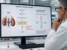
The high-dose dexamethasone suppression test is an older dynamic hormone test used after ACTH-dependent Cushing syndrome has been established. It asks whether a large dose of dexamethasone can suppress cortisol production. A substantial fall in cortisol has traditionally favored Cushing disease, which is caused by an ACTH-secreting pituitary tumor, while little or no suppression has favored ectopic ACTH production. The distinction is imperfect: some pituitary tumors do not suppress, and some ectopic tumors do. For that reason, the test should not be used to screen for Cushing syndrome or to make the source diagnosis by itself. Modern evaluation combines ACTH measurement, pituitary MRI, clinical features, other dynamic tests, and often inferior petrosal sinus sampling when results disagree. Dexamethasone metabolism, estrogen therapy, medicines, illness, alcohol use, and laboratory method can all affect results. Interpretation belongs within a specialist-directed sequence rather than as a stand-alone positive or negative answer.
- The high-dose test is a source-localization test, not an initial screening test for Cushing syndrome.
- A cortisol fall of about 50% or more has historically supported pituitary Cushing disease, but it is not definitive.
- Failure to suppress does not rule out Cushing disease, because a meaningful minority of pituitary tumors show little response.
- Dexamethasone-altering medicines and estrogen can distort results, so preparation and medication review are essential.
- Discordant results often require pituitary imaging and inferior petrosal sinus sampling, not repeated high-dose testing alone.
Table of Contents
- Where the High-Dose Test Fits
- How the Test Works
- High-Dose Dexamethasone Protocols
- How Results Are Calculated
- Suppression and Cushing Disease
- False-Positive and False-Negative Results
- Comparison With Other Localization Tests
- What Happens After the Test
Where the High-Dose Test Fits
Cushing syndrome describes sustained exposure to excessive cortisol from any cause. Endogenous Cushing syndrome is first confirmed with tests designed to detect cortisol excess, such as late-night salivary cortisol, 24-hour urine free cortisol, or the 1-mg overnight low-dose dexamethasone suppression test. The Cushing syndrome test panel uses more than one approach because no single screening result is perfect.
Once cortisol excess is confirmed, plasma ACTH divides the problem into ACTH-independent and ACTH-dependent forms. A suppressed ACTH points toward an adrenal source. A normal or elevated ACTH suggests either a pituitary corticotroph tumor, called Cushing disease, or ACTH secretion from a nonpituitary tumor, called ectopic ACTH syndrome. The high-dose dexamethasone test was developed to help distinguish those two ACTH-dependent causes.
Pituitary corticotroph adenomas often retain some sensitivity to glucocorticoid feedback. Ectopic ACTH-producing tumors usually do not. Giving a high dose of dexamethasone therefore may reduce ACTH and cortisol in Cushing disease while leaving ectopic ACTH secretion relatively unchanged. This biological tendency is real, but the response overlaps too much for a single result to establish the source with certainty.
The test has a smaller role than it once did. Pituitary MRI has improved, corticotropin-releasing hormone and desmopressin tests can provide complementary information, and inferior petrosal sinus sampling can directly compare central and peripheral ACTH. Current specialist practice may use the high-dose test when it adds information, but many centers do not rely on it routinely.
The test is inappropriate before hypercortisolism has been confirmed. A person with suggestive symptoms but normal first-line testing should not proceed directly to high-dose dexamethasone. Likewise, the test does not distinguish exogenous steroid exposure from endogenous Cushing syndrome and cannot localize an adrenal cortisol-producing tumor.
How the Test Works
Dexamethasone is a potent synthetic glucocorticoid. In a healthy hypothalamic-pituitary-adrenal axis, it signals the pituitary to reduce ACTH, which lowers adrenal cortisol production. Standard cortisol assays generally do not measure dexamethasone as cortisol, allowing the laboratory to observe the suppression of endogenous hormone.
In pituitary Cushing disease, the tumor secretes ACTH autonomously but often retains partial feedback sensitivity. A low dexamethasone dose may not suppress it, while a much larger dose sometimes does. In ectopic ACTH syndrome, tumor cells usually lack this feedback response. The test therefore compares cortisol before and after dexamethasone rather than relying only on the absolute post-dose value.
This mechanism explains both the value and the weakness of the test. Pituitary tumors vary biologically. Some are highly suppressible; others are resistant. Ectopic tumors are also diverse, and occasional neuroendocrine tumors can suppress. Severe hypercortisolism itself may alter metabolism and receptor signaling. The result is a continuum rather than a clean division.
Dexamethasone concentration can be measured at the same time as cortisol in some laboratories. A low dexamethasone level suggests poor absorption, missed dosing, or rapid metabolism and can explain apparent failure to suppress. This measurement is especially useful when the result conflicts with the broader clinical picture.
The test evaluates biochemical response, not tumor visibility. A tiny pituitary adenoma can produce a strong suppressive response, while a visible pituitary lesion may be incidental. Small pituitary abnormalities are common in the general population, so MRI findings must be interpreted with endocrine evidence.
High-Dose Dexamethasone Protocols
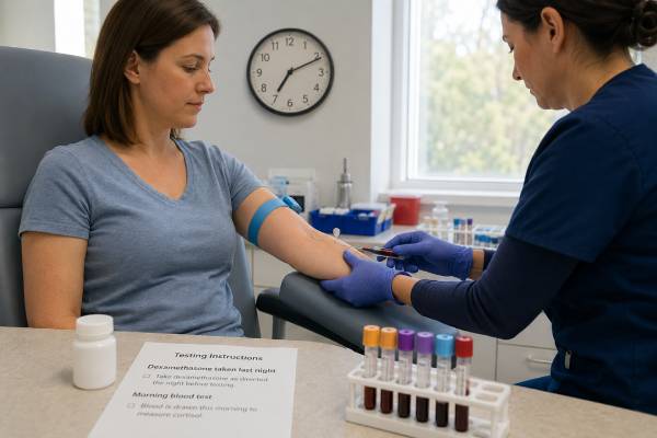
Two classic protocols are used. The overnight test gives 8 mg of dexamethasone late in the evening, commonly around 11 p.m., followed by serum cortisol the next morning around 8 a.m. A baseline cortisol is obtained before dosing, either that morning or shortly before the test, according to the center’s protocol.
The longer protocol gives 2 mg of dexamethasone every six hours for 48 hours, for a total of eight doses. Cortisol may be measured in blood, and 24-hour urine free cortisol may be collected before and during dexamethasone. This approach provides more observations but is less convenient and increases the chance of missed or mistimed doses.
A typical overnight sequence is:
- Confirm that endogenous ACTH-dependent Cushing syndrome has already been established.
- Review medicines, estrogen exposure, alcohol use, acute illness, and factors affecting dexamethasone metabolism.
- Obtain the required baseline cortisol and sometimes ACTH.
- Take 8 mg dexamethasone at the exact instructed time.
- Return for morning cortisol at the scheduled time.
- Measure serum dexamethasone when the laboratory or clinical question warrants it.
Patients should follow the center’s written instructions precisely. Taking the dose early or late changes the exposure interval. Vomiting after the dose, gastrointestinal malabsorption, or forgetting a tablet can invalidate the test. The ordering team should be contacted if any of these occur.
Fasting requirements vary. The morning blood draw is often performed before breakfast because other tests may be collected at the same time. Usual medicines should not be stopped unless the clinician gives specific directions. Abrupt withdrawal of glucocorticoids can be dangerous and can also make interpretation impossible.
Short exposure to dexamethasone is generally well tolerated. Temporary insomnia, restlessness, mood change, increased appetite, indigestion, or a rise in glucose can occur. People with diabetes may need a plan for glucose monitoring. Severe psychiatric reactions are uncommon after a single dose but deserve prompt attention, especially in someone with a history of steroid-induced mania or psychosis.
How Results Are Calculated
High-dose results are commonly expressed as the percentage decrease in serum cortisol or urine free cortisol from baseline. The calculation is:
Percentage suppression = [(baseline cortisol − post-dexamethasone cortisol) ÷ baseline cortisol] × 100
For example, if baseline serum cortisol is 24 mcg/dL and the next-morning value is 10 mcg/dL, the decrease is 14 mcg/dL. Dividing 14 by 24 and multiplying by 100 gives about 58% suppression. Under the traditional rule, that pattern supports Cushing disease.
A threshold of at least 50% suppression is most widely recognized. Some protocols or studies use 80% suppression to improve specificity, but a stricter threshold misses more pituitary cases. Laboratories and endocrine centers may define response differently depending on whether serum cortisol, urine free cortisol, or another endpoint is used.
| Response pattern | Traditional interpretation | Important limitation |
|---|---|---|
| At least 50% cortisol reduction | Supports pituitary Cushing disease | Some ectopic tumors also suppress |
| Less than 50% reduction | Raises concern for ectopic ACTH | Many pituitary tumors fail to suppress |
| Unexpectedly low dexamethasone level | Test exposure may have been inadequate | Result may be uninterpretable |
| Discordant serum and urine responses | No secure localization | Requires integration with other tests |
The percentage calculation can be unstable when baseline cortisol varies. Cushing syndrome may be cyclic, and cortisol secretion changes from day to day. A baseline taken during a lower phase can make the percentage response misleading. This is one reason specialists review the complete sequence of cortisol measurements rather than a single ratio.
An absolute post-dexamethasone cortisol may also be considered, but no universal value reliably separates pituitary from ectopic disease. Assay-specific differences further limit fixed thresholds. The result should be labeled “supportive” or “not supportive,” not “diagnostic” or “excluded.”
Suppression and Cushing Disease
Substantial suppression increases the probability of Cushing disease when the patient already has confirmed ACTH-dependent hypercortisolism. It becomes more persuasive when other findings agree: a pituitary lesion on high-quality MRI, a rise in ACTH or cortisol after CRH or desmopressin, and a clinical course typical of a pituitary source.
Even then, the high-dose response cannot prove that a visible pituitary lesion is the source. Incidental pituitary lesions occur in people without Cushing disease. A small lung, thymic, pancreatic, or other neuroendocrine tumor could be producing ACTH while an unrelated pituitary spot appears on MRI.
Failure to suppress does not exclude Cushing disease. Depending on protocol and study population, sensitivity is often only moderate. A substantial minority of pituitary adenomas do not show the traditional 50% reduction. Treating every nonsuppressor as ectopic disease would misclassify many patients.
Ectopic ACTH syndrome is also heterogeneous. Some tumors grow slowly and cause relatively mild cortisol excess; others produce severe, rapid hypercortisolism with low potassium, muscle weakness, infections, diabetes, and psychiatric symptoms. Rare ectopic tumors show dexamethasone suppression and can imitate pituitary disease.
The pretest probability matters. Cushing disease is the most common source of endogenous ACTH-dependent Cushing syndrome in many adult populations. A supportive high-dose test therefore adds to an already meaningful probability, whereas the same response in a population enriched for ectopic tumors may be less convincing.
No result should delay treatment of severe cortisol excess. Marked hypokalemia, uncontrolled diabetes, infection, thrombosis, heart failure, or psychosis may require urgent cortisol-lowering therapy while localization continues. The source investigation and control of immediate risk can proceed in parallel.
False-Positive and False-Negative Results
A false-positive localization result occurs when an ectopic ACTH source suppresses enough to resemble Cushing disease. A false-negative occurs when a pituitary tumor fails to suppress. Biological overlap is the main limitation, but technical and medication factors add further uncertainty.
Dexamethasone is metabolized mainly through hepatic CYP3A4 pathways. Medicines that accelerate this pathway can lower dexamethasone exposure and create apparent nonsuppression. Examples include some antiseizure drugs, rifampin, and other enzyme inducers. Medicines that slow metabolism can increase exposure and exaggerate suppression. The exact interaction list is long, so a pharmacist or endocrinologist should review all medicines rather than relying on a few remembered examples.
Oral estrogen raises cortisol-binding globulin. This increases total serum cortisol even when free cortisol is not increased and can make suppression appear inadequate. Depending on urgency and clinical circumstances, a clinician may arrange a supervised pause in oral estrogen before serum-based dexamethasone testing. Transdermal estrogen generally has less effect on cortisol-binding globulin, but treatment changes still require individualized advice.
Other sources of error include:
- Missed, delayed, or vomited dexamethasone doses.
- Malabsorption after gastrointestinal surgery or active intestinal disease.
- Incorrect blood-draw timing.
- Acute illness, major stress, severe sleep disruption, or hospitalization.
- Heavy alcohol use, major depression, or other states that can activate the cortisol axis.
- Cyclic Cushing syndrome, with testing during a lower-cortisol phase.
- Assay interference or a change in laboratory method between baseline and follow-up.
The high-dose test should generally be performed after a stable diagnosis of endogenous hypercortisolism, not during an acute confounding illness. Measuring dexamethasone can identify inadequate exposure, but a normal drug level cannot fix the test’s biological overlap.
Comparison With Other Localization Tests
Pituitary MRI is usually part of the ACTH-dependent workup. It can identify a corticotroph adenoma, but many tumors are only a few millimeters and may be missed. Conversely, small incidental lesions can be seen in people whose ACTH comes from elsewhere. MRI is strongest when the lesion size, endocrine testing, and clinical findings agree.
CRH and desmopressin stimulation tests use the tendency of pituitary corticotroph tumors to respond to these signals. Ectopic tumors usually respond less, but overlap remains. Combining several noninvasive tests may improve confidence when all point in the same direction; conflicting results lower confidence.
Inferior petrosal sinus sampling, or IPSS, compares ACTH in veins draining the pituitary with ACTH in peripheral blood, usually before and after CRH or desmopressin. A central-to-peripheral ACTH gradient supports a pituitary source. IPSS is invasive and should be performed by an experienced team, but it is generally the reference localization test when MRI is negative, shows a small lesion, or conflicts with biochemistry.
A high-dose suppression result should not be used to avoid IPSS when current criteria indicate it. Likewise, IPSS should not be used to prove hypercortisolism; Cushing syndrome must be established first because normal people can show central ACTH gradients.
Imaging for ectopic sources may include chest and abdominal CT, MRI, and functional imaging selected according to the biochemical pattern and suspected tumor type. Starting extensive imaging before confirming ACTH-dependent Cushing syndrome can uncover incidental findings and complicate rather than clarify the diagnosis.
| Test | Main role | Main limitation |
|---|---|---|
| High-dose dexamethasone | Noninvasive supportive localization | Moderate sensitivity and specificity |
| Pituitary MRI | Finds a possible pituitary lesion | Small tumors can be missed; incidental lesions occur |
| CRH or desmopressin test | Assesses pituitary-type responsiveness | Biological overlap |
| IPSS | Determines central versus peripheral ACTH source | Invasive and expertise-dependent |
What Happens After the Test
The endocrinologist reviews the high-dose result alongside ACTH, the screening tests that confirmed Cushing syndrome, MRI findings, clinical severity, and any CRH or desmopressin response. A concordant pituitary pattern may lead toward transsphenoidal surgery. A discordant or uncertain pattern often leads to IPSS before surgery is considered.
If ectopic ACTH is more likely, imaging focuses on common neuroendocrine tumor sites, particularly the chest and abdomen. Some tumors remain occult at first. During that search, treatment may be needed to reduce cortisol and protect against infection, blood clots, fractures, hypertension, diabetes, and psychiatric complications.
If the test appears invalid because dexamethasone exposure was inadequate, the team may repeat it under controlled conditions or choose another localization method. Repeating a technically valid but biologically inconclusive test several times usually adds less value than moving to a more discriminating test.
Questions worth asking at follow-up include:
- Has endogenous Cushing syndrome been firmly confirmed with more than one appropriate test?
- Is ACTH clearly in the dependent range?
- Was the dexamethasone level adequate, if measured?
- How much did cortisol fall, and which endpoint was used?
- Does MRI show a lesion large enough to change the need for IPSS?
- Are severe cortisol complications being treated while localization continues?
A high-dose dexamethasone result is one piece of probabilistic evidence. The safest decisions come from agreement among several independent findings, interpreted by a team experienced in Cushing syndrome.
The result also does not predict surgical cure. After pituitary surgery, clinicians use early postoperative cortisol, the need for glucocorticoid replacement, pathology, and later surveillance to judge remission. A patient can have a suppressive high-dose test before surgery and still have persistent disease, while a nonsuppressive test does not mean pituitary surgery will fail when IPSS and imaging establish the source.
People being evaluated for Cushing syndrome often have substantial medical risk before the source is settled. High cortisol increases susceptibility to infection, venous thromboembolism, osteoporosis, muscle weakness, high blood pressure, and difficult-to-control glucose. Clinicians may prescribe potassium, blood-pressure treatment, diabetes therapy, anticoagulation in selected high-risk situations, infection prevention, or a cortisol-lowering medicine. These measures do not invalidate the need for localization; they reduce preventable harm during a complex workup.
The high dexamethasone dose itself should be documented in the medical record because it can temporarily affect glucose, sleep, and subsequent hormone measurements. If another cortisol test is planned immediately afterward, the endocrine team should decide the interval needed for dexamethasone to clear. Patients should not independently repeat the dose because a result seems confusing.
The test may be particularly difficult to interpret in cyclic Cushing syndrome. During a low-output phase, baseline cortisol can approach normal and percentage suppression can look large even though the tumor source has not changed. Specialists may first document that the patient is in an active hypercortisolemic phase with late-night salivary cortisol or urine free cortisol, then perform localization testing. A symptom diary paired with repeated biochemical testing can help identify those phases.
Pregnancy, severe kidney or liver disease, and hospitalization also alter the ordinary testing framework. Pregnancy raises cortisol-binding globulin and changes normal cortisol physiology. Liver disease can affect dexamethasone metabolism, while kidney disease may limit urine-based endpoints. In such settings, an experienced endocrinology laboratory should select the protocol and interpretive method.
Why modern centers use the result selectively
The high-dose test remains attractive because it is noninvasive, but its overlapping responses can create false confidence. Centers that use it often place greatest weight on concordance: suppression, a pituitary-type response to CRH or desmopressin, and a matching MRI lesion. When those findings conflict, IPSS generally provides stronger source evidence than choosing whichever noninvasive result seems most convenient.
A measured dexamethasone concentration can confirm exposure, but it cannot solve biological overlap between pituitary and ectopic tumors. Adequate drug levels make the test technically valid, not diagnostically perfect.
Because localization errors can lead to the wrong operation, a supportive result should never outweigh clearly discordant ACTH, imaging, or petrosal sinus evidence.
References
- Consensus on diagnosis and management of Cushing’s disease: a guideline update 2021 (Guideline)
- Dynamic Testing for Differential Diagnosis of ACTH-Dependent Cushing Syndrome: A Systematic Review and Meta-Analysis 2023 (Systematic Review)
- Cushing Syndrome: A Review 2023 (Review)
- Diagnostic workup of Cushing’s syndrome 2022 (Review)
- Dexamethasone Suppression Test 2024 (Review)
Disclaimer
This article is for general education and does not replace interpretation by an endocrinologist. High-dose dexamethasone testing is not an appropriate self-ordered screening test, and its result cannot by itself locate the source of ACTH. Medicine changes, especially involving glucocorticoids or estrogen, should be made only with medical guidance.