
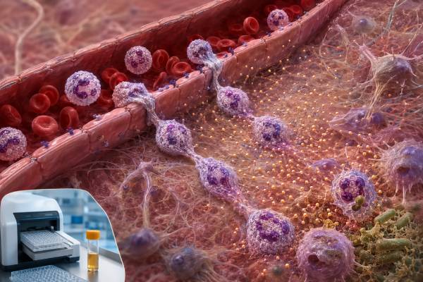
An interleukin-8 (IL-8) test measures a chemokine—also named CXCL8—that directs neutrophils toward infection, tissue injury, and other inflammatory signals. IL-8 can rise quickly when epithelial cells, macrophages, endothelial cells, or other tissues detect microbes or damage. That makes it biologically important in sepsis, lung inflammation, wounds, burns, inflammatory disease, and cancer, but it also makes the result highly nonspecific. A blood IL-8 value cannot tell whether inflammation is bacterial, viral, sterile, autoimmune, or tumor-related without other evidence. Direct testing is most common in specialty care, severe-illness panels, and research rather than routine outpatient diagnosis. Levels vary by specimen, collection method, assay platform, age, timing, and treatment. A high result supports active neutrophil-recruiting signaling but does not prove that neutrophil function is effective or that infection is present. A low result may be normal, may occur after the brief peak has passed, and does not rule out localized inflammation. The useful interpretation combines the exact assay with symptoms, vital signs, blood counts, cultures, CRP, procalcitonin, organ function, imaging, and the clinical timeline.
- IL-8 and CXCL8 are two names for the same major human neutrophil-attracting chemokine.
- CXCR1 and CXCR2 are its principal receptors, with overlapping but distinct roles in neutrophil movement and activation.
- Serum and plasma IL-8 values may differ and should not be compared across platforms.
- High IL-8 is a pattern of inflammation, not a diagnosis of sepsis or cancer.
- Tissue IL-8 can be high while a blood result remains low or undetectable.
Table of Contents
- CXCL8 and Neutrophil Traffic
- IL-8 Test Formats and Specimens
- Infection, Sepsis, and Critical Illness
- Noninfectious Inflammation and Cancer
- Interpreting a High IL-8 Value
- Low Values, Timing, and Test Limitations
- Useful Follow-Up Tests and Next Steps
CXCL8 and Neutrophil Traffic
IL-8 belongs to the CXC chemokine family and is officially designated CXCL8. Chemokines are directional signals: they form concentration gradients that guide cells bearing matching receptors. CXCL8 is produced by monocytes and macrophages, epithelial cells, endothelial cells, fibroblasts, neutrophils, and some tumor cells after stimulation by microbial products, IL-1, tumor necrosis factor, oxidative stress, or tissue damage.
Its principal receptors are CXCR1 and CXCR2, both G-protein-coupled receptors expressed strongly on neutrophils. CXCR2 responds to several ELR-positive CXC chemokines and is important in early recruitment from blood toward an inflammatory site. CXCR1 binds a narrower ligand set and can contribute strongly to activation closer to high local chemokine concentrations. Receptor expression changes as neutrophils encounter inflammatory signals, so the same circulating CXCL8 concentration does not guarantee the same cellular response in every patient.
When CXCL8 binds these receptors, neutrophils change shape, adhere to vessel walls, migrate through endothelium, and follow the gradient into tissue. Signaling can promote granule release, respiratory-burst priming, integrin activation, and cooperation with other chemoattractants. CXCL8 can contribute to neutrophil extracellular trap formation in some contexts, but this effect depends on additional signals and remains more complicated than a simple direct switch.
Neutrophils are essential for controlling many bacterial and fungal infections. They engulf microbes, generate reactive oxygen species, release enzymes and antimicrobial proteins, and organize inflammatory responses. The same mechanisms can injure lung, vessel, kidney, skin, or other tissue when recruitment is excessive or prolonged. CXCL8 therefore sits at the boundary between protective defense and collateral damage.
The pathway is redundant. CXCL1, CXCL2, CXCL5, CXCL6, leukotriene B4, complement fragments, and bacterial peptides can also attract neutrophils. A low IL-8 result does not mean neutrophil recruitment has stopped, and blocking one chemokine does not always eliminate tissue infiltration.
A useful way to think about CXCL8 is as a local road sign rather than a circulating fuel level. Endothelial cells display chemokines on vessel surfaces, where passing neutrophils can detect them. Tissue proteases can process CXCL8 into forms with altered activity, and glycosaminoglycans can hold it in place to preserve a gradient. These local interactions are not visible in a routine serum number. Two people with the same blood concentration may therefore have very different neutrophil accumulation in a wound, airway, joint, or tumor. Receptor desensitization can also make cells less responsive after intense exposure, so a high ligand value does not automatically mean stronger migration.
CXCL8 has no direct mouse equivalent that perfectly reproduces human biology, which complicates translation from animal models. Human assay findings must therefore be validated in the actual disease and clinical population before being used as diagnostic cutoffs.
IL-8 Test Formats and Specimens
A direct IL-8 immunoassay can be performed on serum, plasma, cerebrospinal fluid, bronchoalveolar lavage fluid, sputum, urine, wound fluid, synovial fluid, or cell-culture supernatant. The specimen is chosen according to the question. Blood reflects systemic spillover, while a local fluid may better represent inflammation in one organ.
Platforms include enzyme-linked immunosorbent assays, chemiluminescent and electrochemiluminescent methods, bead-based multiplex panels, and high-sensitivity digital assays. Results are often expressed in picograms per milliliter. Antibody specificity, calibration, matrix effects, lower detection limit, and reportable range differ, so a number from one method should not be judged by another laboratory’s range.
Serum is collected after clotting; plasma is separated from anticoagulated blood. During clotting and delayed processing, platelets and leukocytes can alter cytokine concentrations. A 2025 comparability study of IL-6, IL-8, and IL-10 reinforced that serum and plasma results require matrix-specific evaluation rather than automatic interchangeability.
Cell-based assays expose blood cells to bacterial ligands, mitogens, antigens, or experimental compounds and measure IL-8 released into culture. These tests evaluate production capacity under defined conditions. A high culture value is not the same as a high circulating level, and a low response requires valid positive controls and viable cells.
Tissue studies may measure CXCL8 messenger RNA, immunostaining, spatial protein expression, or downstream receptor signatures. Tumors and inflamed mucosa can show strong local expression even when serum testing is unremarkable. Gene-expression units, staining intensity, and protein concentration cannot share one reference interval.
IL-8 commonly appears within a chemokine panel or broader cytokine panel. Pattern analysis can be helpful in specialized settings, but multiple testing raises the chance of an incidental out-of-range result. The panel must be validated for the intended diagnostic or monitoring use.
Before interpreting any result, identify whether the report says IL-8, CXCL8, or both; whether the sample was serum, plasma, or local fluid; whether cells were stimulated; and whether the assay is labeled clinical or research use.
Infection, Sepsis, and Critical Illness
Microbial products can induce CXCL8 quickly, often before slower downstream markers reach their peak. This has made IL-8 an attractive candidate biomarker for early infection and sepsis. Studies frequently find that higher concentrations are associated with greater illness severity or poorer outcomes in selected cohorts. Association, however, does not create a universally reliable diagnostic threshold.
Sepsis is organ dysfunction caused by a dysregulated response to infection. Diagnosis requires evidence and judgment about infection, circulation, breathing, mental status, kidney and liver function, coagulation, and tissue perfusion. A high IL-8 value can fit sepsis but can also occur after major surgery, burns, trauma, pancreatitis, or immune therapy. A low value cannot safely rule sepsis out.
Neonatal and pediatric studies have examined IL-8 because newborn inflammatory responses and reference ranges differ from adult patterns. Some algorithms combine IL-8 with CRP or other markers to reduce unnecessary antibiotics, but performance depends on age, timing, setting, prevalence, and assay. A local validated protocol should not be generalized to all children.
In newborn care, even a promising early marker must be judged against the consequences of missing infection and the harms of unnecessary antibiotics. Gestational age, delivery stress, maternal infection, membrane rupture, respiratory distress, and sampling time can all influence the probability of sepsis and the inflammatory response. A cutoff that performs well in one neonatal intensive-care unit may perform poorly where disease prevalence or laboratory methods differ. Clinicians therefore use serial examinations and validated local pathways rather than applying a published IL-8 number in isolation. Parents should not interpret a neonatal cytokine result without the team that knows the infant’s complete course.
In pneumonia and acute respiratory distress syndrome, airway epithelial cells and macrophages can produce CXCL8, drawing neutrophils into the lungs. Bronchoalveolar or sputum concentrations may relate more closely to airway inflammation than serum levels. Sampling the lower respiratory tract is invasive and is performed only when clinically justified.
Burns and major trauma can generate substantial sterile CXCL8 release while also increasing infection risk. A rising value may reflect tissue injury, infection, or both. Serial examination, cultures, wound assessment, imaging, and organ trends are needed to distinguish these possibilities.
Cytokine release syndrome and macrophage activation syndromes can include IL-8 elevations, but their cytokine patterns and clinical triggers differ from ordinary bacterial sepsis. The patient can also have overlapping infection and immune dysregulation. Treatment should not be delayed while awaiting a specialty cytokine panel when urgent stabilization or antimicrobials are indicated.
A procalcitonin test, CRP, lactate, cultures, and molecular pathogen testing can add evidence, but no single biomarker is definitive. The most reliable approach integrates host-response markers with source-directed evaluation.
Noninfectious Inflammation and Cancer
CXCL8 is released in many sterile inflammatory conditions. Chronic obstructive pulmonary disease, severe asthma with neutrophilic features, inflammatory bowel disease, rheumatoid arthritis, gout, psoriasis, vasculitis, metabolic inflammation, and tissue ischemia can involve the pathway. The importance of CXCL8 differs by organ and disease phase.
In the airways, cigarette smoke, pollutants, infection, and damaged epithelium can stimulate CXCL8. Sputum neutrophils and local chemokines may remain elevated even when blood neutrophil counts are normal. A serum IL-8 test cannot diagnose COPD or distinguish a bacterial exacerbation from every noninfectious flare.
In joints, synovial cells can produce CXCL8 and recruit neutrophils, particularly in acute inflammatory arthritis. Crystal analysis, culture, imaging, and disease-specific evaluation are more clinically decisive. Septic arthritis and crystal arthritis can look similar and both can generate strong neutrophil signals; joint aspiration should not be replaced by serum cytokines.
Tumors may produce CXCL8 directly or induce stromal and immune cells to release it. The CXCL8-CXCR1/2 axis can recruit tumor-associated neutrophils and myeloid-derived suppressor cells, support angiogenesis, alter immune therapy response, and contribute to invasion. Elevated blood IL-8 has been studied as a prognostic or treatment-response biomarker in several cancers, but it is not a screening test and cannot identify tumor location.
Cancer treatment can change IL-8 through tumor response, infection, corticosteroids, immune activation, or tissue injury. A research association with survival should not be interpreted as proof that a person’s treatment is failing. Imaging, pathology, validated tumor markers where applicable, and clinical response remain primary.
The cancer setting also illustrates why cause and consequence are difficult to separate. A growing tumor may release CXCL8, but infection, recent surgery, smoking-related lung inflammation, or treatment toxicity can raise the same marker. Neutrophils recruited into tumors can have antitumor or tumor-supporting functions depending on their state. A single blood value cannot determine which population is present. Longitudinal studies may eventually define useful response patterns for specific therapies, yet those patterns require prospective validation before they can guide an individual decision.
Cardiovascular disease, kidney dysfunction, obesity, and aging can be associated with chronic low-grade elevations. These relationships may reflect broader inflammatory and vascular biology. They do not justify CXCR1/2-directed therapy outside evidence-based indications or clinical trials.
Interpreting a High IL-8 Value
A high result means the measured concentration exceeded the laboratory’s reference or decision limit. First confirm the specimen and units. Local fluids can contain concentrations far above blood values, and research studies may report normalized expression rather than pg/mL.
Next examine timing. CXCL8 can rise rapidly after exposure and decline as the trigger resolves or receptors and tissues consume the signal. A single high value may capture a brief peak. Serial values are interpretable only when collection timing and assay conditions are consistent.
Look at the neutrophil pattern. High CXCL8 with neutrophilia may reflect marrow release and recruitment, but severe infection can also produce normal or low circulating neutrophils because cells are consumed or have moved into tissue. An elevated neutrophil count does not prove bacterial infection, and a normal count does not negate a high-risk clinical picture.
Consider organ evidence. Respiratory symptoms require oxygen assessment, examination, and often imaging. Abdominal pain may require liver, pancreas, bowel, or surgical evaluation. Joint swelling may require aspiration. The cytokine does not locate the source.
The magnitude may correlate with severity in a validated population, but there is no universal scale. A cutoff developed in neonatal sepsis, burn intensive care, or immunotherapy cannot be transferred to an otherwise stable outpatient. Clinicians should ask whether the test was intended for diagnosis, prognosis, research stratification, or treatment monitoring.
Technical causes include heterophile antibodies, rheumatoid factor, calibration error, sample contamination, hemolysis, delayed processing, values beyond the assay’s linear range, and serum-plasma differences. A surprising result may be repeated or confirmed, but only if the answer could alter management.
The direction of change should also be linked to the patient’s course. Falling IL-8 with improving blood pressure and organ function can support recovery, but a decline after neutrophils have already entered tissue may not mean local injury has resolved. Persistent elevation can reflect an uncontrolled source, ongoing sterile damage, or delayed clearance in organ dysfunction. The clinician should avoid treating the graph rather than the patient. A trend is useful when it agrees with clinical evidence and prompts a specific action, such as reassessing source control or specimen timing.
A high IL-8 level should never prompt unsupervised antibiotics, corticosteroids, or “anti-inflammatory” supplements. The correct intervention targets the cause and protects threatened organs.
Low Values, Timing, and Test Limitations
Low or undetectable IL-8 is common in resting blood. Chemokines are designed to form local gradients, not to remain continuously high throughout circulation. “Below detection” means the assay could not quantify the protein above its threshold; it does not mean the body cannot make CXCL8.
A low result may occur before production begins, after a short peak, after source control, or during corticosteroid and anti-inflammatory treatment. It can also coexist with strong localized production in a lung, joint, wound, tumor, or intestinal lesion.
Serious infection can occur with a muted cytokine response in newborns, older adults, people receiving immune suppression, or patients with bone marrow dysfunction. Clinical deterioration outweighs a reassuring laboratory number. Treatment decisions should not wait for a rise.
A low stimulated response may reflect poor cell viability, delayed transport, an ineffective stimulus, too few monocytes, or a genuine signaling defect. Positive controls and responses to other cytokines are needed before considering immune deficiency. Direct serum IL-8 is not a screening test for neutrophil function.
Neutrophil quantity and function are separate. A person can have a normal count but an oxidative-burst defect, adhesion disorder, or medication-related dysfunction. Conversely, high neutrophils can be present without effective migration into tissue. A neutrophil oxidative burst test answers a different question from an IL-8 concentration.
No therapy is needed to raise a low IL-8 value. Artificially increasing neutrophil recruitment could damage tissue. Healthy immune support does not require targeting a single chemokine.
Medication history can explain both low and high findings. Corticosteroids may reduce some inflammatory signals while increasing circulating neutrophils by changing cell movement. Colony-stimulating factors can raise neutrophil counts without proving that CXCL8 is elevated. CXCR1 or CXCR2 inhibitors used in research can alter cell trafficking even when the measured ligand remains present. Antibiotics do not lower IL-8 directly; they reduce a microbial trigger when the chosen drug reaches and controls the infection. Anti-inflammatory treatment can lower symptoms yet obscure fever or laboratory trends. These effects are why a result should be interpreted with exact dosing and timing.
People with recurrent deep bacterial or fungal infections may need evaluation of neutrophil number and function, but a low IL-8 level is not enough to begin that workup. Age at onset, infection organisms, wound healing, mouth ulcers, abscess formation, family history, and prior blood counts help determine whether a specialist assessment and carefully selected functional testing are appropriate for the individual clinical pattern, exposure history, and current treatment context overall clinically.
Useful Follow-Up Tests and Next Steps
For suspected infection or systemic inflammation, common follow-up includes a complete blood count with differential, metabolic panel, liver tests, lactate, CRP, procalcitonin, urinalysis, blood cultures, and cultures or molecular testing from the likely source. Imaging and direct examination often determine where inflammation originates.
A CRP test is a slower but more standardized marker. Ferritin, fibrinogen, D-dimer, triglycerides, and soluble IL-2 receptor may be added when hyperinflammation is suspected. Results should be interpreted as a pattern rather than a competition to find the “most abnormal” marker.
Persistent neutrophilia or unexplained systemic symptoms may require review of medicines, smoking, corticosteroid exposure, chronic infection, inflammatory disease, and hematologic causes. Peripheral smear, flow cytometry, bone marrow testing, or molecular studies are reserved for appropriate findings.
For cancer, follow-up follows the diagnosed tumor: pathology, staging imaging, genomic testing, and validated response measures. IL-8-targeted therapies and CXCR1/2 inhibitors are under investigation, but a high cytokine value does not establish that an experimental treatment will help.
For chronic airway or bowel disease, organ-specific markers and outcomes are more useful than repeated serum IL-8. Spirometry, sputum analysis, exhaled markers, endoscopy, fecal calprotectin, and imaging are selected according to the condition.
Urgent medical care is appropriate for confusion, low blood pressure, severe breathing difficulty, rapidly worsening fever, reduced urine, bluish skin, severe localized pain, or signs of organ failure. The IL-8 result is secondary to immediate stabilization.
The most responsible conclusion is often limited but useful: the test shows that a neutrophil-recruiting signal was detectable at a particular time. Determining why requires the clinical setting, source evaluation, and more established tests.
References
- A narrative review of chemokine receptors CXCR1 and CXCR2 in neutrophil function and disease 2024 (Review)
- Serum cytokine panels in pediatric clinical practice 2024 (Review)
- Biomarkers to guide sepsis management 2025 (Review)
- CXCL8 and its cognate receptors CXCR1/CXCR2 in hematological malignancies 2024 (Review)
- The Role of Interleukin-8 (IL-8) in Treatment-Resistant Cancer 2025 (Review)
- Structural basis of CXC chemokine receptor 1 ligand recognition and activation 2023 (Research Article)
Disclaimer
This article is for general educational use and does not diagnose infection, sepsis, inflammatory disease, or cancer. IL-8 testing is specialized and method-dependent, and urgent treatment decisions must be based on the patient’s condition and established clinical evaluation. Seek immediate care for severe breathing difficulty, confusion, low blood pressure, reduced urine, or rapidly worsening illness.





