
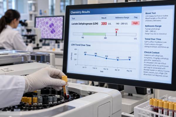
A lactate dehydrogenase (LDH) test measures an enzyme that is released when cells are damaged or turn over rapidly. In blood cancers, LDH can rise when leukemia, lymphoma, or myeloma is highly active, when there is a large disease burden, or when treatment causes rapid tumor-cell breakdown. However, LDH is not a blood cancer test by itself. The same result can rise from hemolysis, liver injury, muscle injury, infection, inflammation, or other noncancer conditions. There is also no single universal LDH cutoff that means cancer is present or progressing because reference ranges differ among laboratories. Clinicians usually interpret LDH relative to the laboratory’s upper limit of normal and compare it with prior values. LDH has an established role in several lymphoma prognostic scores and contributes to risk assessment in myeloma and tumor lysis settings. A trend can be useful when it matches the rest of the disease picture, but diagnosis, remission, and relapse still require disease-specific tests, examination, imaging, and pathology.
- High LDH is nonspecific: it can reflect fast tumor turnover, but it can also rise from hemolysis, liver disease, muscle injury, infection, and other conditions.
- Use the laboratory’s own reference range or upper limit of normal; there is no single LDH number that diagnoses blood cancer.
- LDH is an established prognostic factor in several lymphomas and is often interpreted with stage, performance status, age, blood counts, and other markers.
- A hemolyzed blood sample can falsely raise LDH because red blood cells contain substantial LDH.
- A rapid LDH rise in a high-burden leukemia or lymphoma can increase concern for tumor lysis, especially when uric acid, potassium, phosphate, creatinine, or calcium are also abnormal.
Table of Contents
- What the LDH Test Measures
- LDH Reference Ranges and Test Preparation
- Why LDH Can Be High in Blood Cancer
- LDH in Lymphoma, Leukemia, and Myeloma
- Noncancer Causes and False-High Results
- Using LDH for Prognosis and Disease Trends
- When High LDH Needs Urgent Evaluation
What the LDH Test Measures
LDH is an intracellular enzyme found in many tissues, including blood cells, liver, heart, skeletal muscle, kidneys, lungs, and other organs. It helps convert lactate and pyruvate during energy metabolism. Small amounts normally circulate in blood. When cells are injured, destroyed, or turning over rapidly, more LDH can enter the bloodstream.
This broad tissue distribution explains why the test is sensitive to cell injury but not specific to its source. A high result can be a clue that something is happening, yet LDH alone usually cannot tell where the problem is or whether it is malignant.
In hematologic cancers, several mechanisms can raise LDH:
- a large number of rapidly dividing tumor cells;
- spontaneous death of fragile leukemia or lymphoma cells;
- tissue infiltration and organ injury;
- hemolysis or ineffective blood-cell production;
- treatment-related tumor-cell breakdown; and
- complications such as tumor lysis syndrome.
LDH should therefore be viewed as a context marker. In a newly diagnosed aggressive lymphoma with bulky disease, elevated LDH may reflect high tumor activity and carry prognostic weight. In a person without a cancer diagnosis who has a mildly high LDH, it is much more useful to review the blood sample, liver enzymes, blood count, symptoms, and other tests than to assume cancer.
The test is usually ordered as total serum or plasma LDH. Older approaches sometimes measured LDH isoenzymes to help localize tissue injury, but routine hematologic cancer management more often uses total LDH together with disease-specific studies.
LDH Reference Ranges and Test Preparation
LDH has no universal normal range. Laboratories use different instruments, reagents, temperatures, and measurement methods, so the upper limit of normal can vary substantially. A value that is high at one laboratory may fall within the reference interval at another.
For that reason, the most useful report elements are:
- the measured LDH value;
- the laboratory’s reference interval;
- the upper limit of normal (ULN); and
- the trend over time using the same or a comparable laboratory method.
Clinical research and prognostic scores often express LDH as normal versus above ULN, or as a multiple of ULN, instead of relying on one fixed number. If a laboratory’s ULN is 250 U/L, for example, an LDH of 500 U/L is 2 times ULN. That ratio is more transferable than saying that 500 U/L is always “very high.”
The degree of elevation can still help set priorities. A value just above ULN in a stable patient may be repeated or interpreted alongside routine labs, while a result several-fold above ULN in a patient with rapidly enlarging lymphoma, high blast counts, or new kidney dysfunction deserves faster assessment. The magnitude does not identify the cause, but it can increase concern when it agrees with a high-risk clinical picture. Conversely, a dramatic isolated result from a visibly hemolyzed specimen may reflect the sample rather than sudden cancer acceleration.
Most LDH blood tests require no special fasting. The main practical issue is specimen quality. Hemolysis during or after the blood draw can falsely elevate LDH because red blood cells release LDH when they rupture. A difficult draw, prolonged tourniquet use, rough specimen handling, or delayed processing can contribute to hemolysis.
If LDH is unexpectedly high and other findings do not fit, the laboratory may flag the specimen as hemolyzed or the clinician may repeat the test. Repeating an isolated abnormal result can be especially important before interpreting a sudden change as cancer progression.
Exercise and muscle injury can also raise LDH, so recent strenuous activity may be relevant when the increase is otherwise unexplained. The clinical history should guide whether the result needs immediate action or simple confirmation.
Why LDH Can Be High in Blood Cancer
Blood cancers can produce high LDH because malignant cells may grow, divide, and die faster than normal cells. The effect is especially noticeable in aggressive lymphomas and acute leukemias with a large number of rapidly proliferating cells.
LDH is related to cell turnover, not to one specific cancer pathway. This is why it can roughly track disease burden in some patients without being a tumor-specific biomarker. A bulky lymphoma can produce a high LDH, but so can severe hemolytic anemia. A high-grade leukemia can cause a sharp rise, but so can ischemic tissue injury.
Several patterns are clinically useful:
| LDH pattern | Possible blood cancer interpretation | What else should be checked |
|---|---|---|
| High at new lymphoma diagnosis | May reflect tumor burden or aggressive biology and contribute to prognostic scoring | Stage, PET/CT or other imaging, pathology, performance status, blood counts |
| Very high with acute leukemia | May reflect high blast turnover or a large proliferative burden | Blast count, uric acid, potassium, phosphate, calcium, creatinine |
| Falls with effective treatment | Can support an overall response trend | Disease-specific response testing |
| Rises during follow-up | May raise concern for recurrence or progression if other evidence agrees | Symptoms, exam, imaging, blood counts, marrow or molecular testing |
The key limitation is that LDH does not identify the malignant clone. It cannot replace a lymphoma biopsy, leukemia flow cytometry, molecular testing, or plasma-cell studies. It is best used as one signal among several.
LDH in Lymphoma, Leukemia, and Myeloma
The meaning of LDH changes by disease.
Lymphoma
LDH has its clearest long-standing prognostic role in lymphoma. In diffuse large B-cell lymphoma (DLBCL), LDH above the ULN is part of the International Prognostic Index and related models. Modern studies continue to show that elevated LDH can identify groups with worse outcomes when combined with other variables.
A 2025 DLBCL study developed a Laboratory Prognostic Index using LDH, hemoglobin, and beta-2 microglobulin. Elevated LDH contributed one point, and the combined score separated patients into groups with different 5-year event-free survival. This does not mean the LDH value alone predicts an individual outcome; it shows that LDH remains useful when combined with other objective markers.
LDH also appears in prognostic systems for other lymphomas. A 2024 marginal zone lymphoma index included LDH above ULN among five independent adverse factors. A newer follicular lymphoma model, FLIPI24, includes LDH together with age, hemoglobin, beta-2 microglobulin, and white blood cell count.
An aggressive molecular diagnosis such as double-hit lymphoma cannot be inferred from LDH. Those diagnoses require tissue pathology and specific genetic testing even when LDH is markedly elevated.
Leukemia
In acute leukemia, LDH often rises with a high white blood cell or blast burden and rapid cellular turnover. It can help identify patients at higher risk for metabolic complications such as tumor lysis, but leukemia diagnosis and response rely on morphology, flow cytometry, cytogenetics, and molecular testing.
LDH is especially poor as a substitute for measurable residual disease. A patient can have persistent microscopic leukemia with normal LDH, or high LDH from another cause while the leukemia is in remission.
Myeloma
In multiple myeloma, LDH contributes to risk assessment and staging frameworks because high LDH can reflect more aggressive disease biology. It is interpreted with beta-2 microglobulin, albumin, cytogenetic/FISH risk features, and clinical disease burden. A beta-2 microglobulin test provides different information and is strongly influenced by both tumor burden and kidney function.
LDH cannot replace the core multiple myeloma test panel, which uses monoclonal protein studies, serum free light chains, bone marrow plasma-cell assessment, imaging, and myeloma-specific FISH.
Noncancer Causes and False-High Results
A high LDH result is common enough outside cancer that nonmalignant causes must always be considered. The test rises whenever tissues release significant LDH into the circulation.
Common possibilities include:
- sample hemolysis, which can create a false laboratory elevation;
- hemolytic anemia or other in-body red-cell destruction;
- liver injury or hepatitis;
- skeletal muscle injury, intense exercise, or rhabdomyolysis;
- infection and systemic inflammation;
- tissue ischemia or infarction;
- lung injury;
- kidney or other organ injury; and
- some nonhematologic cancers.
Other laboratory tests help localize the source. A high bilirubin, low haptoglobin, reticulocytosis, and anemia can support hemolysis. High AST, ALT, alkaline phosphatase, or bilirubin may point toward liver or biliary disease. Marked creatine kinase supports skeletal muscle injury. Symptoms, physical examination, and imaging can provide further clues.
The magnitude of LDH does not solve the specificity problem. A very high value deserves attention, but there is no level at which LDH becomes a cancer diagnosis. Severe hemolysis, liver injury, tissue ischemia, and other medical emergencies can also produce large elevations.
An isolated mild elevation is often less informative than a persistent or rapidly changing pattern. If the blood sample was hemolyzed, repeating LDH from a clean specimen may prevent unnecessary cancer-focused investigation.
Using LDH for Prognosis and Disease Trends
LDH is strongest as a prognostic and contextual marker, not as a stand-alone response test. Prognostic models use it because elevated LDH at diagnosis often identifies patients with more active or extensive disease. The value becomes more meaningful when combined with stage, age, performance status, blood counts, beta-2 microglobulin, and disease-specific biology.
For serial monitoring, the personal baseline matters. Suppose a patient’s LDH was high at lymphoma diagnosis, normalized with treatment, and then rises again months later. That pattern may justify a closer look for recurrence, especially if symptoms, imaging, or blood counts also change. But LDH alone cannot confirm relapse because infection, hemolysis, and tissue injury can create the same pattern.
The reverse is also true. A normal LDH does not guarantee remission. Small-volume lymphoma, marrow-only disease, or molecular residual leukemia may produce little or no LDH elevation. Disease-specific tests are more sensitive and specific for those purposes.
Timing matters when evaluating a trend. LDH can change within hours to days as cell injury changes, whereas structural tumor shrinkage on imaging may take longer to document. After effective treatment of a highly proliferative lymphoma or leukemia, LDH may transiently rise as cells break down before it falls. That early rise should be interpreted with uric acid, electrolytes, kidney function, symptoms, and the treatment timeline rather than being labeled progression automatically. Later in follow-up, a sustained rise across several properly collected samples is more persuasive than one isolated spike.
When comparing results over time, clinicians should consider whether the same laboratory method and reference range were used. A change from 220 to 280 U/L may look like a rise, but if the laboratories have different ULNs, the biological interpretation may be unchanged. Expressing each result relative to ULN can make trends easier to compare.
LDH can be particularly useful for risk communication when framed correctly: it is one factor associated with disease activity and outcome, not a clock predicting how long someone will live. Modern treatment can substantially change outcomes even among patients who start with high-risk laboratory features.
When High LDH Needs Urgent Evaluation
A high LDH value by itself is rarely the emergency. Urgency comes from the clinical setting and accompanying abnormalities.
One important blood cancer emergency is tumor lysis syndrome (TLS). Rapid breakdown of leukemia or lymphoma cells releases intracellular potassium, phosphate, and nucleic acids. Uric acid can rise, calcium can fall, and acute kidney injury or dangerous heart rhythm problems can develop. TLS can occur after treatment and, in highly proliferative disease, sometimes before therapy begins.
LDH is not part of the core laboratory definition of TLS, but a high or rapidly rising LDH can signal high tumor turnover and help identify a high-risk setting. Clinicians look much more directly at potassium, phosphate, uric acid, calcium, creatinine, urine output, and symptoms.
Urgent evaluation is warranted when a high LDH occurs with findings such as:
- rapidly increasing white blood cell or blast counts;
- new kidney dysfunction or reduced urine output;
- high potassium or phosphate;
- low calcium;
- severe weakness, muscle cramps, confusion, or seizures;
- palpitations or abnormal heart rhythm;
- rapidly enlarging lymph nodes or bulky aggressive lymphoma; or
- fever, low blood pressure, shortness of breath, or other signs of acute illness.
Patients at high TLS risk may receive hydration, frequent laboratory monitoring, uric-acid-lowering therapy, and treatment in a setting where electrolyte and kidney complications can be managed quickly.
For a stable outpatient with an unexpected isolated LDH elevation, the appropriate response is usually more measured: confirm the result if needed, check for hemolysis, review other labs and symptoms, and interpret it against the known cancer status.
The core message is simple: LDH can help show how active or biologically aggressive a blood cancer may be, but it is never specific enough to diagnose progression, remission, or relapse on its own.
When discussing an LDH result, the most useful questions are what the laboratory ULN is, whether the sample was hemolyzed, how the value compares with prior measurements, and which disease-specific findings changed at the same time. Those four points usually provide more clinical meaning than the raw LDH number alone.
References
- FLIPI24: A Modern Prognostic Model and Clinical Trial Enrichment Tool for Newly Diagnosed Follicular Lymphoma 2026
- Laboratory Prognostic Index (LAB-PI) in diffuse large B-cell lymphoma: a single blood analysis predicts outcomes as good as IPI, NCCN-IPI, and GELTAMO-IPI 2025
- Marginal zone lymphoma international prognostic index: a unifying prognostic index for marginal zone lymphomas requiring systemic treatment 2024
- Prognostic value of lactate dehydrogenase, serum albumin and the lactate dehydrogenase/albumin ratio in patients with diffuse large B-cell lymphoma 2024
- Rasburicase in treating tumor lysis syndrome: An umbrella review 2023 (Review)
Disclaimer
LDH is a nonspecific marker of cell and tissue injury and cannot diagnose blood cancer, progression, remission, or relapse by itself. An unexpected high result should be interpreted with the laboratory reference range, specimen quality, symptoms, and disease-specific testing. Rapidly changing LDH with electrolyte abnormalities, kidney dysfunction, or a high-burden leukemia or lymphoma requires prompt clinical assessment for complications such as tumor lysis syndrome.





