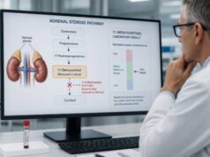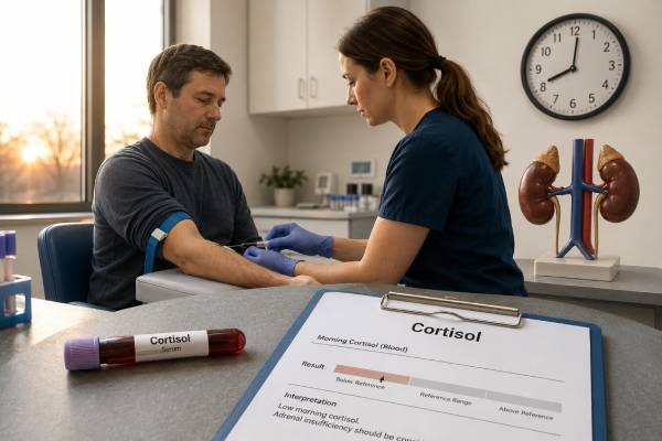
A morning cortisol test measures cortisol near the time it should naturally be highest, usually between 8 and 9 a.m. It is often the first blood test used when adrenal insufficiency is suspected, but the result is rarely interpreted as a simple high-or-low flag. A very low value strongly supports inadequate cortisol production, while a clearly high value usually makes adrenal insufficiency unlikely. Many results fall in an intermediate range and require an ACTH stimulation test. The exact thresholds depend on the cortisol assay, the time of collection, steroid medicines, estrogen status, sleep schedule, recent illness, and whether the person is critically ill. Cortisol also needs to be interpreted with ACTH, sodium, potassium, blood pressure, and symptoms. A person with severe vomiting, fainting, confusion, low blood pressure, or known adrenal disease may need immediate treatment before laboratory confirmation because adrenal crisis can be life-threatening.
- Morning cortisol is best collected around 8–9 a.m., close to the normal daily peak.
- A very low result, often below about 3 mcg/dL, strongly suggests adrenal insufficiency, but assay-specific cutoffs apply.
- A clearly high value, often above about 15–18 mcg/dL, usually excludes adrenal insufficiency in a stable untreated outpatient.
- Intermediate results commonly require an ACTH stimulation test rather than diagnosis from one sample.
- Steroid medicines, oral estrogen, acute illness, and night-shift sleep schedules can make the result misleading.
Table of Contents
- What Morning Cortisol Shows
- Symptoms and Reasons for Testing
- Timing, Preparation, and Medication Review
- Low, Intermediate, and Normal Results
- Primary, Secondary, and Tertiary Adrenal Insufficiency
- Factors That Change Cortisol Results
- ACTH Stimulation and Related Tests
- Urgent Care, Treatment, and Follow-Up
What Morning Cortisol Shows
Cortisol is a glucocorticoid made by the adrenal cortex. It helps maintain blood pressure, blood glucose, energy availability, immune regulation, and the body’s response to illness or injury. Production is controlled by the hypothalamus and pituitary through corticotropin-releasing hormone and ACTH.
Cortisol follows a circadian rhythm. In people who sleep at night, it begins rising before waking, reaches a peak in the early morning, and falls through the day. Testing during the morning peak improves the chance of detecting inadequate production. A random afternoon cortisol is much harder to interpret because a low value may be normal for that time.
The test usually measures total serum cortisol. Most circulating cortisol is bound to cortisol-binding globulin and albumin, while a smaller free fraction is biologically active. Conditions that change binding proteins can alter total cortisol without changing the free fraction proportionally. Oral estrogen is a common example because it raises cortisol-binding globulin.
Morning cortisol is a screening and triage test. It can sometimes make adrenal insufficiency very likely or very unlikely, but it cannot always establish the diagnosis. The intermediate zone is wide because cortisol secretion is pulsatile and assays differ. Dynamic testing is often needed.
The test also cannot determine the cause by itself. A low result may reflect primary adrenal gland failure, reduced pituitary ACTH, suppression after glucocorticoid treatment, severe illness, or a poorly timed sample. Measuring ACTH at the same time helps separate these possibilities.
A normal morning cortisol should not be interpreted after dexamethasone or another suppression test as if it were an untreated baseline. Likewise, a result drawn immediately after exogenous hydrocortisone may largely reflect the medication. The clinical context and exact timing are part of the test.
Symptoms and Reasons for Testing
Adrenal insufficiency can develop gradually or present suddenly. Chronic symptoms are often nonspecific, which is why the test is usually ordered when several findings occur together or when there is a clear risk factor.
Possible symptoms include persistent fatigue, muscle weakness, reduced appetite, unintentional weight loss, nausea, abdominal discomfort, dizziness on standing, low blood pressure, salt craving, and episodes of low blood glucose. Primary adrenal insufficiency may cause skin darkening, particularly in skin creases, scars, gums, and pressure areas, because ACTH is elevated.
Laboratory clues include low sodium, high potassium, unexplained low glucose, eosinophilia, or worsening kidney markers from dehydration. High potassium is more characteristic of primary adrenal failure because aldosterone is lost; it is usually absent in secondary or tertiary disease.
Common reasons to order morning cortisol include:
- Symptoms and electrolytes suggesting adrenal insufficiency.
- A history of autoimmune disease that raises the risk of autoimmune adrenalitis.
- Pituitary or hypothalamic disease, surgery, radiation, or head trauma.
- Long-term or repeated glucocorticoid exposure followed by dose reduction.
- Unexplained low blood pressure or recurrent faintness.
- Follow-up during recovery from glucocorticoid-induced adrenal suppression.
- Evaluation after treatment for Cushing syndrome or pituitary surgery.
A morning cortisol test is not a general measure of stress or “adrenal fatigue.” Fatigue can arise from anemia, thyroid disease, sleep disorders, depression, infection, medication effects, heart or lung disease, and many other causes. A broad clinical assessment remains necessary.
In a person with known adrenal insufficiency, routine morning cortisol is not always useful while taking replacement hydrocortisone because the medicine can be measured as cortisol. The treating endocrinologist may use symptoms, dosing history, electrolytes, renin, and carefully timed testing instead.
Timing, Preparation, and Medication Review
For a conventional sleep schedule, blood is usually drawn between 8 and 9 a.m. Some laboratories accept a slightly wider morning window, but values collected later become harder to compare with standard cutoffs. Record the exact time.
People who work permanent night shifts or sleep during the day may not have a normal clock-time peak. Their cortisol rhythm follows sleep and light exposure imperfectly and can be disrupted. An endocrinologist may time testing relative to awakening or choose a dynamic test rather than applying an 8 a.m. reference interval automatically.
Fasting is not always essential, but it may be requested because ACTH, glucose, electrolytes, or other tests are collected at the same time. Water is generally allowed. Strenuous exercise, acute sleep deprivation, and severe emotional stress can increase cortisol and potentially mask a mild deficiency.
The medication review is critical. Tell the clinician about every glucocorticoid route:
- Oral prednisone, prednisolone, dexamethasone, methylprednisolone, or hydrocortisone.
- Steroid injections into joints, muscles, the spine, or soft tissues.
- Inhaled or nasal steroids.
- Potent skin creams, scalp solutions, and rectal preparations.
- Compounded products and unregulated “adrenal support” supplements.
Do not stop glucocorticoids suddenly. The clinician may switch a short-acting preparation, hold a dose for a carefully defined interval, or use an assay less affected by the specific drug. Dexamethasone usually does not cross-react with many cortisol assays, but it strongly suppresses ACTH and still changes physiology.
Oral estrogen and pregnancy raise cortisol-binding globulin and total cortisol. A value may look reassuringly high even when free cortisol reserve is less robust. Low albumin or low binding protein in critical illness, liver disease, or nephrotic syndrome can make total cortisol look lower than free cortisol.
Biotin and assay interference are less common but should be considered when the result is incompatible with the clinical picture. Repeat testing with another method may be needed.
Whenever possible, draw ACTH in the same session before giving glucocorticoid treatment. ACTH requires careful handling, often in a chilled tube with rapid processing. In an emergency, treatment should not be delayed for perfect specimen collection.
Low, Intermediate, and Normal Results
No single set of cutoffs applies to every assay. Older thresholds were developed with less specific immunoassays, while newer assays and LC-MS/MS often read lower. The laboratory and endocrine service should use method-validated limits.
In a stable, untreated outpatient, a morning cortisol below about 3 mcg/dL, or 83 nmol/L, strongly suggests adrenal insufficiency. A value above roughly 15–18 mcg/dL, or 414–497 nmol/L, usually indicates adequate basal reserve. Many laboratories use different upper thresholds, and some modern methods support lower exclusion cutoffs.
Values between the low and high thresholds form an indeterminate zone. A result of 7 or 10 mcg/dL cannot safely diagnose or exclude adrenal insufficiency without context. Dynamic testing is usually required.
| Morning cortisol pattern | General interpretation | Common next step |
|---|---|---|
| Very low, such as below 3 mcg/dL | Adrenal insufficiency strongly suspected | Measure ACTH and evaluate urgently if symptomatic |
| Intermediate | Basal result is inconclusive | ACTH stimulation test or specialist-directed assessment |
| Clearly high, often above 15–18 mcg/dL | Adrenal insufficiency unlikely in a stable untreated outpatient | Look for other causes of symptoms |
| Unexpected result with steroid or estrogen exposure | Medication or binding effect possible | Review timing and choose an appropriate repeat strategy |
A low cortisol during severe illness can be difficult to interpret. Critical illness changes cortisol production, metabolism, and binding proteins. Standard outpatient cutoffs should not be applied mechanically in an intensive-care setting. Treatment decisions may depend on blood pressure, shock, infection, and the overall clinical course.
A high morning cortisol does not diagnose Cushing syndrome. Stress, pain, illness, pregnancy, and oral estrogen can raise total cortisol. Cushing screening uses loss of nighttime suppression, urine free cortisol, or dexamethasone feedback rather than a single high morning value.
Repeated baseline testing can be helpful when a specimen was collected late or under confounded conditions, but repeating borderline values without moving to dynamic testing may delay diagnosis.
Primary, Secondary, and Tertiary Adrenal Insufficiency
Primary adrenal insufficiency occurs when the adrenal cortex itself is damaged. Autoimmune adrenalitis is a common cause in many regions. Other causes include infections, bilateral adrenal hemorrhage, metastatic disease, genetic disorders, infiltrative disease, and surgical removal of both adrenal glands.
In primary disease, cortisol is low and ACTH is usually markedly high because the pituitary is trying to stimulate failing glands. Aldosterone may also be low, producing low sodium, high potassium, salt craving, dehydration, and elevated renin. The adrenal insufficiency test panel helps document this multi-hormone pattern.
Secondary adrenal insufficiency results from inadequate pituitary ACTH. Causes include pituitary tumors, surgery, radiation, apoplexy, inflammation, and head trauma. ACTH is low or inappropriately normal, cortisol is low, and aldosterone is usually preserved because it is controlled mainly by the renin-angiotensin system. Other pituitary hormones may also be deficient.
Tertiary adrenal insufficiency reflects reduced hypothalamic signaling, most commonly after prolonged glucocorticoid treatment suppresses the axis. Recovery can take weeks to many months and varies widely. Even inhaled, injected, or topical steroids can contribute when exposure is substantial.
ACTH interpretation must be paired with cortisol. A “normal” ACTH is not appropriate when cortisol is very low; in that setting, it may indicate central insufficiency. A high ACTH with normal cortisol can appear early in primary adrenal disease and warrants specialist assessment.
DHEA-S may be low in both primary and central forms, but it is only supportive. Renin and aldosterone are more useful for mineralocorticoid involvement. Adrenal antibodies may support autoimmune disease, while pituitary MRI or adrenal imaging is selected according to the suspected cause rather than ordered reflexively.
Factors That Change Cortisol Results
Exogenous glucocorticoids are the most common interpretive problem. Some drugs cross-react with the assay and falsely elevate measured cortisol; others do not cross-react but suppress ACTH and lower endogenous production. The timing of the last dose, route, duration, and assay all matter.
Binding proteins can shift total cortisol. Oral estrogen, pregnancy, and some rare conditions raise cortisol-binding globulin. Low albumin, cirrhosis, nephrotic syndrome, and critical illness can lower it. Free cortisol or a carefully chosen dynamic test may be more informative in selected cases.
Sleep and circadian disruption also matter. A sample drawn at 8 a.m. after an all-night shift may not represent a physiologic peak. Jet lag, severe insomnia, and irregular schedules can widen variability.
Acute stress raises ACTH and cortisol. Pain, fever, hypoglycemia, vigorous exercise, and anxiety around venipuncture can produce a higher value and may mask mild adrenal insufficiency. Conversely, very severe prolonged illness can alter metabolism and binding in complex ways.
Other pitfalls include:
- Drawing the sample after 10 or 11 a.m. and applying an 8 a.m. cutoff.
- Interpreting a post-dexamethasone value as an untreated morning level.
- Testing immediately after hydrocortisone without accounting for the dose.
- Using a cutoff from a different assay.
- Ignoring pregnancy or oral estrogen.
- Assuming normal electrolytes exclude central adrenal insufficiency.
- Assuming low DHEA-S proves cortisol deficiency.
Laboratory results should be reconciled with the clinical picture. An unexpectedly normal cortisol in someone with severe symptoms and recent steroid withdrawal deserves a careful assay and timing review. An unexpectedly low value in a healthy person may need repeat collection before a lifelong diagnosis is assigned.
ACTH Stimulation and Related Tests
The standard ACTH stimulation test gives synthetic ACTH, also called cosyntropin, and measures cortisol before and after the injection, commonly at 30 and 60 minutes. A sufficient rise shows that the adrenal glands can respond. Modern assay-specific peak cutoffs may be around 14–15 mcg/dL rather than the older 18–20 mcg/dL thresholds, but the laboratory’s validated standard is essential.
The ACTH stimulation test is especially useful for an intermediate morning cortisol. It performs well for established primary adrenal insufficiency. Early central adrenal insufficiency can occasionally produce a normal response because the glands have not yet atrophied, so timing and clinical history matter.
Other tests may include:
- Plasma ACTH to distinguish primary from central disease.
- Sodium, potassium, glucose, and kidney function.
- Plasma renin and aldosterone for suspected primary adrenal failure.
- 21-hydroxylase antibodies for autoimmune Addison disease.
- Other pituitary hormones when central disease is possible.
- Insulin tolerance or metyrapone testing in specialized centers when standard testing remains inconclusive.
Morning cortisol may also be used during glucocorticoid tapering. Some guidelines recommend testing after the dose has reached a physiologic range and after an appropriate hold of the short-acting glucocorticoid. A clearly recovered morning value may allow discontinuation, while an intermediate value leads to continued physiologic dosing and later reassessment.
No dynamic test should be attempted without considering safety. The insulin tolerance test intentionally causes hypoglycemia and is contraindicated in several conditions. Specialist supervision is required.
Urgent Care, Treatment, and Follow-Up
Adrenal crisis is a medical emergency. Symptoms can include severe weakness, repeated vomiting or diarrhea, abdominal or back pain, confusion, fever, dehydration, very low blood pressure, fainting, and shock. Low sodium, high potassium, and low glucose may occur. A person with known adrenal insufficiency, recent steroid withdrawal, or major illness is at particular risk.
When adrenal crisis is suspected, clinicians draw cortisol and ACTH if this can be done immediately, then give intravenous hydrocortisone and fluids without waiting for results. Delaying treatment is more dangerous than temporarily affecting later testing.
Confirmed adrenal insufficiency is treated with glucocorticoid replacement. Primary disease often also requires fludrocortisone to replace mineralocorticoid activity. Patients need education about stress dosing during fever, surgery, vomiting, or injury; an emergency steroid injection kit; and medical identification.
Follow-up after an abnormal morning cortisol should answer:
- Was the sample truly collected near the morning peak?
- Was any glucocorticoid or estrogen affecting the result?
- What was the ACTH level at the same time?
- Are sodium, potassium, glucose, and blood pressure abnormal?
- Is dynamic testing needed, and which assay cutoff will be used?
- Is the pattern primary, central, or medication-induced?
People recovering from glucocorticoid-induced suppression should receive a taper and retesting plan. Recovery is not determined by symptoms alone because steroid withdrawal can resemble adrenal insufficiency even as cortisol production improves. Conversely, feeling well at rest does not guarantee adequate reserve during severe illness.
A properly timed morning cortisol is a useful first filter. Its safest use is to identify clearly low or clearly reassuring results and direct everyone in the middle toward a more definitive assessment.
Interpretation after pituitary surgery deserves special attention. ACTH deficiency may be temporary or permanent, and an early postoperative morning cortisol can help guide replacement. The surgical center sets its own thresholds because timing, perioperative dexamethasone, assay method, and the type of pituitary operation affect the result. Patients should leave with clear instructions about hydrocortisone, emergency dosing, and the date of reassessment.
After successful treatment for Cushing syndrome, the cortisol axis is often suppressed because normal ACTH-producing cells have been exposed to high cortisol for a long time. A low morning cortisol may then be expected and can support remission, but the person still needs glucocorticoid replacement until recovery. Recovery can take months or longer, and stopping replacement prematurely creates risk during illness.
In autoimmune primary adrenal insufficiency, other autoimmune conditions may coexist. Thyroid disease, type 1 diabetes, celiac disease, premature ovarian insufficiency, and vitamin B12 deficiency may be assessed according to symptoms and family history. Starting thyroid hormone in an unrecognized cortisol-deficient person can worsen adrenal stress, which is another reason to evaluate the full endocrine pattern.
Morning cortisol is also affected by kidney and liver disease through changes in binding proteins and metabolism. A low total value in a person with very low albumin may not represent the same free cortisol exposure as an identical value in a healthy outpatient. Specialized free serum cortisol or salivary cortisol measurements can occasionally help, but they are not universally available and require expert interpretation.
Children have age-related physiology and different clinical clues. Poor weight gain, recurrent low glucose, prolonged jaundice in infancy, or growth and pubertal abnormalities may accompany central hormone deficiencies. Pediatric endocrinology should select the assay thresholds and dynamic protocol.
A home blood-pressure log, weight trend, medication timeline, and symptom history can make the follow-up visit more productive. Recording the exact steroid product, dose, route, and last use is particularly valuable because “a steroid shot months ago” can mean very different exposures.
Using the result during recovery
Recovery from glucocorticoid suppression is gradual. A rising morning cortisol trend can be reassuring, but the person may still need stress-dose coverage until the treating clinician confirms adequate reserve. The timing of the last dose and the assay method must remain consistent across measurements.
References
- Adrenal insufficiency: identification and management 2024 (Guideline)
- European Society of Endocrinology and Endocrine Society Joint Clinical Guideline: diagnosis and therapy of glucocorticoid-induced adrenal insufficiency 2024 (Guideline)
- New Cutoffs for the Biochemical Diagnosis of Adrenal Insufficiency after ACTH Stimulation using Specific Cortisol Assays 2021
- Redefining the Diagnostic Approach to Adrenal Insufficiency: Re-Assessment of Baseline and Cortisol Increment Cut-Offs with the 1 µg Synacthen Test 2025 (Clinical Study)
- Diagnosis and Treatment of Primary Adrenal Insufficiency: An Endocrine Society Clinical Practice Guideline 2016 (Guideline)
Disclaimer
This article provides general education and cannot diagnose or exclude adrenal insufficiency in an individual. Morning cortisol thresholds vary by assay, medication exposure, binding proteins, illness, and collection time. Severe symptoms or possible adrenal crisis require emergency medical care, and prescribed glucocorticoids should never be stopped abruptly without medical supervision.