OncoBEAM is a highly sensitive liquid-biopsy approach that uses BEAMing digital PCR to detect selected tumor mutations in circulating tumor DNA (ctDNA) from plasma. Its best-established clinical application is RAS testing in metastatic colorectal cancer, where blood-based KRAS and NRAS results can help assess whether anti-EGFR therapy is appropriate and can track resistant RAS clones over time. Unlike broad next-generation sequencing panels that survey hundreds of genes, OncoBEAM assays are designed around predefined mutation targets. That narrow design allows very sensitive detection of low-frequency variants, but it also means the test cannot discover alterations outside the assay menu. A positive result can provide actionable information without another tissue biopsy. A negative result needs more caution because some cancers release little ctDNA into blood, particularly when tumor burden is low or disease is concentrated in sites that shed poorly. Tissue testing may still be needed after an uninformative plasma result.
- OncoBEAM uses BEAMing digital PCR, a method that amplifies individual DNA molecules on magnetic beads and counts mutant versus wild-type signals.
- The OncoBEAM RAS CRC assay targets predefined KRAS and NRAS mutations, rather than performing broad genome-wide profiling.
- A detected RAS mutation in metastatic colorectal cancer can affect anti-EGFR treatment eligibility; the exact clinical decision depends on current guidelines and treatment history.
- A negative plasma result may be false negative when ctDNA shedding is low, especially with small-volume or certain metastatic patterns.
- Serial OncoBEAM testing can track emerging or declining resistant clones, but mutation trends should be interpreted with imaging and the overall clinical picture.
Table of Contents
- What OncoBEAM Is
- How BEAMing Digital PCR Works
- OncoBEAM RAS CRC and Colorectal Cancer
- How to Interpret OncoBEAM Results
- OncoBEAM for Treatment Monitoring and Resistance
- OncoBEAM Versus NGS Liquid Biopsy
- Limitations, Sample Timing, and Follow-Up
What OncoBEAM Is
OncoBEAM is a branded implementation of BEAMing, which stands for beads, emulsion, amplification, and magnetics. The technology was developed to detect rare mutant DNA molecules within a much larger background of normal DNA. In oncology, the sample is usually plasma, where tumor-derived DNA may represent only a small fraction of total cell-free DNA.
The platform is not one universal cancer test. Different OncoBEAM assays have been developed for specific mutation sets and clinical or research applications. The most widely documented is OncoBEAM RAS CRC, used to evaluate RAS mutations in metastatic colorectal cancer. Current Sysmex materials describe the RAS CRC kit as testing 34 predefined alterations—16 in KRAS and 18 in NRAS—across clinically relevant codons in exons 2, 3, and 4.
That targeted design is important. OncoBEAM is closer to an ultra-sensitive molecular question—“Is one of these specific mutations present in this blood sample?”—than to a broad ctDNA mutation panel that surveys many genes at once.
OncoBEAM has also been used to examine alterations such as EGFR in lung cancer and other hotspot mutations in research or laboratory-service settings. Availability, regulatory status, and exact assay menus vary by country and laboratory. A patient’s report should therefore be interpreted according to the named OncoBEAM assay actually performed, not according to the platform name alone.
How BEAMing Digital PCR Works
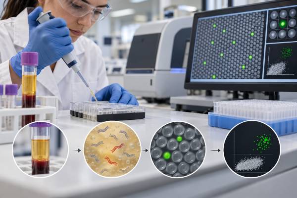
BEAMing combines emulsion PCR with bead-based detection and flow cytometry. The goal is to convert very rare mutant DNA molecules into individually countable fluorescent bead signals.
A simplified workflow looks like this:
- Cell-free DNA is isolated from plasma. The specimen contains DNA from normal cells plus any ctDNA shed by the tumor.
- Target regions are pre-amplified. The assay enriches the gene segments that contain the mutations of interest.
- DNA molecules are partitioned into tiny emulsion droplets. Ideally, individual template molecules are amplified separately.
- PCR products attach to magnetic beads. Each bead becomes associated with copies derived from a small number of original molecules.
- Mutation-specific fluorescent probes are added. Mutant and wild-type sequences generate different signals.
- Flow cytometry counts the beads. The laboratory calculates the proportion carrying the mutant sequence.
This digital counting strategy can detect very low mutant fractions. Sysmex performance materials for certain OncoBEAM mutation assays report analytical cutoffs around hundredths of a percent, although the exact limit varies by gene, mutation, DNA input, and assay version. A quoted analytical sensitivity should never be treated as a guaranteed clinical sensitivity in every patient. If the bloodstream contains no tumor DNA from a particular lesion, even a technically excellent assay cannot detect it.
BEAMing is one of several highly sensitive techniques used in liquid biopsy testing. Digital droplet PCR uses a different partitioning format, while NGS uses massively parallel sequencing and error-correction methods. Each technology trades breadth, sensitivity, speed, and complexity differently.
OncoBEAM RAS CRC and Colorectal Cancer
The clearest clinical role for OncoBEAM is RAS genotyping in metastatic colorectal cancer. KRAS and NRAS status matters because activating RAS mutations predict resistance to anti-EGFR monoclonal antibodies such as cetuximab and panitumumab in settings where those drugs require RAS-wild-type disease.
Traditionally, RAS testing is performed on tumor tissue. Plasma testing offers an alternative when tissue is unavailable, insufficient, difficult to retrieve, or no longer representative of the cancer after years of therapy.
Several studies have compared OncoBEAM plasma results with tumor-tissue RAS testing. Concordance has generally been high, but not perfect. The differences are clinically instructive because they show where blood testing works best and where caution is needed.
Tumor burden and metastatic site affect detection
Patients with substantial liver metastases often have higher ctDNA shedding. By contrast, studies have found lower plasma-tissue concordance in patients with lung-only metastases, particularly when lesions are small or few. Peritoneal disease can also be challenging in some cohorts.
This means a plasma RAS-negative result is more convincing when there is clear evidence that enough tumor DNA is circulating. A “wild-type” call in a specimen with little detectable ctDNA may actually be a non-informative negative.
One practical question is therefore not only “Was RAS detected?” but also “Was there enough ctDNA in this sample to trust a negative result?” Some reports provide mutant allele fraction or other quality indicators that help the oncology team judge this.
Blood can show tumor evolution
The molecular profile of colorectal cancer can change during therapy. A tumor that was RAS wild type before anti-EGFR treatment may develop resistant RAS-mutant clones. Those clones can later decline after EGFR blockade is stopped. Serial plasma testing can capture this dynamic in a way that a single archived tissue biopsy cannot.
For a broader explanation of how RAS results influence treatment, a KRAS liquid biopsy result should be interpreted together with NRAS, BRAF, treatment history, and other relevant markers rather than in isolation.
How to Interpret OncoBEAM Results
OncoBEAM reports are usually easier to interpret than very broad genomic panels because the assay asks about a defined mutation set. The challenge is understanding what the result means clinically.
Mutation detected
A positive result means the assay found one of its targeted mutations above the reporting threshold. The report may include the exact variant and mutant allele fraction (MAF).
In metastatic colorectal cancer, a detected activating KRAS or NRAS mutation can make anti-EGFR treatment inappropriate in settings that require RAS-wild-type disease. If the mutation appears after prior anti-EGFR therapy, it may represent an acquired resistance clone.
MAF is the percentage of measured DNA molecules carrying the mutation, not the percentage of tumor cells in the body. A MAF of 0.5% does not mean that 0.5% of the cancer is mutant. The value is influenced by total cfDNA, disease burden, metastatic site, treatment, and how much DNA the tumor releases.
No mutation detected
A negative result means the assay did not find any of the targeted mutations above its detection threshold. It does not necessarily prove that the tumor is RAS wild type.
A negative result may occur because:
- The tumor truly lacks the tested RAS mutations.
- The cancer is shedding too little ctDNA into plasma.
- The mutation is present below the test’s limit of detection.
- The tumor carries a RAS alteration outside the assay’s predefined mutation list.
- Recent treatment reduced the amount of circulating tumor DNA.
- Sample quantity or quality limited analytical sensitivity.
When a negative result would determine whether a patient receives anti-EGFR therapy, clinicians may use tissue genotyping or a broader assay if the plasma result appears biologically non-informative.
Discordant blood and tissue results
A blood result and an old tissue result can differ for legitimate reasons. Tumor heterogeneity means not every lesion contains the same clones. Therapy can also select new resistant populations after the original biopsy. On the other hand, low shedding can make plasma miss a tissue mutation.
A discordant result should therefore trigger investigation rather than an assumption that one test is “wrong.” The timing of each specimen, disease sites, prior therapies, and assay coverage can often explain the difference.
OncoBEAM for Treatment Monitoring and Resistance
Serial blood sampling is where a sensitive digital assay becomes especially interesting. Instead of asking only whether a RAS mutation exists at one time point, clinicians and researchers can watch resistant clones appear, expand, and sometimes decline.
During anti-EGFR treatment in metastatic colorectal cancer, RAS or other pathway alterations can emerge under drug pressure. This is a classic example of liquid-biopsy resistance testing.
The reverse process can also occur. When anti-EGFR therapy is withdrawn, resistant RAS-mutant clones may lose their selective advantage and decrease. This observation led to trials of anti-EGFR rechallenge, in which patients previously treated with cetuximab or panitumumab are screened again in plasma before restarting EGFR blockade.
Recent studies have supported the idea that ctDNA can help select patients for this strategy, but the details matter. The required washout interval, which resistance genes are tested, how low the mutation fraction must be, and which treatment combinations are used vary across trials and guidelines. “RAS not detected” should not be interpreted as an automatic guarantee that rechallenge will work.
Serial OncoBEAM testing has also been studied as a response marker. Falling mutant allele fractions can parallel tumor response, while rising values may precede or accompany progression. These trends can be biologically informative, but they should not replace imaging on their own unless a particular clinical protocol has validated that use.
OncoBEAM Versus NGS Liquid Biopsy
OncoBEAM and NGS answer overlapping but different questions.
| Feature | OncoBEAM / targeted digital PCR | Broad NGS liquid biopsy |
|---|---|---|
| Primary strength | Very sensitive detection of predefined mutations | Broad profiling across many genes and alteration types |
| Mutation discovery | Limited to assay targets | Can find unexpected or multiple coexisting alterations |
| Low-frequency hotspot detection | Often excellent | Depends on sequencing depth, error suppression, and DNA input |
| Resistance profiling | Strong when known resistance hotspots are targeted | Better for complex or multiple possible resistance mechanisms |
| Report complexity | Relatively focused | May include many pathogenic, uncertain, and incidental findings |
The right choice depends on the question. If the clinical decision hinges on a small number of known RAS hotspots, a highly sensitive targeted test may be ideal. If the patient is progressing on therapy and many possible resistance pathways need to be surveyed, broad NGS may provide more useful breadth.
These approaches can also be complementary. Studies comparing OncoBEAM with NGS have shown that targeted digital PCR may detect lower-frequency hotspot mutations, while NGS can uncover rare variants or alteration classes outside the digital assay’s menu.
A practical example helps show the difference. Suppose a patient with metastatic colorectal cancer has an old tissue specimen reported as RAS wild type and now has progression after several therapies. If the immediate question is whether a very low-level RAS-resistant clone is still present before an anti-EGFR rechallenge, a highly sensitive RAS-focused assay can be well matched to the decision. If the question is instead why the cancer is progressing and whether any new targeted option exists, a broad NGS panel may be preferable because resistance could involve BRAF, EGFR, MET, ERBB2, a fusion, or another pathway that a RAS-only assay cannot see.
The meaning of very low mutant fractions also requires discipline. Detecting a mutation at a fraction such as 0.05% can be analytically impressive, but clinical action thresholds are not automatically identical to the laboratory limit of detection. An assay may be capable of detecting a rare molecular signal long before its clinical significance is fully established. For serial monitoring, clinicians therefore look at the specific trial or guideline that defines how the result should be used, rather than treating every detectable molecule as a mandate to change therapy.
Finally, assay performance statistics from validation studies are population averages. Sensitivity and concordance can be excellent across a cohort while still being poor for an individual patient with low-shedding disease. The most useful report interpretation combines the laboratory’s analytical capability with biological evidence that the patient’s cancer is actually represented in plasma.
Limitations, Sample Timing, and Follow-Up
OncoBEAM’s major strength—very sensitive targeted detection—is also its main limitation. The assay can only detect what it was designed to detect. A negative result cannot exclude a mutation outside the covered hotspots, another resistance gene, a gene fusion not included in the assay, or a non-genetic resistance mechanism.
The biological limitation of ctDNA shedding is just as important. Small tumors, recent treatment response, and some metastatic patterns may yield too little ctDNA. No molecular technology can recover tumor mutations from plasma if the relevant DNA molecules are not present in the tube.
No fasting is generally required. Practical quality depends more on using the correct collection tube, adequate blood volume, timely plasma processing, and minimizing contamination by genomic DNA from normal blood cells.
Testing is often most informative:
- Before treatment when a baseline RAS status is needed
- At progression when acquired resistance is suspected
- Before a planned anti-EGFR rechallenge in a validated clinical strategy
- At serial time points when a clinician or trial protocol is tracking a known mutation
Questions worth asking after an OncoBEAM result include which exact mutation set was tested, what the assay’s detection threshold was for that sample, whether ctDNA quantity was adequate, whether the result agrees with recent tissue data, and whether broader genomic testing is still needed.
The key distinction is simple: OncoBEAM is a highly sensitive targeted liquid-biopsy technology, not a comprehensive cancer-genome test. Its value is greatest when the clinical question matches the mutations it was built to detect.
That fit between assay design and clinical question is more important than choosing a platform simply because it advertises the lowest analytical detection limit.
References
- ESMO recommendations on the use of circulating tumour DNA assays for patients with cancer: a report from the ESMO Precision Medicine Working Group 2022 (Guideline)
- Baseline Liquid Biopsy in Relation to Tissue-Based Parameters in Metastatic Colorectal Cancer: Results From the Randomized FIRE-4 (AIO-KRK-0114) Study 2025
- Sensitive cell-free DNA assay for accurate detection of RAS and BRAF mutations in liquid biopsies of patients with metastatic colorectal cancer: Final results of the multicentric ColoBEAM study 2025
- A multi-institutional observational study evaluating the incidence and the clinicopathological characteristics of NeoRAS wild-type metastatic colorectal cancer 2023
- Evaluation of RAS Mutational Status in Liquid Biopsy to Monitor Disease Progression in Metastatic Colorectal Cancer Patients 2023
- KRAS and NRAS mutational analysis in plasma ctDNA from patients with metastatic colorectal cancer by real-time PCR and digital PCR 2022
Disclaimer
OncoBEAM results should be interpreted by an oncology team using the exact assay version, tumor type, tissue findings, treatment history, imaging, and evidence of adequate ctDNA shedding. A negative plasma result does not exclude a clinically important tumor mutation, and treatment decisions should not be made from this article alone.