
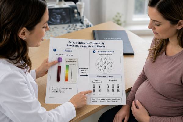
Patau syndrome genetic testing looks for an extra copy of chromosome 13, a finding called trisomy 13. Prenatal screening can estimate the chance that a pregnancy is affected, while chorionic villus sampling or amniocentesis can provide a diagnostic chromosome result. The distinction is critical: a “high-risk” cell-free DNA result is not the same as a confirmed fetal diagnosis, and a normal screening result cannot exclude every form of trisomy 13.
Most cases arise from a random chromosome-separation error in an egg or sperm. Less often, trisomy 13 results from a Robertsonian translocation that may be inherited from a healthy parent, or from mosaicism in which only some cells carry the extra chromosome. These forms can change recurrence counseling and sometimes the expected range of medical effects. Ultrasound findings may raise concern, but imaging alone cannot identify the chromosome result with certainty. When testing suggests trisomy 13, prompt referral to maternal-fetal medicine and genetic counseling helps families understand confirmation options, pregnancy care, prognosis, and choices without assuming that every case will follow the same course.
- Cell-free DNA is a screening test for trisomy 13; a positive result should be confirmed with CVS or amniocentesis before irreversible decisions.
- Diagnostic testing can identify full trisomy 13, mosaic trisomy 13, and many translocation forms.
- A low-risk screen greatly lowers the chance of trisomy 13 but does not make the chance zero.
- Ultrasound abnormalities such as holoprosencephaly, heart defects, growth restriction, or extra fingers or toes can increase suspicion.
- CVS is generally performed from about 10 weeks; amniocentesis is usually performed from 15 weeks.
- A translocation result may lead to parental karyotyping because recurrence risk can differ from the usual age-related risk.
Table of Contents
- What Trisomy 13 Testing Detects
- Screening Tests and Their Results
- Diagnostic Tests for Patau Syndrome
- How to Interpret a Positive or Negative Result
- Ultrasound Findings and Pregnancy Care
- Prognosis and Care After Diagnosis
- Recurrence Risk and Future Pregnancies
What Trisomy 13 Testing Detects
Patau syndrome occurs when cells contain extra genetic material from chromosome 13. A typical human cell has two copies of chromosome 13. In full trisomy 13, there are three separate copies in nearly all tested cells. The standard chromosome notation is usually 47,XX,+13 or 47,XY,+13.
Three main chromosome patterns can cause the condition:
- Full trisomy 13: every or almost every cell has a complete extra chromosome 13. This is the most common form and usually results from nondisjunction, a random error during egg or sperm formation.
- Translocation trisomy 13: extra chromosome 13 material is attached to another chromosome, often through a Robertsonian translocation. The total chromosome count may be 46 rather than 47, even though extra chromosome 13 material is present.
- Mosaic trisomy 13: some cells have trisomy 13 and others have the usual chromosome number. The proportion can differ across blood, placenta, and fetal tissues, so a percentage from one sample does not precisely predict medical severity.
Partial trisomy 13 is rarer. Only a segment of chromosome 13 is duplicated, often because of a parental rearrangement. Effects depend on the size and location of the duplicated segment and whether another chromosome segment is missing.
Different tests answer different questions. Prenatal cell-free DNA analyzes placental DNA fragments circulating in maternal blood and estimates risk. A karyotype visually counts and examines chromosomes. Chromosomal microarray measures gains and losses of DNA at higher resolution, although it may not show how a translocation is physically arranged. Rapid tests such as fluorescence in situ hybridization or quantitative fluorescent PCR may provide preliminary information for chromosome 13, but a complete laboratory workup is often needed to define the final chromosome pattern.
The extra chromosome is usually not caused by anything a pregnant person did. Full trisomy 13 is generally sporadic, and its frequency increases with maternal age because chromosome-separation errors become more common in eggs over time. A translocation form raises a different question: whether one parent carries a balanced rearrangement without having Patau syndrome.
Screening Tests and Their Results
Prenatal screening estimates probability; it does not prove whether fetal cells have trisomy 13. All pregnant patients should have an opportunity to discuss screening and diagnostic testing, regardless of age or baseline risk. The available choices vary by gestational age, whether the pregnancy is singleton or multiple, local practice, and personal preferences.
Cell-free DNA screening
Cell-free DNA screening, also called cfDNA or NIPT, can usually be performed from about 10 weeks of pregnancy. It measures fragments released mainly by placental cells. For common trisomies, it is more accurate than traditional serum screening, but performance for trisomy 13 is lower than for trisomy 21 because the condition is rarer and biological factors can create discordant results.
A report may say “high risk,” “screen positive,” “increased chance,” or provide a probability. The positive predictive value—the chance that a high-risk result reflects an affected pregnancy—depends on maternal age, gestational age, ultrasound findings, the laboratory method, and the condition’s prevalence in the tested population. It is not the same as the test’s sensitivity or a marketing claim of “99% accuracy.”
False-positive trisomy 13 results can occur because the placenta contains an abnormal cell line while the fetus does not, a phenomenon called confined placental mosaicism. Other causes include a vanished twin, maternal chromosome variation, maternal medical conditions in rare cases, or technical limitations. Because cfDNA reflects placenta more directly than fetus, a diagnostic sample is needed for confirmation.
A “no-call,” “insufficient fetal fraction,” or inconclusive result is not automatically reassuring. Low fetal fraction can result from early gestational age, higher maternal weight, sample handling, certain medications, placental biology, or fetal chromosome conditions. The next step may be repeat cfDNA, detailed ultrasound, or diagnostic testing, depending on gestational age and the complete clinical picture.
First- and second-trimester serum screening
First-trimester combined screening uses maternal age, ultrasound measurement of nuchal translucency, pregnancy-associated plasma protein A, and free or total beta-hCG. A second-trimester quad screen measures AFP, hCG, unconjugated estriol, and inhibin A. These methods can identify a higher chance of trisomy 13, but they are less sensitive and specific than cfDNA for common aneuploidies.
Serum-screen risk estimates may group trisomy 13 with trisomy 18 or may not report trisomy 13 separately, depending on the program. An abnormal screen can also reflect inaccurate dating, placental dysfunction, multiple pregnancy, or another fetal condition. Screening should be interpreted with ultrasound and the exact laboratory report.
Ultrasound as a screening tool
Ultrasound can detect structural differences commonly associated with trisomy 13, but a normal scan does not rule it out. A first-trimester scan may show increased nuchal translucency or major anomalies. The detailed anatomy scan later in pregnancy evaluates the brain, heart, face, kidneys, limbs, growth, placenta, and amniotic fluid.
A patient should generally avoid taking multiple independent screening tests that answer the same question without a coordinated plan. Sequential screens can create conflicting probabilities and confusion. A clinician or genetic counselor can explain whether a follow-up test is a better screen or a true diagnostic test. More detail about the broader choices appears in prenatal genetic screening guidance.
Diagnostic Tests for Patau Syndrome
Diagnostic testing examines cells from the placenta or amniotic fluid. It is offered after a high-risk screen, suspicious ultrasound, previous affected pregnancy, parental chromosome rearrangement, or simply because a patient wants a definitive chromosome result rather than a probability.
Chorionic villus sampling
Chorionic villus sampling, or CVS, removes a small sample of placental tissue, usually between 10 and 13 weeks. The clinician reaches the placenta through the abdomen or cervix under ultrasound guidance. The route depends on placental position and clinical factors.
CVS provides earlier results than amniocentesis, but placental mosaicism can complicate interpretation. If CVS shows mosaic trisomy 13, the fetus may or may not be affected. Amniocentesis may be recommended because amniotic-fluid cells are generally a closer reflection of fetal chromosome status. Direct-preparation and cultured-cell results may also differ, so the laboratory and maternal-fetal medicine specialist should explain exactly which cell populations were abnormal.
Amniocentesis
Amniocentesis is usually performed at or after 15 weeks. A thin needle passes through the abdomen under ultrasound guidance to remove amniotic fluid. The fluid contains fetal cells that can be tested by karyotype, rapid aneuploidy testing, microarray, or other methods.
Procedure-related pregnancy-loss risk is low in experienced hands, but it is not zero. The best estimate depends on the center, operator, patient factors, and the comparison population. Counseling should separate the background chance of miscarriage from the additional procedure-related risk.
Karyotype, rapid testing, and microarray
A prenatal karyotype test can confirm an extra chromosome 13 and show whether it is free-standing or part of a large translocation. Culture commonly takes about 1 to 2 weeks, although timing varies.
Rapid FISH or QF-PCR may return a preliminary result within several days for chromosomes 13, 18, 21, X, and Y. A rapid result is useful but does not replace full analysis when the chromosome structure, mosaicism, or an unexpected finding needs clarification.
Chromosomal microarray detects chromosome gains and losses at much higher resolution than a karyotype. It will detect full trisomy 13 and many partial duplications, but it may not reveal the balanced structure in a parent. When ultrasound shows multiple anomalies and standard chromosome testing is normal, microarray may identify another diagnosis. The laboratory may recommend both methods when a translocation is possible.
A positive screening result should not be treated as diagnostic. Confirmatory testing is particularly important before pregnancy termination, major changes in obstetric management, or assigning a prognosis based solely on the chromosome screen.
How to Interpret a Positive or Negative Result
The meaning of a result begins with the test type. “Positive” on a screening report means increased probability. “Positive” on a diagnostic karyotype means extra chromosome 13 material was identified in the sampled cells, but the exact form still matters.
| Result | What it means | Typical next step |
|---|---|---|
| Low-risk cfDNA | The chance of trisomy 13 is substantially reduced, not eliminated. | Continue routine ultrasound care; consider diagnostic testing if major anomalies appear or definitive information is desired. |
| High-risk cfDNA | Placental DNA pattern suggests trisomy 13, but false positives occur. | Genetic counseling, detailed ultrasound, and CVS or amniocentesis. |
| No-call cfDNA | The laboratory could not issue a reliable result. | Review gestational age and fetal fraction; discuss repeat screening versus diagnostic testing. |
| Full trisomy 13 on amniocentesis | Fetal cells have three copies of chromosome 13. | Maternal-fetal medicine, genetics, pediatric, and values-based counseling. |
| Mosaic trisomy 13 | Both normal and trisomy 13 cell lines were found. | Clarify sample and percentage, consider confirmatory testing, and interpret with imaging. |
| Translocation trisomy 13 | Extra chromosome 13 material is attached to another chromosome. | Parental karyotypes and family-specific recurrence counseling. |
A negative CVS or amniocentesis result usually rules out full fetal trisomy 13 in the tested sample with high confidence. It does not rule out every genetic condition or every cause of an abnormal ultrasound. Very low-level mosaicism can be difficult to detect, and placental and fetal tissues may differ.
Mosaic results require special care. The percentage of abnormal cells in a culture can be affected by sampling and cell growth. A result of “20% mosaic” does not mean exactly 20% of every fetal organ is affected or that symptoms will be 20% as severe. Ultrasound findings, sample source, laboratory method, and follow-up testing all contribute to interpretation.
Translocation notation should identify the chromosomes and breakpoints. If a parent carries the balanced form, siblings and other relatives may also be carriers. If neither parent carries it, the rearrangement is likely de novo, and recurrence is usually lower, though not zero because germline mosaicism cannot always be excluded.
Ultrasound Findings and Pregnancy Care
Trisomy 13 can affect many organ systems. Common prenatal findings include holoprosencephaly, facial clefts, congenital heart defects, enlarged or echogenic kidneys, omphalocele, growth restriction, clenched hands, and polydactyly. Not every fetus has every feature, and some abnormalities become visible only later.
After diagnosis, care often includes:
- A detailed anatomic ultrasound by maternal-fetal medicine
- Fetal echocardiography to define heart anatomy
- Serial growth and amniotic-fluid assessment when results will guide care
- Discussion of stillbirth risk and possible pregnancy complications
- Neonatology or pediatric consultation before delivery
- A delivery plan that reflects maternal safety and the family’s goals for fetal monitoring and newborn treatment
- Palliative-care involvement when comfort-focused care is being considered
Testing and counseling should not reduce the pregnancy to a chromosome label. Families may choose pregnancy continuation with intensive newborn treatment, pregnancy continuation with comfort-focused care, adoption planning in some settings, or pregnancy termination where legal and accessible. The clinician’s role is to provide accurate information, acknowledge uncertainty, and support informed choices without coercion.
A high-risk screening result can cause intense anxiety during the interval before confirmation. Whenever possible, diagnostic testing should be arranged promptly and the expected turnaround explained. Emergency symptoms such as heavy bleeding, severe pain, fever, fluid leakage, or reduced fetal movement later in pregnancy require urgent obstetric assessment, whether or not trisomy 13 is suspected.
Prognosis and Care After Diagnosis
Full trisomy 13 is a serious, life-limiting condition associated with a high rate of miscarriage, stillbirth, and death in infancy. Many liveborn infants have major neurologic, cardiac, breathing, feeding, vision, hearing, and developmental needs. Survival statistics vary because studies include different eras, treatment approaches, mosaic or partial cases, and infants selected for surgery or intensive care.
It is more accurate to describe a range than to predict an exact lifespan from the chromosome result alone. Some infants die within hours or days, many do not survive the first year, and a smaller group lives for years. Recent data suggest survival has improved in some health systems, partly because selected children receive cardiac surgery, respiratory support, feeding support, and other interventions. Longer survival does not remove the likelihood of profound developmental disability and complex medical care.
Parents should be offered balanced information about both burdens and possible benefits of treatment. Decisions may include resuscitation at birth, respiratory support, feeding tubes, heart surgery, seizure treatment, and management of infections or apnea. A treatment may prolong life but also require hospitalization and repeated procedures. Comfort-focused care can include warmth, holding, feeding as tolerated, symptom relief, memory-making, spiritual support, and hospice services.
Mosaic or partial trisomy 13 may have a broader range of outcomes, but neither label guarantees mild effects. The duplicated chromosome segment, tissue distribution, structural anomalies, and clinical course are more informative than the category alone.
Families often benefit from meeting neonatology, cardiology, palliative care, social work, and genetics before delivery. Written care plans reduce the need to make every decision during a crisis and can be revised as new information emerges.
Recurrence Risk and Future Pregnancies
After full trisomy 13 caused by nondisjunction, recurrence risk is usually low but higher than the population risk for someone of the same age. Counseling often uses the greater of an age-related risk estimate or a small added recurrence estimate, but the exact figure should come from a genetics professional familiar with the laboratory result and reproductive history.
Translocation trisomy 13 requires a different calculation. Both parents should usually have a blood karyotype. A healthy parent may carry a balanced Robertsonian or reciprocal translocation and have an increased chance of miscarriage or another pregnancy with an unbalanced chromosome result. The risk differs by the specific translocation and which parent carries it; broad internet percentages should not replace family-specific counseling. A parental karyotype can show whether the rearrangement is inherited.
Future-pregnancy options may include:
- Early ultrasound and cfDNA screening
- CVS or amniocentesis for definitive prenatal diagnosis
- IVF with preimplantation genetic testing for structural rearrangements when a parent is a carrier
- Donor egg, donor sperm, or donor embryo
- Natural conception without testing
Even after a prior affected pregnancy, screening remains optional. Some families prefer early diagnostic testing because they do not want another period of screening uncertainty. Others choose noninvasive screening first. The choice should account for the exact chromosome mechanism, maternal age, fertility, values, access, and how the information would be used.
A trisomy 13 result can be medically and emotionally overwhelming. Clear separation of screening from diagnosis, precise definition of the chromosome pattern, and compassionate counseling allow families to make decisions based on confirmed information rather than fear or assumptions.
References
- Noninvasive prenatal screening (NIPS) for fetal chromosome abnormalities in a general-risk population: an evidence-based clinical guideline of the American College of Medical Genetics and Genomics (ACMG) 2023 (Guideline)
- Society for Maternal-Fetal Medicine Consult Series #74: Cell-free DNA screening for aneuploidies: Updated guidance 2025 (Guideline)
- Guidance for Caring for Infants and Children With Trisomy 13 and Trisomy 18: Clinical Report 2025 (Guideline)
- Prenatal diagnosis after high chance non-invasive prenatal testing for trisomies 21, 18 and 13, chorionic villus sampling or amniocentesis? An audit of clinical practice 2023 (Review)
- Patau syndrome (trisomy 13) 2025 (Review)
- Trends in the survival of patients with trisomy 13 from 1995 to 2021: A population-based study 2024 (Review)
Disclaimer
This article provides general education and does not replace prenatal diagnosis, maternal-fetal medicine care, or genetic counseling. Screening probabilities, diagnostic options, prognosis, and recurrence risk depend on the exact test, chromosome result, ultrasound findings, gestational age, and individual pregnancy. Seek urgent obstetric care for concerning pregnancy symptoms rather than relying on genetic test information alone.





