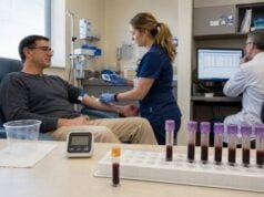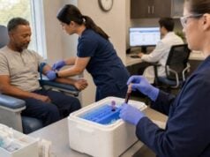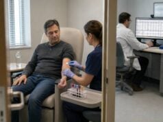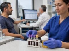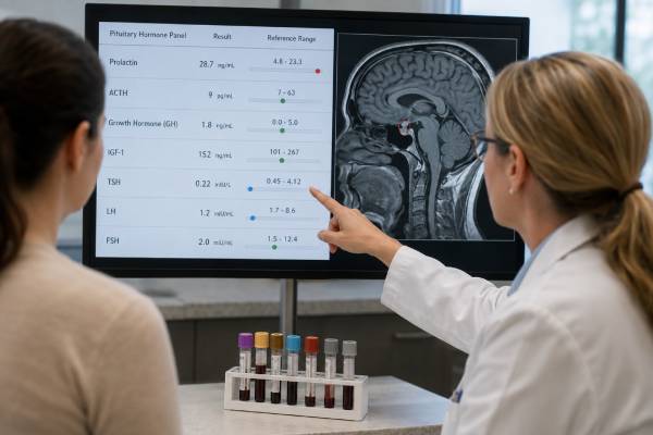
A pituitary tumor hormone panel determines whether a sellar lesion is secreting excess hormone, compressing normal pituitary tissue, or both. The panel commonly measures prolactin, IGF-1, ACTH and cortisol, TSH and free T4, LH and FSH with estradiol or testosterone, and sometimes growth hormone. The exact tests depend on symptoms and imaging. Prolactinomas, GH-secreting adenomas, ACTH-secreting adenomas, and rare TSH-secreting tumors produce different biochemical patterns. Clinically nonfunctioning tumors do not cause a recognizable hormone-excess syndrome, but they can still raise prolactin through stalk compression and cause adrenal, thyroid, reproductive, or growth hormone deficiencies. Tumor size does not reliably predict hormone activity: a tiny corticotroph tumor can cause severe Cushing disease, while a large nonfunctioning adenoma may present through vision loss or hypopituitarism. Results should be reviewed with a dedicated pituitary MRI and visual assessment.
- Every pituitary mass needs evaluation for hormone excess and clinically important hormone deficiency.
- Prolactin should be diluted when a large mass has only a modest result, to exclude the hook effect.
- IGF-1 is the preferred screen for GH excess; random GH alone is unreliable.
- Low free T4 with a normal TSH can indicate central hypothyroidism from tumor compression.
- Low morning cortisol may signal dangerous ACTH deficiency and requires prompt action.
Table of Contents
- Goals of a Pituitary Tumor Hormone Panel
- Prolactin and Prolactin-Secreting Tumors
- ACTH, Cortisol, and Cushing Disease
- GH, IGF-1, Acromegaly, and Gigantism
- TSH, Free T4, LH, FSH, and Sex Hormones
- Nonfunctioning, Mixed, and Rare Tumors
- MRI, Vision Testing, and Laboratory Pitfalls
- Treatment, Follow-Up, and Urgent Signs
Goals of a Pituitary Tumor Hormone Panel
Pituitary adenomas arise from hormone-producing cells in the anterior pituitary. They are usually benign by pathology, but they can cause major illness through hormone excess, pressure on nearby structures, or loss of normal pituitary function. Other sellar lesions—including Rathke cleft cysts, craniopharyngiomas, meningiomas, metastases, inflammation, and aneurysms—can produce similar compression patterns.
The hormone panel answers four separate questions:
- Is the lesion secreting prolactin, growth hormone, ACTH, or TSH?
- Has it impaired cortisol, thyroid, reproductive, or GH function?
- Is a result altered by stalk compression rather than direct secretion?
- Is there an urgent deficiency or tumor effect that needs immediate treatment?
The first-line profile often includes morning cortisol, ACTH, TSH, free T4, prolactin, IGF-1, LH, FSH, and a sex hormone appropriate to the person’s physiology. Sodium and osmolality are added when thirst, high urine output, or postoperative water-balance problems are present.
A random GH level may appear on the panel, but it is not a reliable screen because normal GH is pulsatile. IGF-1 is more stable. Suspected acromegaly may need an oral glucose suppression test; suspected GH deficiency may need stimulation.
Similarly, one cortisol measurement does not fully evaluate cortisol excess. Cushing disease screening uses late-night salivary cortisol, 24-hour urinary free cortisol, or overnight dexamethasone suppression. ACTH helps classify the source after hypercortisolism is confirmed.
Tumors are often described by size. A microadenoma is less than 10 mm, while a macroadenoma is 10 mm or larger. Size affects the risk of compression but does not define secretion. Prolactinomas often show some relation between tumor size and prolactin, but assay artifacts and cystic components can disrupt that relationship.
Incidental lesions found on unrelated imaging still need clinical and laboratory evaluation. “Incidental” describes how the mass was discovered, not whether it is harmless.
Prolactin and Prolactin-Secreting Tumors
Prolactin is usually measured in every pituitary mass because prolactinomas are common and often respond well to medicine. Elevated prolactin also occurs when a non-prolactin-secreting lesion compresses the pituitary stalk and interrupts dopamine delivery. Dopamine normally restrains prolactin release.
Symptoms of hyperprolactinemia include irregular or absent periods, infertility, galactorrhea, low libido, erectile dysfunction, low testosterone, reduced bone density, and delayed puberty. Large tumors can also cause headache and loss of peripheral vision.
A clearly elevated monomeric prolactin that broadly matches tumor size supports prolactinoma. However, no single numerical threshold is perfect across assays. Pregnancy, breastfeeding, stress, hypothyroidism, kidney disease, and many medications can elevate the result.
Two laboratory issues are essential:
Hook effect
In some two-site immunoassays, extremely high prolactin saturates the test and produces a falsely modest result. A large pituitary mass with prolactin only mildly or moderately elevated should prompt measurement after serial dilution. Missing the hook effect can lead to a giant prolactinoma being mislabeled as a nonfunctioning tumor.
Macroprolactin
Macroprolactin is a large prolactin-immunoglobulin complex that can elevate total prolactin with less biological activity. PEG precipitation estimates the monomeric fraction. A macroprolactin test can prevent unnecessary imaging in the right setting, but it does not explain mass-related symptoms or exclude coexisting true hyperprolactinemia.
Stalk-effect prolactin elevation is often lower than levels produced by a large prolactinoma, but overlap exists. Imaging anatomy, dilution studies, symptoms, and treatment response contribute to classification.
Dopamine agonists such as cabergoline are first-line treatment for most prolactinomas. They often lower prolactin and shrink the tumor. Surgery may be considered for medication intolerance or resistance, acute compression, cerebrospinal fluid leakage after shrinkage, or a preference-sensitive situation in an expert center.
ACTH, Cortisol, and Cushing Disease
ACTH-secreting pituitary adenomas cause Cushing disease, one form of Cushing syndrome. These tumors are often small and can be difficult to see on MRI. The severity of cortisol excess does not depend on a large tumor.
Possible features include easy bruising, facial rounding, proximal muscle weakness, wide purple stretch marks, high blood pressure, diabetes, osteoporosis, mood change, infections, menstrual disruption, and weight gain concentrated around the trunk. Many features are common in the general population, so biochemical confirmation is required.
Random cortisol and ACTH are not adequate screening tests for Cushing disease. Recommended first tests usually include one or more of:
- Late-night salivary cortisol on repeated nights
- 24-hour urinary free cortisol, often repeated
- Overnight 1 mg dexamethasone suppression
After endogenous hypercortisolism is confirmed, ACTH determines whether the process is ACTH-dependent. A low suppressed ACTH suggests an adrenal cause. A detectable or elevated ACTH points toward a pituitary or ectopic ACTH source.
Further tests can include pituitary MRI, corticotropin-releasing hormone or desmopressin testing, high-dose dexamethasone in selected settings, and inferior petrosal sinus sampling when imaging and biochemistry do not establish the source. Inferior petrosal sinus sampling is an invasive specialist procedure and is not a general screening test.
The same tumor can also impair normal ACTH secretion after surgery, radiation, apoplexy, or compression. Low morning cortisol with low or inappropriately normal ACTH suggests central adrenal insufficiency. Symptoms include fatigue, nausea, low blood pressure, low sodium, weakness, and low glucose.
Cortisol deficiency has immediate safety implications. If adrenal crisis is suspected, clinicians draw samples when feasible and give glucocorticoid treatment without waiting for all results. Thyroid hormone replacement should not be started or increased in untreated central adrenal insufficiency without a cortisol plan.
After Cushing disease surgery, low cortisol may be an expected sign of remission, but it requires temporary glucocorticoid replacement and careful follow-up. Recovery of the axis can take months or longer.
GH, IGF-1, Acromegaly, and Gigantism
Growth hormone-secreting adenomas cause acromegaly after the growth plates close and pituitary gigantism before they close. IGF-1 is the preferred screening test because it reflects integrated GH action and is more stable than GH.
Acromegaly may cause enlarged hands and feet, facial and jaw changes, sweating, skin thickening, headaches, joint disease, carpal tunnel syndrome, sleep apnea, high blood pressure, cardiomyopathy, and glucose intolerance. In children, rapid height gain accompanies enlargement of soft tissues and extremities.
An IGF-1 result must use an accurate age-adjusted range. Puberty, pregnancy, liver disease, kidney disease, undernutrition, diabetes, and oral estrogen can alter interpretation. A clearly elevated IGF-1 with typical features strongly supports the diagnosis. Borderline results should be repeated using the same validated assay.
When confirmation is needed, the oral glucose growth hormone suppression test measures serial GH after a 75 g glucose drink. Normal GH suppresses to a low nadir. Failure to suppress below an assay- and BMI-appropriate cutoff supports autonomous secretion.
Random GH alone can mislead. A healthy person may be sampled during a pulse, while someone with mild acromegaly may have a low baseline. Pegvisomant treatment raises circulating GH while blocking its receptor, so IGF-1—not GH—is used to monitor that therapy.
Some pituitary adenomas secrete both GH and prolactin. A high prolactin result may represent mixed secretion, stalk effect, or a separate assay issue. Pathology can identify cell lineage after surgery, but biochemical testing defines the clinical syndromes.
After surgery, IGF-1 and GH are reassessed at appropriate intervals. IGF-1 may take weeks to stabilize. Persistent elevation can lead to repeat surgery, somatostatin receptor ligands, pegvisomant, cabergoline, radiation, or combination therapy.
TSH, Free T4, LH, FSH, and Sex Hormones
A pituitary mass commonly affects thyroid and reproductive function through compression. Rarely, a tumor secretes TSH.
TSH and free T4
Central hypothyroidism produces low free T4 with a TSH that is low, normal, or mildly elevated but biologically inappropriate. TSH alone can look reassuring and should never be the only thyroid test in a person with a pituitary mass.
Symptoms include fatigue, cold intolerance, constipation, dry skin, slowed thinking, and weight gain, but these overlap with other conditions. Treatment uses levothyroxine and is monitored mainly with free T4, not a TSH target. Cortisol status should be secured first.
A TSH-secreting pituitary tumor is rare. It typically causes elevated free T4 and free T3 with TSH that is not suppressed. Before diagnosing a TSHoma, clinicians exclude assay interference, biotin, irregular thyroid medication use, and thyroid hormone resistance. SHBG, alpha-subunit, family testing, imaging, and dynamic studies may contribute.
LH, FSH, estradiol, and testosterone
Compression of gonadotroph function can cause low sex hormones with low or inappropriately normal LH and FSH. In premenopausal adults this may cause absent periods, infertility, hot flashes, or low bone density. In adult males it may cause low libido, erectile dysfunction, reduced muscle mass, infertility, or anemia.
High LH and FSH with low sex hormones usually indicate primary ovarian or testicular failure rather than pituitary compression. Menopause normally raises gonadotropins.
Prolactin excess can secondarily suppress LH and FSH. Treatment of a prolactinoma may restore cycles, testosterone, and fertility, sometimes quickly. Pregnancy planning requires discussion because tumor monitoring and medication decisions change.
In children, pituitary tumors can cause delayed puberty through deficiency or early puberty through specific lesions and hypothalamic effects. Evaluation includes growth, bone age, pubertal stage, gonadotropins, and sex hormones.
Nonfunctioning, Mixed, and Rare Tumors
Clinically nonfunctioning pituitary adenomas do not produce a recognizable hormone-excess syndrome. Many arise from gonadotroph lineage and may make hormone subunits that do not create a clear clinical effect. They often present incidentally, through headache or vision change, or through hypopituitarism.
A nonfunctioning label is reached only after appropriate excess-hormone screening. A lesion should not be called nonfunctioning because the person lacks obvious symptoms; acromegaly and Cushing disease can be subtle.
Larger nonfunctioning tumors may cause:
- Central adrenal insufficiency
- Central hypothyroidism
- Hypogonadotropic hypogonadism
- GH deficiency
- Mild prolactin elevation from stalk effect
- Visual-field loss from optic chiasm compression
Mixed or plurihormonal tumors can produce more than one biochemical syndrome. GH-prolactin co-secretion is the most familiar. Rare tumors may secrete TSH, gonadotropins, or multiple transcription-factor lineages.
Functional gonadotroph adenomas are uncommon. They may produce ovarian hyperstimulation, menstrual disturbance, enlarged ovaries, or testicular effects, but many FSH/LH-staining tumors are clinically silent. Interpretation requires sex hormones, gonadotropin subunits, imaging, and specialist expertise.
Aggressive pituitary tumors are defined by clinical behavior rather than size alone. Rapid growth, invasion, repeated recurrence, and resistance to standard treatment raise concern. Pituitary carcinoma is very rare and requires metastasis for diagnosis.
Other sellar masses can mimic adenomas. Craniopharyngiomas and stalk lesions more often disrupt water balance. Hypophysitis may occur in pregnancy, postpartum, autoimmune disease, or with immune checkpoint inhibitors. Metastases may cause rapid symptoms and diabetes insipidus. The hormone panel helps show functional impact but does not identify pathology by itself.
MRI, Vision Testing, and Laboratory Pitfalls
A dedicated pituitary MRI with thin slices and contrast provides the best anatomical detail in most cases. It shows size, cystic or hemorrhagic components, cavernous sinus involvement, stalk deviation, and proximity to the optic nerves. CT may be used when MRI is unavailable or contraindicated.
Hormones generally guide imaging rather than the reverse. A small MRI lesion can be incidental, so biochemical evidence is necessary before attributing a hormone syndrome to it. Conversely, a hormone-secreting microadenoma may be too small for routine MRI to detect.
Formal visual-field testing is recommended when a lesion contacts or compresses the optic chiasm, or when the person reports peripheral vision loss. Acuity alone may remain normal while side vision narrows.
Laboratory pitfalls include:
- Prolactin hook effect in a large prolactinoma
- Macroprolactin causing false total prolactin elevation
- Random GH sampling
- TSH-only testing that misses central hypothyroidism
- Oral estrogen changing cortisol, GH, and IGF-1 interpretation
- Biotin interference
- Acute illness suppressing reproductive and thyroid axes
- Glucocorticoid exposure suppressing ACTH and cortisol
- Assay-specific GH and IGF-1 ranges
- Drawing ACTH without correct chilled handling
A mismatch between results and clinical findings should trigger a laboratory discussion. Repeating on another platform, serial dilution, equilibrium dialysis, PEG precipitation, or review of medication timing may resolve the discrepancy.
The tumor’s relationship to hormone values can also change with hemorrhage, cystic degeneration, medication, and prior surgery. Historical results and serial imaging are often more informative than one snapshot.
Before and after pituitary surgery
Preoperative testing establishes which hormones the tumor secretes and which normal axes are already impaired. This changes anesthesia planning and postoperative care. Possible ACTH deficiency is especially important because cortisol coverage may be lifesaving. Hypercortisolism, acromegaly, and hyperthyroidism can also increase surgical risk through hypertension, diabetes, sleep apnea, arrhythmia, infection risk, or heart disease. Correcting severe hormone excess before surgery may reduce complications.
After transsphenoidal surgery, hormone priorities change by the hour and day. Excessive urine output with intense thirst and rising sodium can indicate central diabetes insipidus. Several days later, nausea, headache, confusion, or seizures with low sodium may signal delayed antidiuretic hormone release. Morning cortisol helps guide steroid needs, but interpretation depends on perioperative glucocorticoids and the center’s protocol. Free T4 may not fall immediately even when TSH secretion changes, and IGF-1 can take weeks or months to reflect the new GH state.
Early biochemical remission criteria differ by tumor type. Prolactin can fall rapidly after complete prolactinoma removal. ACTH and cortisol may become very low after successful Cushing disease surgery because normal corticotrophs were suppressed. GH can be measured early, but formal remission assessment also uses age-adjusted IGF-1 after sufficient time. TSHoma follow-up requires free T4, free T3, TSH, symptoms, and imaging rather than TSH alone. A single postoperative panel should therefore not be used as a universal cure test.
When results and imaging disagree
A visible lesion does not prove hormone secretion, and normal imaging does not fully exclude a tiny functional tumor. Incidental microadenomas are common enough that the biochemical syndrome must come first. Repeating abnormal hormones, checking assay interference, and using the correct dynamic test can prevent surgery for an unrelated lesion. Conversely, a large mass with normal random GH or a mildly abnormal prolactin should not be dismissed; pulsatile secretion and the prolactin hook effect are classic pitfalls.
Mixed tumors can produce more than one hormone, especially GH with prolactin or TSH. Tumor compression can simultaneously create excess of one hormone and deficiency of another. For example, a GH-secreting macroadenoma may elevate IGF-1 while lowering LH, FSH, testosterone, or free T4 through pressure on normal tissue. The panel should be read as a map of both secretion and remaining reserve.
Long-term surveillance remains necessary even after apparently complete treatment. Residual tumor can regrow, hormone excess can recur, and radiation can cause new deficiencies years later. The follow-up schedule is based on pathology, residual MRI findings, the original hormone syndrome, treatment type, and symptoms. Reproductive plans, bone health, cardiovascular risk, vision, and quality of life are part of endocrine follow-up, not separate from it.
Treatment, Follow-Up, and Urgent Signs
Treatment depends on tumor type, size, anatomy, symptoms, and hormone effects. Prolactinomas usually begin with dopamine agonist medicine. Many GH-, ACTH-, TSH-secreting, and symptomatic nonfunctioning tumors are treated with transsphenoidal surgery. Medication and radiation are added when disease persists or surgery is unsuitable.
Before surgery, adrenal and thyroid deficiencies must be recognized. Visual status, sodium, and comorbidities related to hormone excess are assessed. An experienced pituitary team improves coordination of endocrine, surgical, imaging, and eye care.
After surgery, monitoring may include:
- Early morning cortisol and glucocorticoid needs
- Sodium and urine output for diabetes insipidus or delayed hyponatremia
- Free T4 and TSH after an appropriate interval
- Prolactin, IGF-1, GH, or cortisol-remission testing based on tumor type
- Gonadal hormones and fertility goals
- MRI and visual fields
- Long-term testing after radiation
Seek emergency care for sudden severe headache, new vision loss, double vision, vomiting, confusion, fainting, neck stiffness, or severe weakness. Pituitary apoplexy can cause acute hemorrhage or infarction and rapidly threaten vision and cortisol production.
Symptoms of adrenal crisis include severe weakness, low blood pressure, vomiting, abdominal pain, low glucose, confusion, and collapse. A person with known ACTH deficiency should follow sick-day steroid instructions and carry emergency identification and medication as advised.
Very high urine output and intense thirst after surgery may indicate diabetes insipidus. Headache, nausea, confusion, and seizures several days later can signal low sodium. Both require urgent testing.
Long-term follow-up continues even after successful treatment because tumors can recur and pituitary deficits can evolve. A structured pituitary hormone test panel is repeated according to the tumor and treatment rather than on a fixed one-size-fits-all schedule.
The most reliable follow-up compares current results with the original pre-treatment biochemical signature, not only with a generic reference range.
New symptoms should trigger earlier reassessment rather than waiting for the routine surveillance date.
References
- Pituitary incidentaloma: a Pituitary Society international consensus guideline statement 2025 (Consensus Statement)
- Management of pituitary incidentalomas 2024 (Review)
- A Consensus for Pituitary Adenoma Diagnosis in Indonesia 2025 (Consensus Statement)
- Consensus guideline for the diagnosis and management of pituitary adenomas in childhood and adolescence: Part 1, general recommendations 2024 (Consensus Guideline)
- Consensus guideline for the diagnosis and management of pituitary adenomas in childhood and adolescence: Part 2, specific diseases 2024 (Consensus Guideline)
Disclaimer
A pituitary tumor hormone panel must be interpreted with symptoms, medicines, dynamic tests, MRI findings, and laboratory methods. This article is educational and cannot classify a sellar mass or determine treatment. Sudden severe headache, vision change, adrenal-crisis symptoms, or postoperative water-balance changes require urgent medical care.