
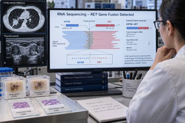
A RET fusion test looks for an abnormal rearrangement that joins part of the RET gene to another gene, creating a continuously active growth signal in some cancers. RET fusions are best established in non-small cell lung cancer (NSCLC), especially adenocarcinoma, and in a subset of papillary and other thyroid cancers. A confirmed RET fusion can be both a diagnostic molecular finding and a treatment-selection biomarker because selective RET inhibitors are available for appropriate patients with advanced RET-driven cancers. The result is different from a RET point mutation: activating RET mutations are especially important in medullary thyroid cancer, whereas RET fusions are more typical of papillary thyroid carcinoma and RET-rearranged NSCLC. Testing method matters because some DNA-only panels can miss fusions, particularly when breakpoints occur in difficult regions. RNA-based next-generation sequencing is often highly effective for detecting expressed RET fusion transcripts. A positive result should name the fusion partner when possible and be interpreted with tumor type, stage, specimen quality, and treatment history.
- A positive RET fusion means a functional RET rearrangement was detected and may identify a cancer driven by RET signaling.
- RET fusions are clinically important in NSCLC and thyroid cancer because selective RET-targeted therapy may be available.
- A RET fusion is not the same as a RET point mutation; the tumor types, testing strategy, and treatment implications can differ.
- RNA-based fusion testing can detect some RET fusions that DNA-only testing misses, especially when intronic breakpoints are hard to capture.
- A negative result is most reliable when the assay is validated for RET fusions and the sample contains enough viable tumor.
Table of Contents
- What a RET Fusion Is
- Cancers Where RET Fusion Testing Matters
- How RET Fusions Are Tested
- How to Interpret RET Fusion Results
- RET Fusions and Targeted Treatment
- Resistance and Repeat Testing
- Limitations and Practical Next Steps
What a RET Fusion Is
RET encodes a receptor tyrosine kinase that normally transmits growth and survival signals after controlled activation. In a RET fusion, the kinase-containing portion of RET becomes joined to part of another gene. Common partners in lung cancer include KIF5B and CCDC6, although many partners have been described. The partner often contributes a domain that promotes dimerization, allowing the RET kinase to remain active without its usual external signal.
This is a classic oncogenic driver: the fusion can provide a dominant signal that helps a cancer cell grow. In many RET fusion-positive tumors, other major drivers such as EGFR mutations, ALK fusions, or ROS1 fusions are absent, although co-alterations can occur, especially after treatment.
RET fusions must be distinguished from other RET abnormalities. A point mutation changes one or a few DNA bases within RET and can activate the protein in a different way. Germline activating RET mutations cause multiple endocrine neoplasia type 2 and hereditary medullary thyroid cancer. Somatic RET mutations are also common drivers of sporadic medullary thyroid carcinoma. Those are not “RET fusion-positive” tumors.
A report that simply says “RET altered” is therefore incomplete for clinical interpretation. The important details are the alteration class—fusion, mutation, amplification, or another change—the exact partner or variant, the specimen tested, and the laboratory’s evidence that the alteration is functional.
Cancers Where RET Fusion Testing Matters
RET fusion testing is most routinely relevant in lung and thyroid cancer.
In NSCLC, RET fusions occur in a small percentage of cases, most often in adenocarcinoma. Because several different targetable drivers are possible, RET is usually tested as part of a broad lung cancer biomarker panel rather than as a single-gene test. Broad profiling is especially useful in advanced non-squamous NSCLC because tissue can be limited and sequential single-gene testing may consume the sample before all actionable drivers are assessed.
RET fusion-positive lung cancers can occur in people with little or no smoking history, but smoking history cannot be used to decide whether testing is necessary. Clinical characteristics enrich probability; they do not replace molecular testing.
In thyroid cancer, RET fusions are particularly associated with papillary thyroid carcinoma and can occur in both adults and children. Radiation-associated papillary thyroid cancers historically showed a higher frequency of RET rearrangements. In advanced radioactive iodine-refractory thyroid cancer, identifying a RET fusion can open a selective targeted-treatment option.
RET can also be fused in other rare solid tumors. Whether a fusion is actionable outside lung or thyroid cancer depends on current regulatory approvals, clinical guidelines, and trial access. Broad sequencing may uncover these uncommon events, but the oncology team should confirm that the fusion is known or strongly predicted to activate RET before treating it as a driver.
If a lung tumor is being profiled broadly, a lung cancer NGS panel can often assess RET alongside EGFR, ALK, ROS1, BRAF, MET, NTRK, KRAS, ERBB2, and other clinically relevant alterations in one workflow.
How RET Fusions Are Tested
Several laboratory methods can detect RET rearrangements, but they answer slightly different questions.
RNA-based next-generation sequencing
RNA sequencing directly looks for the fusion transcript produced after two genes are joined. This is often highly efficient for kinase fusions because it avoids the need to capture large or repetitive introns where DNA breakpoints may occur. Anchored multiplex PCR and other targeted RNA approaches can detect fusions even when the partner is not known in advance, depending on panel design.
RNA has one important weakness: it degrades more easily than DNA, especially in old or poorly fixed formalin-fixed tissue. A failed RNA assay is not the same as a true negative result.
DNA-based next-generation sequencing
DNA panels can detect RET rearrangements while simultaneously assessing mutations and copy-number changes. Performance depends heavily on whether the assay adequately covers RET introns where rearrangements occur. Large introns, repetitive sequences, and breakpoints outside captured regions can lead to false-negative fusion results.
When a DNA panel is negative but clinical suspicion is high, or when the report states that fusion detection is limited, reflex RNA testing may be appropriate. This is one reason “no RET fusion detected” should always be read alongside the technical limitations section.
FISH and other methods
Break-apart fluorescence in situ hybridization (FISH) can show that the RET locus is rearranged without identifying the partner transcript. It can be useful when sequencing is unavailable or when tissue is very limited, but interpretation requires validated cutoffs and experienced review. Reverse-transcription PCR can be sensitive for known fusion transcripts but may miss uncommon partners if primers are too specific.
Immunohistochemistry is not generally considered a stand-alone substitute for molecular confirmation of RET fusion status in the same way that highly validated IHC assays are used for some other drivers.
How to Interpret RET Fusion Results
A positive RET fusion result should ideally identify the partner and breakpoint or transcript. Examples include KIF5B::RET and CCDC6::RET. The report may classify the fusion as pathogenic, oncogenic, or clinically significant. In a cancer type where RET is an established driver, this result can have immediate treatment relevance.
Not every rearrangement involving RET is automatically actionable. Some genomic rearrangements may be out-of-frame, non-expressed, or biologically uncertain. RNA evidence that an in-frame transcript is expressed strengthens confidence. Many clinical laboratories filter these details before issuing a report, but unusual fusions may receive a cautious interpretation.
A negative result means no reportable fusion was detected within the assay’s validated range. It does not mean the cancer has no targetable driver. Other genes may be responsible, and a technically limited assay may not exclude every RET rearrangement. Sample adequacy matters: low tumor content can dilute the abnormal signal below the detection threshold.
An indeterminate or quantity not sufficient result should not be treated as negative. The laboratory may have had too little tissue, degraded RNA, low nucleic-acid yield, or failed quality-control metrics. A new tissue block, repeat biopsy, or blood-based assay may be considered depending on the cancer and clinical urgency.
Liquid biopsy can sometimes detect RET fusions in circulating tumor DNA, particularly in advanced disease with sufficient tumor shedding. A positive plasma finding can be useful, but a negative plasma result is less reassuring because some tumors release little DNA into blood. When plasma is negative and a targetable driver is still needed, tissue testing remains important.
RET Fusions and Targeted Treatment
The major clinical reason to identify a RET fusion is the availability of selective RET inhibitors. Selpercatinib and pralsetinib were developed to inhibit RET more specifically than older multikinase drugs. Their use depends on cancer type, disease stage, prior treatment, country-specific approval, patient factors, and current guidelines.
In advanced RET fusion-positive NSCLC, randomized phase 3 evidence has shown strong activity for first-line selpercatinib compared with platinum-based chemotherapy with or without pembrolizumab. Selective RET inhibition is therefore a central treatment option for appropriately selected patients. RET inhibitors also have meaningful intracranial activity, which matters because brain metastases are common in oncogene-driven NSCLC.
In advanced RET fusion-positive thyroid cancer, selective RET inhibition can also produce substantial responses. Testing is especially relevant when systemic therapy is being considered for progressive disease that is not adequately controlled by surgery, radioactive iodine, or other standard approaches.
The fusion result does not determine the entire treatment plan. Stage, symptoms, pace of progression, brain involvement, organ function, prior therapies, drug interactions, and toxicity risks remain important. A patient with a localized, surgically curable tumor may not need systemic RET therapy simply because the fusion is present.
PD-L1 expression also should not be used to dismiss a targetable RET fusion. In metastatic driver-positive NSCLC, the presence of an established oncogenic fusion often has greater treatment-selection importance than PD-L1 alone. This is why comprehensive molecular testing is ideally completed before first-line systemic therapy whenever the clinical situation allows.
Resistance and Repeat Testing
RET fusion-positive cancers can eventually develop resistance to selective RET inhibitors. Resistance may occur through changes in RET itself or through alternative signaling pathways that let the tumor bypass RET dependence.
On-target resistance includes kinase-domain changes that reduce drug binding, such as solvent-front mutations. Off-target resistance can involve activation of MET, KRAS, or other pathways. The exact pattern varies and remains an active area of research.
When a cancer progresses on a RET inhibitor, repeat molecular profiling may help identify the resistance mechanism and possible next options. Depending on disease location and safety, this can involve a new tissue biopsy or circulating tumor DNA testing. A blood test is often easier, but tissue can provide additional information about histologic transformation, tumor architecture, and alterations that are not well captured in plasma.
Repeat testing is not automatically necessary after every scan change. It is most useful when the result could alter treatment, qualify the patient for a clinical trial, or clarify unexpected resistance.
The original fusion usually remains biologically important even after resistance develops. A later report may therefore show the same RET fusion plus one or more new alterations. The added findings can explain why a previously effective drug stopped controlling the disease.
Limitations and Practical Next Steps
The biggest interpretive error is assuming that every negative test equally excludes a RET fusion. A negative high-quality RNA fusion assay on an adequate tumor sample is more informative than a negative small DNA panel with limited intronic coverage. The report’s methodology and limitations section should be reviewed before deciding that RET is truly absent.
A second error is confusing RET fusion with RET mutation. In thyroid cancer especially, this can send interpretation in the wrong direction. The treatment and hereditary implications of an activating RET mutation in medullary thyroid cancer differ from those of a RET fusion in papillary thyroid carcinoma.
A third error is ignoring tissue quality. Decalcified bone samples, tiny biopsies, low-tumor specimens, and heavily necrotic tissue can reduce sensitivity. If no driver is found in a patient who is highly likely to have an actionable alteration, re-testing with a better specimen or an orthogonal method may be worthwhile.
After a positive RET fusion result, useful questions for the oncology team include:
- Is this fusion an established oncogenic driver in my tumor type?
- Is a selective RET inhibitor approved or guideline-recommended for my disease stage?
- Was the fusion confirmed as expressed and in-frame?
- Do I need brain imaging or other staging before treatment selection?
- If I later develop resistance, would repeat tissue or liquid-biopsy profiling be useful?
A RET fusion test is therefore more than a yes/no molecular label. Its value comes from matching a technically reliable fusion result to the correct cancer context and using it at the point where it can guide diagnosis or treatment.
References
- First-Line Selpercatinib or Chemotherapy and Pembrolizumab in RET Fusion-Positive NSCLC 2023 (RCT)
- RET Fusion-Positive Non-small Cell Lung Cancer: The Evolving Treatment Landscape 2023 (Review)
- Central Nervous System Disease in Patients With RET Fusion-Positive NSCLC Treated With Selpercatinib 2023
- Predictive molecular pathology in metastatic thyroid cancer: the role of RET fusions 2022 (Review)
- Selpercatinib in Early-Stage RET Fusion-Positive Non-Small-Cell Lung Cancer 2026 (RCT)
Disclaimer
RET fusion results should be interpreted by the treating oncology and pathology team because assay design, cancer type, stage, and current drug approvals determine clinical significance. This article is educational and does not replace professional diagnosis, treatment selection, or molecular pathology review.





