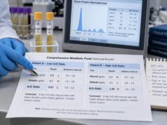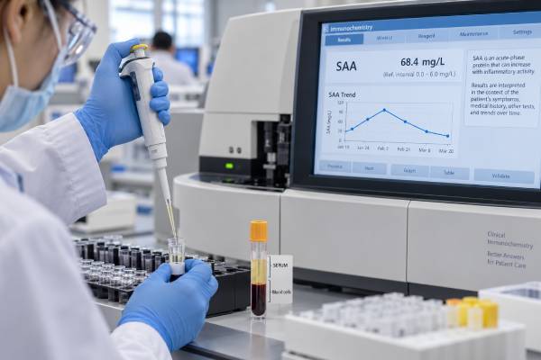
Serum amyloid A (SAA) is a group of acute-phase proteins that can rise sharply when the body responds to infection, tissue injury, or inflammatory disease. The liver produces most circulating SAA after stimulation by inflammatory cytokines, and levels may change faster and over a wider range than several traditional markers. Clinicians sometimes use an SAA blood test to assess active inflammation, monitor autoinflammatory or rheumatic disease, and estimate whether long-term inflammation is adequately suppressed in people at risk for AA amyloidosis. A high value is sensitive but nonspecific: it does not identify the source of inflammation or prove that amyloid has deposited in organs. A single temporary spike during an infection is not the same as years of persistent elevation. Assays and reference intervals also vary, and SAA is not as routinely available or standardized as C-reactive protein. Interpretation should consider the laboratory range, symptoms, diagnosis, treatment, related markers, kidney findings, and the pattern across repeated measurements.
- SAA is a rapidly changing acute-phase protein produced mainly by the liver.
- High levels indicate an inflammatory response but do not reveal its cause.
- Persistent elevation—not one isolated result—is the concern for AA amyloidosis risk.
- SAA can help monitor selected autoinflammatory, rheumatic, infectious, and intestinal diseases.
- Reference ranges and results vary between assays, so trends are best followed in the same laboratory.
Table of Contents
- What Serum Amyloid A Is and What the Test Measures
- Reference Ranges, Timing, and Changes During Inflammation
- Common Causes of High SAA Levels
- How SAA Is Used to Monitor Disease Activity
- Persistent SAA and the Risk of AA Amyloidosis
- Limitations, Confounders, and Comparison With Other Markers
- Interpreting Results and Planning Follow-Up
What Serum Amyloid A Is and What the Test Measures
Serum amyloid A refers to a family of related proteins. SAA1 and SAA2 are the main acute-phase forms in humans. During health, they circulate at low concentrations. When inflammatory cytokines such as interleukin-1, interleukin-6, and tumor necrosis factor signal the liver, production can increase dramatically.
Most circulating SAA attaches to high-density lipoprotein (HDL). In the acute response, it participates in transport of lipids, recruitment of immune cells, and tissue-repair signaling. These functions are part of a normal defense response. Problems arise when the inflammatory stimulus persists and SAA remains elevated for long periods.
The blood test quantifies SAA protein, usually in serum, and reports a concentration in milligrams per liter (mg/L). It measures the body’s response rather than a specific disease. The test does not identify bacteria, demonstrate an autoimmune antibody, or show where inflammation is located.
SAA is sometimes confused with several unrelated uses of the word amyloid. It is not the same as:
- Amyloid beta: a protein fragment associated with plaques in Alzheimer disease;
- Light-chain amyloid: the material deposited in AL amyloidosis from abnormal immunoglobulin light chains;
- Transthyretin amyloid: the protein deposited in hereditary or wild-type ATTR amyloidosis;
- A tissue amyloid stain: a biopsy method used to show amyloid deposits.
SAA is the circulating precursor from which AA amyloid fibrils can form in the setting of prolonged inflammation. Measuring SAA can estimate inflammatory exposure, but it cannot diagnose tissue deposition. Diagnosis of amyloidosis generally requires clinical evidence plus tissue confirmation and accurate amyloid typing.
The test requires a routine venous blood sample. Fasting is usually unnecessary unless other ordered tests require it. Because concentrations can change quickly, the date of symptom onset, medication changes, infection, and timing relative to an inflammatory attack are important.
Reference Ranges, Timing, and Changes During Inflammation
Many laboratories use an upper reference limit near 10 mg/L, but this is not universal. Different antibodies, calibrators, analyzers, and populations can produce different ranges. Some reports provide an interval rather than a single upper limit, and specialist centers may use treatment targets that are not identical to the laboratory’s healthy reference range.
Always interpret the number beside the range printed on the report. A result of 12 mg/L may be only slightly above one laboratory’s cutoff, while another method may classify it differently. Comparing numbers from different platforms can create an artificial trend.
SAA is capable of increasing several hundredfold during a strong acute-phase response. Its general time course is:
- Initial rise: production may increase within hours after an inflammatory stimulus.
- Peak response: substantial elevation can develop during the first one to several days, depending on the trigger.
- Decline: levels often fall quickly when the stimulus resolves or effective treatment suppresses inflammation.
- Persistent elevation: ongoing infection or uncontrolled inflammatory disease can maintain a raised baseline between symptomatic episodes.
This dynamic behavior makes timing central to interpretation. A normal sample drawn between attacks may miss a brief inflammatory episode. A high sample taken during influenza, pneumonia, or another acute illness may not represent the person’s usual disease control. For chronic monitoring, clinicians often choose planned intervals and may repeat a surprising result after intercurrent infection has cleared.
A value cannot be translated directly into a percentage of inflammation. SAA of 100 mg/L does not mean the body is “ten times more inflamed” than at 10 mg/L. The relation between concentration, tissue activity, and clinical severity differs by disease and individual response.
Low SAA is generally reassuring when the purpose is to assess active systemic inflammation. It does not rule out every inflammatory condition. Localized disease, mild activity, immunosuppressive treatment, or timing may produce a value within range. Nor does a low current value erase previous years of inflammatory exposure in someone already being evaluated for AA amyloidosis.
Common Causes of High SAA Levels
SAA responds to many types of injury and immune activation. A high result is therefore a starting point, not a diagnosis.
Acute infection
Bacterial infections can produce large increases, especially when systemic. Viral infections can also raise SAA, sometimes markedly. Fungal, parasitic, and other infections are possible triggers. The concentration cannot reliably distinguish the organism or prove that antibiotics are needed.
Autoinflammatory disease
Familial Mediterranean fever (FMF), cryopyrin-associated periodic syndromes, tumor necrosis factor receptor-associated periodic syndrome, mevalonate kinase deficiency, and other inherited autoinflammatory disorders can cause episodic or continuous SAA production. Some patients have elevated SAA even between obvious attacks, called subclinical inflammation.
Autoimmune and rheumatic disease
Rheumatoid arthritis, vasculitis, juvenile idiopathic arthritis, spondyloarthritis, and other inflammatory rheumatic diseases may increase SAA. The result may parallel disease activity in some people but not in all. Symptoms, physical findings, imaging, and disease-specific scores remain necessary.
Inflammatory bowel disease
Crohn disease and ulcerative colitis can raise SAA during active inflammation. A blood result cannot specify which bowel segment is involved or replace stool markers, endoscopy, imaging, and clinical assessment.
Tissue injury and other systemic conditions
Surgery, trauma, burns, myocardial injury, and other tissue damage can activate the acute-phase response. Some cancers are associated with high SAA because of tumor-related inflammation, infection, tissue injury, or treatment. Kidney and liver conditions may alter the clinical context, although the liver’s capacity to produce acute-phase proteins varies with severe hepatic dysfunction.
Lower-grade influences
Age, sex, adiposity, smoking, metabolic disease, and chronic comorbidities may influence baseline values, particularly near the upper reference limit. These factors are more relevant to modest elevation than to a dramatic acute spike.
The following pattern can help frame possibilities:
| Result pattern | Possible setting | What still needs clarification |
|---|---|---|
| Sudden large increase with fever | Acute infection or inflammatory attack | Source, organism, organ involvement, severity |
| Repeated elevation between attacks | Subclinical autoinflammation or ongoing disease | Treatment adherence, hidden infection, disease control |
| Modest chronic elevation | Low-grade inflammation or confounding factors | Baseline, assay, obesity, smoking, comorbid illness |
| High SAA with protein in urine | Active inflammation with possible kidney involvement | Cause of proteinuria and whether amyloid is present |
How SAA Is Used to Monitor Disease Activity
SAA is most useful when there is a defined clinical reason to follow it. It is not routinely recommended as a general wellness screen for silent inflammation. Its availability and clinical adoption vary by country and specialty.
In periodic fever syndromes, serial SAA can help answer whether inflammation is fully suppressed between attacks. For FMF, this may support assessment of colchicine adherence and response. Persistent elevation despite symptom improvement can lead clinicians to review dosing, diagnosis, complications, or the need for additional therapy. Treatment changes remain specialist decisions rather than responses to one value.
In rheumatic disease, SAA may complement examination findings and standard activity measures. It can be particularly helpful when a clinician has established that SAA tracks reliably with a specific patient’s disease. In another person, joint swelling, imaging, or other biomarkers may correlate better.
SAA may also be measured during infection or inflammatory bowel disease, but it is not the default marker in many health systems. More widely available tests, including C-reactive protein, are often sufficient. SAA may be chosen when rapid sensitivity, a specific autoinflammatory diagnosis, or AA amyloidosis risk makes it clinically relevant.
A useful monitoring plan defines the question before testing:
- Is the patient having an acute inflammatory attack?
- Has the value normalized between episodes?
- Is treatment suppressing inflammation over months?
- Is persistent inflammatory exposure increasing AA amyloidosis risk?
- Does the trend agree with symptoms, examination, and organ tests?
Serial testing should use the same laboratory when possible. A graph of SAA alongside symptoms, medication adherence, infection dates, CRP, urine protein, and kidney function can reveal patterns that one isolated result cannot.
Disease activity is not identical to symptom intensity. Some autoinflammatory disorders produce significant biochemical inflammation with relatively few symptoms, while pain or fatigue can persist after acute-phase markers normalize. SAA is objective but not complete.
Persistent SAA and the Risk of AA Amyloidosis
AA amyloidosis is a complication of prolonged inflammatory disease. Persistently high SAA provides the precursor protein that can be cleaved and deposited as insoluble amyloid A fibrils in tissues. Genetic susceptibility, duration, concentration, and effectiveness of inflammatory control all influence risk. Not everyone with high SAA develops amyloidosis.
The kidneys are commonly affected. Early findings may include protein in the urine, followed by heavy proteinuria, low blood albumin, swelling, and declining kidney function. The liver, spleen, gastrointestinal tract, and other organs can also be involved.
A single high SAA during an ordinary infection does not mean amyloid is forming. Concern focuses on sustained or recurrent elevation over months or years, particularly in a person with an established chronic inflammatory or autoinflammatory disease. The cumulative inflammatory burden matters more than one peak.
In specialist follow-up of known AA amyloidosis, maintaining SAA as low as possible is associated with stabilization or regression of deposits and better outcomes. Some centers use targets below approximately 10 mg/L, but goals must be individualized and assay-specific. A threshold intended for monitoring established AA amyloidosis should not be treated as a universal diagnostic boundary.
SAA cannot confirm AA amyloidosis. Evaluation may include:
- urine albumin or protein measurement;
- serum creatinine and estimated glomerular filtration rate;
- albumin and other organ-function tests;
- imaging or specialist assessment;
- biopsy with Congo red staining;
- amyloid typing by immunohistochemistry or mass spectrometry.
Typing is essential because treatment differs for AA, AL, ATTR, and other forms. AA management centers on controlling the underlying inflammatory trigger. AL amyloidosis requires evaluation for a plasma cell disorder, and ATTR has genetic or transthyretin-directed pathways. The word “amyloid” alone is not enough to choose treatment.
Treatment that suppresses the source of SAA can protect organs. Depending on the disease, this may involve colchicine, biologic therapy targeting interleukin-1 or interleukin-6, disease-modifying antirheumatic drugs, treatment of chronic infection, or control of inflammatory bowel disease. There is no supplement or diet that reliably prevents AA deposition while active inflammation remains untreated.
Limitations, Confounders, and Comparison With Other Markers
SAA’s sensitivity is both a strength and a weakness. It can detect a small or early inflammatory response, but many unrelated events can cause the same elevation. The result has low specificity for any one disease.
Major limitations include:
- Assay variation: Methods differ in calibration, antibody recognition, and reference ranges.
- Limited availability: Many laboratories do not offer SAA routinely or send it to a reference center.
- Biological variation: Acute infection, obesity, smoking, age, and other conditions may shift the baseline.
- No localization: Blood SAA cannot show which tissue is inflamed.
- No amyloid diagnosis: Elevation indicates precursor exposure, not confirmed deposits.
- Treatment effects: Anti-inflammatory drugs may lower SAA even while symptoms have other causes, and infection can raise it despite good control of the underlying disease.
Compared with CRP, SAA may rise earlier, reach a larger relative increase, or remain elevated in some conditions where CRP is less striking. These differences are not consistent enough to make SAA universally superior. CRP is more standardized and widely available.
The erythrocyte sedimentation rate changes more slowly and is affected by anemia, age, pregnancy, and red-cell properties. SAA is a directly measured protein and reacts faster, but it has its own assay and specificity problems.
Ferritin, fibrinogen, white blood cell counts, and albumin reflect different parts of the inflammatory response. An inflammatory marker panel can add context, but more abnormal tests do not automatically create a diagnosis. Clinicians choose tests based on the suspected disease and the decision that results will influence.
Unexpected disagreement between SAA and CRP may be informative, but it should first prompt review of timing, laboratory method, medications, infection, and the patient’s usual pattern. Repeating both without a clinical question may only reproduce uncertainty.
Interpreting Results and Planning Follow-Up
Begin with the laboratory’s range and the purpose of testing. A high SAA during a known infection has a different meaning from persistent elevation during symptom-free FMF follow-up or a result obtained in someone with established AA amyloidosis.
A practical sequence is:
- Confirm context and timing. Note fever, recent vaccination, surgery, injury, flare symptoms, and medication changes.
- Compare with prior values. Look for normalization between episodes and use the same assay when possible.
- Review related markers. CRP, ESR, CBC, albumin, kidney function, and urine protein may clarify the pattern.
- Look for an inflammatory source. History, examination, cultures, imaging, or disease-specific tests are selected according to symptoms.
- Decide whether persistence matters. A repeat after an acute illness may distinguish a temporary response from ongoing inflammation.
Do not attempt to lower SAA directly with unprescribed antibiotics, steroids, supplements, or restrictive diets. Treatment depends on the cause, and suppressing a number without controlling infection or correctly diagnosing inflammatory disease can be harmful.
Contact the treating clinician for persistent elevation, repeated fever attacks, worsening joint or bowel symptoms, new swelling, foamy urine, unexplained weight loss, or declining kidney function. Seek urgent care for severe breathing difficulty, confusion, fainting, signs of sepsis, rapidly progressing swelling, or markedly reduced urine output.
The central interpretation is not simply “high equals amyloidosis.” High SAA means the acute-phase system is active. The important questions are why it is active, whether the elevation resolves, and whether long-term inflammatory exposure is being controlled well enough to protect organs.
Keeping a dated record of SAA results, symptoms, infections, medication changes, urine protein, and kidney function can make serial trends easier for a specialist to interpret.
References
- Serum Amyloid A (SAA) 2025
- Inflammatory Markers 2025
- Advancing Care for AA Amyloidosis with Biomarker-Based Risk Prediction 2024
- Recent Advances in Studies of Serum Amyloid A 2025
- Serum Amyloid A in Inflammatory Rheumatic Diseases: A Compendious Review of a Renowned Biomarker 2021
- EULAR Recommendations for the Management of Familial Mediterranean Fever: 2024 Update 2025
Disclaimer
This article provides general education and cannot diagnose inflammatory disease or amyloidosis. SAA reference intervals, treatment targets, and clinical uses vary by assay and condition, and a single high value does not prove organ deposition. Persistent elevation or kidney symptoms should be reviewed by a qualified clinician.