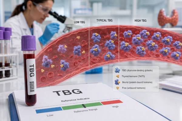
A thyroxine-binding globulin test measures the blood protein that carries most circulating T4 and T3. It is mainly used when total thyroid hormone results look abnormal but TSH, free hormone levels, symptoms, or the clinical examination do not support true thyroid disease. High TBG can raise total T4 and total T3 even when the thyroid is working normally. Low TBG can lower both totals without causing hypothyroidism. Pregnancy, estrogen-containing medicines, inherited variants, liver disease, kidney protein loss, and several medications can change TBG. The test does not directly show how active thyroid hormone is in body tissues, so it should be interpreted with TSH and usually free T4. A laboratory’s reference interval, assay method, age and sex criteria, pregnancy status, and medication list all affect interpretation. Correctly identifying a binding-protein change can prevent an incorrect diagnosis and unnecessary thyroid treatment.
- TBG measures a thyroid hormone carrier protein, not thyroid gland output or hormone action.
- High TBG commonly raises total T4 and total T3 while TSH and free T4 remain normal.
- Low TBG commonly lowers total T4 and total T3 without causing hypothyroid symptoms.
- Reference ranges are laboratory-specific; one major laboratory uses about 12–26 mcg/mL for males and 11–27 mcg/mL for females.
- Pregnancy and oral estrogen often increase TBG, while protein loss, androgens, and some liver disorders can decrease it.
- Unexpected total-hormone results should be checked with TSH, free T4, and the medication and illness history.
Table of Contents
- What the TBG Test Measures
- When a TBG Test Is Useful
- Normal Range and Result Patterns
- Causes of High TBG
- Causes of Low TBG
- Inherited TBG Deficiency and Excess
- Preparation, Limitations, and Next Steps
What the TBG Test Measures
Thyroxine-binding globulin, usually shortened to TBG, is made mainly in the liver. It has a high affinity for thyroid hormones and carries most of the T4 and a substantial share of the T3 circulating in blood. Transthyretin and albumin carry the rest. More than 99% of circulating T4 and T3 is protein-bound at any moment; only a small free fraction can readily enter cells and participate in feedback signaling.
TBG acts as a transport and buffering system. Bound hormone is temporarily unavailable to tissues, but it can detach as free hormone is used. When TBG rises, more hormone becomes bound at first. The pituitary and thyroid then adjust until the free hormone concentration returns toward its usual level. The final result may be a higher total T4 and total T3, because the blood contains more bound hormone, while free T4, free T3, and TSH remain appropriate.
The reverse occurs when TBG falls. Less thyroid hormone can be carried in the bound pool, so total T4 and total T3 may decrease. The feedback system reduces thyroid production enough to preserve the free fraction. A person can therefore have a strikingly low total T4 yet have normal thyroid function.
This compensation explains why TBG is not a direct thyroid function test. It answers a different question: could an abnormal amount of carrier protein explain an unexpected total hormone result? The total T4 test and total T3 include both bound and free hormone, so both are sensitive to binding-protein changes. TSH and free T4 more closely reflect the current thyroid-pituitary relationship in most outpatient settings.
TBG should not be confused with thyroglobulin. Thyroglobulin is a protein made inside the thyroid and is used in selected thyroid cancer follow-up. TBG is a liver-produced transport protein in the bloodstream. It should also not be confused with sex hormone-binding globulin, which carries sex steroids.
Direct measurement versus indirect binding tests
A TBG assay directly measures the concentration of the carrier protein, usually by an immunoassay. Older and still-available thyroid binding tests, such as T3 uptake, estimate the number of unoccupied binding sites rather than measuring TBG itself. The relationship is usually inverse: high TBG tends to produce low T3 uptake, while low TBG tends to produce high T3 uptake. The T3 uptake test does not measure the patient’s T3 concentration.
Total T4 combined with a T3 uptake result can be used to calculate the free thyroxine index. That index was designed to correct for ordinary binding changes. Direct free T4 testing has replaced it in many laboratories, but the index can still help when a direct free T4 result appears unreliable or when a local laboratory has validated the calculation well.
When a TBG Test Is Useful
TBG testing is not needed for routine thyroid screening. TSH is usually the first test in a person without pituitary disease, and free T4 is added when TSH is abnormal or the clinical situation requires it. TBG becomes useful when the numbers do not fit together.
Common reasons to order TBG include:
- High total T4 or total T3 with normal TSH, normal free T4, and no hyperthyroid symptoms
- Low total T4 with normal TSH and no convincing hypothyroid symptoms
- Pregnancy or estrogen treatment with an unexpected total-hormone result
- Suspected inherited TBG deficiency after a low total T4 newborn screen
- Several relatives with unusual total T4 or T3 values but normal thyroid function
- A suspected protein-losing, liver, nutritional, or medication-related binding change
- Discordance among total T4, free T4, TSH, and the clinical examination
The test is most informative when it resolves a specific discrepancy. For example, a person taking an estrogen-containing contraceptive may have high total T4, normal TSH, and normal free T4. A high TBG result explains the high total value and argues against treating hyperthyroidism. In contrast, high total T4 with suppressed TSH and high free T4 cannot be dismissed as a TBG effect; that pattern suggests true thyrotoxicosis.
A low total T4 in a healthy infant can also prompt TBG measurement, especially in screening programs that use total T4. Isolated TBG deficiency does not impair brain development or require levothyroxine, but congenital hypothyroidism does. Confirmatory TSH and free T4 are therefore essential rather than relying on the low total T4 alone.
TBG testing is less useful when TSH and free T4 already give a clear, concordant diagnosis. A high TSH with low free T4 supports primary hypothyroidism regardless of whether TBG is high, normal, or low. A suppressed TSH with high free T4 supports hyperthyroidism. In those settings, measuring TBG may explain the total hormone value but rarely changes the diagnosis.
When free T4 also needs caution
Direct free T4 immunoassays are designed to reduce binding-protein effects, but they are not completely immune to them. Pregnancy, severe illness, unusual albumin variants, very high or low binding proteins, heparin exposure, and certain antibodies can produce method-dependent results. If free T4 conflicts with TSH and the clinical picture, a repeat test on another platform, an equilibrium dialysis or ultrafiltration method, total T4 with a pregnancy-adjusted range, or a free thyroxine index may help.
Normal Range and Result Patterns
There is no single universal TBG range. Laboratories use different antibodies, calibrators, instruments, specimen requirements, and population data. One major U.S. reference laboratory lists adult intervals of 12–26 mcg/mL for males and 11–27 mcg/mL for females, but another laboratory may report a different interval or use mg/L, nmol/L, or a method-specific index. The range printed beside the result is the correct comparison.
Pregnancy requires separate interpretation. Estrogen increases TBG production and slows its clearance, so values may rise well above a nonpregnant adult range. One laboratory observed pregnancy values from about 27 to 66 mcg/mL and cites approximately 47–59 mcg/mL in the third trimester. These figures illustrate the size of the change; they are not universal pregnancy targets.
TBG should be read as part of a pattern:
| Pattern | Total T4 and T3 | Free T4 and TSH | Likely explanation |
|---|---|---|---|
| High TBG | Often high | Usually normal after equilibrium | Estrogen effect, pregnancy, inherited excess, or selected liver conditions |
| Low TBG | Often low | Usually normal | Inherited deficiency, protein loss, androgen effect, severe illness, or liver disease |
| High TBG plus thyroid overactivity | High, sometimes markedly | High free T4 or T3 with low TSH | True hyperthyroidism plus a binding-protein increase |
| Low TBG plus thyroid underactivity | Low | Low free T4 with high TSH in primary disease | True hypothyroidism plus a binding-protein decrease |
| Normal TBG with discordant tests | Variable | Variable | Consider assay interference, illness, medication, pituitary disease, or another binding protein |
A “normal” TBG result does not prove normal thyroid function. It only means the measured TBG concentration is within that assay’s interval. Likewise, a high or low TBG result does not diagnose hyperthyroidism or hypothyroidism. Symptoms caused by thyroid hormone excess or deficiency arise from the free hormone state and tissue response, not simply from the size of the bound reservoir.
Changes over time also need context. TBG may take weeks to reach a new steady state after starting or stopping oral estrogen. Total T4 can move in parallel. Comparing values drawn on different assays or during different medication states can create a false trend, so reports should be reviewed with dates and treatment changes.
Causes of High TBG
High TBG usually reflects increased liver production, slower breakdown, or an inherited variant. The most common physiologic and medication-related cause is estrogen exposure.
Pregnancy and estrogen
Estrogen changes the carbohydrate structure attached to TBG, which slows its clearance from blood. During pregnancy, TBG rises early and often reaches roughly two to three times the pre-pregnancy concentration. Total T4 and total T3 rise with it. This is an expected adaptation, not evidence that every pregnant person is hyperthyroid.
Oral estrogen used in combined contraceptives or menopausal hormone therapy can produce a similar, usually smaller effect. Oral delivery has a strong first-pass effect on the liver. Transdermal estrogen often changes TBG less, although the clinical response varies. Selective estrogen receptor modulators and some fertility treatments may also influence binding proteins.
People taking levothyroxine may need closer TSH monitoring after starting oral estrogen. More hormone becomes bound, and some patients require a dose increase to maintain the same free hormone and TSH. The dose should be adjusted from TSH and free T4 as appropriate, not from TBG alone.
Other acquired causes
High TBG may occur with acute hepatitis, selected chronic liver states, and some inherited or medication effects. Certain drugs, including estrogenic agents, tamoxifen, methadone, and fluorouracil-related therapies, have been associated with increased TBG or altered thyroid hormone binding. The size and clinical relevance of the change depend on dose, route, liver function, and the assay.
Some people with high TBG have both a binding change and genuine thyroid disease. Symptoms and the TSH/free T4 pattern decide whether thyroid treatment is needed. Treating a high total T4 solely because TBG is elevated can cause iatrogenic hypothyroidism if antithyroid medication is prescribed unnecessarily.
Causes of Low TBG
Low TBG can result from reduced liver synthesis, excessive protein loss, accelerated breakdown, hormone or medication effects, severe systemic illness, or an inherited SERPINA7 variant. Total T4 and T3 often fall, but TSH and free T4 can remain normal.
Protein-losing kidney disease is an important acquired cause. In nephrotic syndrome, proteins including TBG and albumin escape into urine. Severe gastrointestinal protein loss and major burns can also reduce circulating carrier proteins. The thyroid may compensate, but people with extensive ongoing loss, limited thyroid reserve, or levothyroxine treatment may develop a true hormone deficit that requires separate assessment.
Advanced liver disease can lower TBG synthesis, although liver disorders can produce complex patterns. Acute hepatitis may raise TBG, while severe synthetic failure may lower it. Albumin, bilirubin, clotting measures, clinical status, and the complete thyroid panel help distinguish these effects.
Androgens and anabolic steroids can lower TBG. Large glucocorticoid doses may also reduce it and alter conversion of T4 to T3. Other associations include severe malnutrition, critical illness, acromegaly, and some medications. In hospitalized patients, low TBG may be only one part of a broader nonthyroidal illness pattern that includes low T3, altered free hormone measurements, and a temporarily abnormal TSH.
A low TBG result should prompt a search for the cause only when clinically appropriate. In a well person with a stable, isolated low total T4, normal TSH and free T4, and a family pattern, inherited deficiency is more likely than severe acquired disease. In a person with edema, heavy urine protein, weight loss, jaundice, or systemic illness, the binding result may point toward an underlying condition that needs attention.
Do low TBG levels cause symptoms?
Isolated low TBG generally causes no symptoms because the free hormone concentration stays normal. Fatigue, cold intolerance, constipation, or weight change should not automatically be attributed to TBG deficiency. Those symptoms require evaluation for true hypothyroidism and other common causes. Levothyroxine should not be prescribed merely to normalize total T4 in someone with normal TSH and free T4.
Inherited TBG Deficiency and Excess
The SERPINA7 gene on the X chromosome provides instructions for making TBG. Variants can cause complete deficiency, partial deficiency, or less commonly TBG excess. Because the condition is X-linked, it often produces a clearer biochemical pattern in males, who have one X chromosome. Females may have normal, mildly low, or variable TBG because each cell inactivates one X chromosome.
Partial inherited TBG deficiency is estimated to occur in roughly 1 in 4,000 newborns, while complete deficiency is rarer. It may be discovered through newborn screening, an insurance examination, fertility testing, or a thyroid panel ordered for unrelated symptoms. A low total T4 can appear alarming, but the infant or adult is usually clinically euthyroid with normal TSH and free T4.
Inherited TBG excess produces high total T4 and total T3. It can be mistaken for familial hyperthyroidism if TSH and free hormones are not reviewed. Affected relatives may show similar results across generations. Genetic testing is usually unnecessary when the biochemical pattern, family history, and clinical state are clear, but it can confirm uncertain cases or support counseling.
Diagnosis commonly rests on:
- Persistently abnormal TBG on repeat testing
- Total T4 and T3 moving in the expected direction
- Normal TSH and a reliable free T4 result
- No medication, pregnancy, protein loss, or liver explanation
- Similar findings in relatives
- Genetic testing when the distinction remains important
Inherited TBG deficiency and excess usually require no treatment. The diagnosis should be documented prominently so future clinicians do not repeatedly interpret the total hormone result as thyroid failure or excess. Family members can be tested selectively, especially when a low total T4 newborn screening result could lead to urgent confusion.
A TBG variant can coexist with Hashimoto thyroiditis, Graves disease, pituitary disease, or another thyroid disorder. Once inherited TBG deficiency is known, future thyroid assessment should still use TSH and free T4 rather than assuming every abnormal result is benign.
Preparation, Limitations, and Next Steps
A TBG test uses a standard blood sample. Fasting is usually unnecessary unless another ordered test requires it. Follow the laboratory’s instructions about supplements. Some immunoassay platforms advise avoiding biotin-containing multivitamins or hair-and-nail products for at least 12 hours, and clinicians may recommend a longer pause after high-dose biotin. Do not stop prescribed medication without medical advice.
Bring or provide a complete list of medicines and supplements, including:
- Estrogen-containing contraceptives or hormone therapy
- Testosterone, anabolic steroids, or antiandrogen therapy
- Glucocorticoids
- Thyroid hormone or antithyroid medication
- Antiseizure drugs and other medicines that alter thyroid hormone metabolism
- Biotin dose and last use
- Recent iodine contrast, amiodarone, or major illness when thyroid tests are also being interpreted
Pregnancy status, recent delivery, kidney or liver disease, protein loss, and family history should also be recorded. A single abnormal TBG result may be repeated if it conflicts with the rest of the picture or if the patient’s medication state is changing.
Analytical interference is uncommon but possible. Heterophile antibodies, human anti-animal antibodies, extreme biotin exposure in susceptible assays, and platform-specific effects can produce an implausible value. Clues include a result that changes dramatically on another method, does not track with total T4 or T3, or contradicts all clinical findings.
A sensible follow-up sequence is:
- Confirm the exact TBG reference interval, units, and assay.
- Review TSH, free T4, total T4, and total T3 drawn at the same time when available.
- Identify pregnancy, estrogen, androgen, glucocorticoid, liver, kidney, nutritional, and protein-loss explanations.
- Repeat discordant tests after an appropriate interval or on another platform.
- Consider inherited TBG variation when the pattern is lifelong, isolated, and familial.
- Treat confirmed thyroid dysfunction or the underlying acquired illness, not the carrier-protein number by itself.
Urgent care is not usually required for an isolated TBG abnormality. Seek prompt assessment for severe thyroid-related symptoms such as chest pain, a very rapid or irregular heartbeat, fainting, confusion, marked agitation, severe weakness, hypothermia, or worsening shortness of breath. Those symptoms are evaluated from the complete clinical picture and thyroid hormone status rather than TBG alone.
The safest interpretation is often simple: total hormone results describe the whole circulating pool, while TSH and a reliable free T4 show whether the thyroid-pituitary system is maintaining an appropriate active fraction. TBG explains why those two views can differ.
References
- National recommendations of the Croatian society of medical biochemistry and laboratory medicine: Thyroid function tests from the laboratory point of view 2025 (Recommendations)
- Thyroid Stimulating Hormone and Thyroid Hormones (Triiodothyronine and Thyroxine): An American Thyroid Association-Commissioned Review of Current Clinical and Laboratory Status 2023 (Review)
- Partial Thyroid Hormone-Binding Globulin Deficiency 2023 (Case Report)
- Inherited thyroxine-binding globulin deficiency 2025 (Official Resource)
- Thyroxine-Binding Globulin (TBG), Serum 2026 (Laboratory Reference)
- Thyroid Function Tests 2024 (Official Resource)
Disclaimer
This article provides general education about TBG testing and cannot diagnose a thyroid, liver, kidney, genetic, or protein-loss disorder. Interpret results with the ordering clinician using the laboratory’s method, reference interval, pregnancy status, medications, symptoms, TSH, and free T4. Do not start, stop, or change thyroid or hormone medication based on a TBG value alone.