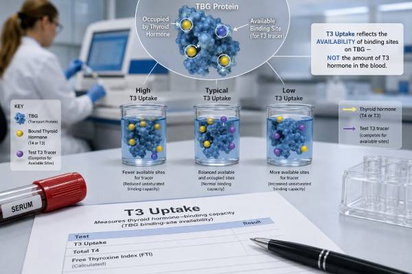
A T3 uptake test is an older thyroid blood test that estimates how many binding sites are available on proteins that carry thyroid hormones. Despite its name, it does not measure the amount of T3 in the blood and does not show how much T3 the thyroid produces. The result mainly reflects thyroid-binding globulin (TBG) and how saturated thyroid hormone carrier proteins are. A high T3 uptake can occur when TBG is low or when high thyroid hormone levels occupy more binding sites. A low uptake can occur when TBG is high or when fewer sites are occupied, as in hypothyroidism. Because those explanations move in opposite clinical directions, T3 uptake should never be read alone. It is usually combined with total T4 to calculate the free thyroxine index (FTI), which estimates free T4 while correcting for binding-protein changes. Direct free T4 assays and TBG tests have replaced T3 uptake in many settings, but older thyroid panels and some current laboratories still report it.
- T3 uptake measures thyroid hormone binding capacity indirectly; it is not a serum T3 concentration.
- High uptake usually means fewer available binding sites from low TBG or greater saturation from hyperthyroidism.
- Low uptake usually means more available binding sites from high TBG or reduced saturation from hypothyroidism.
- Pregnancy and estrogen therapy commonly lower T3 uptake by increasing TBG.
- T3 uptake is interpreted with total T4, often as a free thyroxine index, and should not be used alone.
- A typical adult range may be about 24% to 39%, but methods and reporting formats vary substantially.
Table of Contents
- What the T3 Uptake Test Actually Measures
- How the Test and Free Thyroxine Index Work
- High T3 Uptake Results
- Low T3 Uptake Results
- Reading T3 Uptake With T4 and TSH
- Preparation, Medications, and Interference
- When the Test Is Useful and Modern Alternatives
What the T3 Uptake Test Actually Measures
Most T4 and T3 in blood is attached to transport proteins. TBG carries the largest share, while transthyretin and albumin carry smaller amounts. Only a tiny free fraction can enter cells and participate directly in hormone signaling.
The T3 uptake test estimates the unsaturated binding capacity of these proteins. It may also be called:
- T3 resin uptake
- T3RU
- Thyroid hormone binding ratio
- Thyroxine-binding index
- T-uptake
- Thyroid binding capacity, depending on the laboratory
The terminology can be confusing because some methods report uptake as a percentage and others report a normalized ratio or index. The direction may even be inverted in certain modern assays. The laboratory’s method description and reference interval are therefore essential.
Why it is not a T3 level
In a classic resin-uptake assay, a small amount of labeled T3 is added to the patient’s serum. The tracer first binds to unoccupied sites on TBG and other proteins. A resin or other solid material then captures the tracer left over. The amount captured reflects how many protein-binding sites were already available or occupied.
No meaningful amount of the patient’s own T3 production is measured. A person can have a high total T3 with a normal T3 uptake, or a low total T3 with a high uptake, depending on TBG and thyroid status. For actual hormone concentration, clinicians order total T3 or free T3.
The reciprocal relationship with TBG
In traditional percentage-uptake methods:
- High TBG creates more empty binding sites, so more labeled T3 stays attached to serum proteins and less is taken up by the resin. The reported T3 uptake is low.
- Low TBG creates fewer binding sites, so more tracer remains free for the resin. The reported uptake is high.
This inverse relationship is the reason the test can correct total T4. When TBG alone raises total T4, uptake usually falls. When TBG alone lowers total T4, uptake usually rises.
Binding capacity versus thyroid function
T3 uptake is influenced by both the amount of carrier protein and the amount of thyroid hormone occupying it. Hyperthyroidism can increase uptake because endogenous T4 and T3 saturate more binding sites. Hypothyroidism can decrease uptake because more sites remain unoccupied. Binding-protein disorders can produce the same direction without true thyroid dysfunction.
This dual influence makes the result useful only as part of a pattern.
How the Test and Free Thyroxine Index Work
The main historical purpose of T3 uptake is to interpret total T4 when TBG is abnormal. Total T4 includes free and protein-bound hormone. It rises when TBG rises and falls when TBG falls, even if the free hormone concentration and thyroid function remain normal.
Calculating the FTI
The free thyroxine index combines total T4 with a normalized uptake measurement. The exact equation depends on the laboratory. Common forms include:
- Total T4 × T3 uptake ratio
- Total T4 divided by a thyroid-binding index
- Total T4 × a normalized thyroid hormone binding ratio
A laboratory may calculate the result automatically. Patients should not multiply a percentage by total T4 unless the report specifically provides that formula. For example, an uptake of 30% is not necessarily entered as 30 or 0.30; calibration factors differ.
The dedicated free thyroxine index test explains how the combined value approximates free T4 and why its reference interval is method-specific.
Why the correction works
Consider pregnancy. Estrogen raises TBG, so total T4 rises because more hormone is carried in bound form. T3 uptake falls because more TBG sites are available. When combined, the opposing changes can produce a normal FTI, supporting normal thyroid function.
Now consider inherited TBG deficiency. Total T4 falls because less hormone is protein-bound. T3 uptake rises because fewer binding sites exist. The FTI may remain normal, again indicating that free hormone supply is adequate.
In true hyperthyroidism, total T4 and uptake often move in the same direction: total T4 rises and uptake rises because binding sites are more saturated. The FTI becomes high. In true primary hypothyroidism, total T4 and uptake may both fall, creating a low FTI.
Modern assay variations
Some laboratories no longer use radioactive resin. Automated assays measure binding capacity through competitive immunoassay chemistry and report a T-uptake ratio. In some systems, a high numeric binding index corresponds to high TBG rather than high classic uptake. The report may say that the index is inversely related to traditional T3 uptake.
This is why online charts cannot safely interpret every result. The test name alone does not reveal the direction. The printed comments, equation, and reference range take priority.
High T3 Uptake Results
In a traditional percentage-uptake assay, a high result means that more labeled tracer was captured because fewer serum binding sites were available. This can result from low TBG, saturated TBG, or altered binding affinity.
Hyperthyroidism
Excess T4 and T3 occupy more carrier-protein sites, leaving fewer openings for the tracer. T3 uptake can rise along with total T4. A high FTI and suppressed TSH support true hyperthyroidism. The uptake result alone cannot establish the diagnosis.
Typical symptoms include palpitations, heat intolerance, tremor, sweating, weight loss, frequent bowel movements, anxiety, and menstrual changes. A hyperthyroidism panel relies on TSH, free T4, and T3, with TRAb or TSI when Graves disease is suspected.
Low TBG
Low TBG leaves fewer binding sites and can raise T3 uptake while lowering total T4. Free T4 and TSH may remain normal. Causes include:
- Inherited TBG deficiency
- Androgen or anabolic steroid exposure
- High-dose glucocorticoids in some settings
- Severe liver disease with reduced protein synthesis
- Nephrotic syndrome and other protein-losing states
- Severe systemic illness or malnutrition
- Acromegaly in some patients
An isolated high uptake with low total T4 can therefore look like opposing evidence. A normal FTI or direct free T4 helps show that thyroid function is preserved.
Medication effects
Androgens can lower TBG. Glucocorticoids may lower TBG and also suppress TSH or T4-to-T3 conversion. Certain antiseizure drugs change thyroid hormone binding and metabolism. Medication effects should be judged with dose, duration, total T4, free T4, and TSH.
Familial binding abnormalities
Inherited low TBG is X-linked. Males with a pathogenic variant may have very low TBG and low total T4, while heterozygous females can show variable results. They are usually clinically euthyroid. Thyroid hormone treatment is not needed unless a separate thyroid disorder is present.
When a high result needs follow-up
Useful follow-up tests include TSH, direct free T4, total T4, and sometimes a direct TBG measurement. Liver tests, albumin, urine protein, and medication review may be appropriate when low binding protein is suspected.
Low T3 Uptake Results
In the classic assay, low uptake means fewer labeled molecules reached the resin because more unoccupied binding sites were available in serum. High TBG and low thyroid hormone saturation are the two main explanations.
High TBG
High TBG increases total T4 and total T3 without necessarily increasing free hormone. Causes include:
- Pregnancy
- Estrogen-containing birth control or hormone therapy
- Inherited TBG excess
- Acute hepatitis and some chronic liver conditions
- Selective estrogen receptor modulators
- Certain medicines, including methadone and some fluorouracil-related treatments
The pattern can resemble hyperthyroidism if only total T4 is viewed. Low uptake and a normal FTI or direct free T4 point toward a binding-protein effect instead.
Hypothyroidism
When thyroid hormone production falls, fewer binding sites are occupied. More tracer binds to serum proteins and uptake falls. Total T4 may also be low, making the FTI low. High TSH confirms primary hypothyroidism in the usual setting.
The hypothyroidism blood test panel is more direct than T3 uptake and includes TPOAb when autoimmune thyroiditis is suspected.
Estrogen and pregnancy
Estrogen increases liver production of TBG and reduces its clearance. Total T4 rises over several weeks, while T3 uptake falls. This is a normal binding adaptation, not proof of an overactive or underactive thyroid.
Pregnancy also changes TSH through human chorionic gonadotropin, alters albumin, expands blood volume, and challenges some free T4 immunoassays. A low T3 uptake during pregnancy should not be treated. Pregnancy-specific TSH and thyroid hormone interpretation is required.
Other causes
Low uptake may occur with acute intermittent porphyria, some liver states, or medicines that increase binding proteins. It can also result from assay interference or an incorrectly applied reference interval.
When a low result needs follow-up
TSH and free T4 usually clarify whether hypothyroidism exists. If total T4 is high but TSH and free T4 are normal, pregnancy, estrogen, or TBG excess is likely. If total T4 and free T4 are both low, the clinician considers true hypothyroidism, severe illness, or pituitary disease.
Reading T3 Uptake With T4 and TSH
The most useful interpretation compares T3 uptake with total T4 and TSH. The table below assumes a traditional assay in which high uptake corresponds to low binding capacity.
| Total T4 | T3 uptake | TSH | Likely pattern |
|---|---|---|---|
| High | High | Low | True hyperthyroidism is likely; FTI is usually high |
| Low | Low | High | Primary hypothyroidism is likely; FTI is usually low |
| High | Low | Normal | High TBG from pregnancy, estrogen, or inherited excess; FTI may be normal |
| Low | High | Normal | Low TBG from inherited deficiency, protein loss, liver disease, or androgens; FTI may be normal |
| Normal | Abnormal | Normal | Mild binding change, drug effect, method variation, or compensated thyroid state |
Opposite directions suggest binding effects
When total T4 and traditional uptake move in opposite directions, the change often originates in TBG rather than the thyroid. High total T4 plus low uptake is the classic high-TBG pattern. Low total T4 plus high uptake is the classic low-TBG pattern.
Same direction suggests hormone change
When both move high, hyperthyroidism becomes more likely. When both move low, hypothyroidism becomes more likely. TSH remains the main feedback marker and can overturn a simplistic pattern. For example, severe illness can lower total T4 and alter uptake without producing primary hypothyroidism.
Discordant or impossible combinations
A normal TSH with a very high FTI, no symptoms, and stable prior tests raises concern for assay interference or a binding abnormality not fully corrected by the index. A high TSH with high FTI can occur after taking levothyroxine just before the blood draw, during recovery from hypothyroidism, with inconsistent dosing, or from interference.
Repeat testing with direct free T4 or a different method is often more useful than repeating the same older panel.
Preparation, Medications, and Interference
Fasting is generally not required. The main preparation is to provide an accurate medication, supplement, and pregnancy history.
Tell the clinician about
- Pregnancy, oral contraceptives, or estrogen therapy
- Testosterone, anabolic steroids, or antiandrogen treatment
- Glucocorticoids
- Methadone
- Antiseizure medicines
- Amiodarone and iodine exposure
- Levothyroxine, liothyronine, or desiccated thyroid
- Biotin-containing supplements
- Liver disease, kidney disease, nephrotic syndrome, malnutrition, or major illness
Do not stop prescription medicines solely for a T3 uptake test unless instructed. Their effect may be the reason the test was ordered.
Biotin
Biotin interferes with many immunoassays that use biotin-streptavidin chemistry. The effect on T3 uptake depends on the platform. It can also distort TSH and free T4, producing a misleading overall pattern. Many laboratories advise stopping ordinary non-prescription biotin for at least 48 hours; high-dose therapy may need a longer clinician-directed interval.
Thyroid medication timing
Levothyroxine taken shortly before the draw can raise total and free T4 transiently. For serial comparison, test at a consistent interval after the dose, often before the morning tablet when the clinician prefers. Liothyronine causes a stronger short-term T3 peak, although the uptake test still does not measure that T3 directly.
Acute illness
Hospitalization, inflammation, reduced nutrition, low albumin, kidney protein loss, and liver dysfunction can alter both hormone metabolism and binding proteins. T3 uptake may not correct these complex changes accurately. Unless thyroid disease is strongly suspected, repeating TSH and free T4 after recovery can give a cleaner answer.
Reference ranges
Traditional adult ranges are often near 24% to 39% or 25% to 38%, but a laboratory may report a ratio such as 0.8 to 1.2. Pediatric ranges vary by age. Never interpret a percentage against a ratio interval or compare values from two different methods as though they are identical.
When the Test Is Useful and Modern Alternatives
T3 uptake is now uncommon because direct free T4, TSH, and TBG testing are widely available. It still appears in legacy thyroid profiles and can provide a useful consistency check when total T4 is affected by binding proteins.
Situations where it may help
The test can be useful when:
- A laboratory uses a validated FTI method
- Total T4 is abnormal but TSH and symptoms do not fit
- Pregnancy or estrogen use makes total T4 difficult to interpret
- An inherited or acquired TBG abnormality is suspected
- Direct free T4 immunoassay results appear unreliable
- Historical results were followed with the same method and continuity matters
The FTI can sometimes perform better than a routine direct free T4 immunoassay in unusual binding states, although neither method is perfect.
Direct free T4
Most first-line thyroid evaluations use TSH and direct free T4. Free T4 avoids much of the total-hormone effect from TBG, but routine immunoassays can still be biased by pregnancy, severe illness, abnormal binding proteins, heparin, and antibodies.
Equilibrium dialysis or ultrafiltration followed by mass spectrometry can provide a reference-method free T4 measurement when a major discrepancy remains. These tests are slower and less available.
Direct TBG measurement
A TBG blood test identifies whether the carrier concentration is high or low. It is more intuitive than an uptake result because high TBG is reported as high rather than causing a low traditional uptake. TBG concentration does not measure binding affinity, however, and a normal concentration does not exclude every inherited binding variant.
A practical order of interpretation
When a T3 uptake result appears on a report:
- Confirm whether the assay is traditional uptake or an inverse binding index.
- Read it with total T4 and the calculated FTI.
- Check TSH, which usually provides the strongest evidence for primary thyroid dysfunction.
- Review pregnancy, estrogen, androgen, protein loss, liver disease, and medications.
- Use direct free T4 or TBG testing if the pattern remains unclear.
- Avoid diagnosing hyperthyroidism or hypothyroidism from uptake alone.
T3 uptake remains a binding test with a thyroid-related name. Understanding that distinction prevents the most common error: treating it as though it were a high or low T3 hormone result.
A single abnormal uptake value rarely requires treatment. Treatment is directed at confirmed thyroid dysfunction, an underlying liver or kidney disorder, clinically important protein loss, or another identified cause—not at the uptake percentage itself. Inherited TBG differences usually need documentation rather than medication, so future clinicians do not repeatedly mistake an unusual total T4 for thyroid disease. Keeping the original report, including its method and FTI, can prevent years of unnecessary retesting, referrals, worry, and inappropriate long-term thyroid hormone treatment in otherwise healthy people.
References
– T3RU test 2026 – T3 Uptake 2026 – Free Thyroxine Index (FTI), Serum 2026 – Thyroid Stimulating Hormone and Thyroid Hormones (Triiodothyronine and Thyroxine): An American Thyroid Association-Commissioned Review of Current Clinical and Laboratory Status 2023 (Review) – T3RU test 2025 – Thyroid function tests 2026
Disclaimer
This article provides general education and cannot interpret an individual thyroid panel. T3 uptake methods, directions, formulas, and reference intervals vary, so use the comments on the actual laboratory report. Discuss abnormal or discordant results with a qualified clinician.