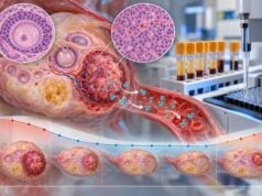
A vulvar cancer biomarker panel commonly uses HPV testing, p16 immunohistochemistry, and p53 immunohistochemistry to classify vulvar squamous cell carcinoma (VSCC) by its underlying pathway. The most useful modern framework separates tumors into HPV-associated cancers, HPV-independent p53-abnormal cancers, and HPV-independent p53-wild-type cancers. p16 “block-positive” staining is a practical surrogate for transforming high-risk HPV activity, while an abnormal p53 pattern suggests TP53-driven, HPV-independent disease. Direct HPV DNA or RNA testing can resolve uncertain or discordant cases. This classification matters because the groups arise from different precursor lesions and have different clinical behavior. HPV-associated tumors generally have a more favorable prognosis than HPV-independent p53-abnormal tumors, although stage, lymph-node status, margin status, age, and treatment remain major outcome factors. Biomarkers do not replace routine pathology. The final interpretation should combine tumor morphology, precursor lesion, p16 pattern, p53 pattern, and direct HPV testing when needed.
- p16 block positivity usually supports HPV-associated vulvar squamous cell carcinoma: strong continuous nuclear and cytoplasmic staining is more meaningful than scattered positive cells.
- Abnormal p53 staining supports an HPV-independent TP53-driven pathway: overexpression, null, or other mutation-type patterns can be seen.
- The main biologic groups are HPV-associated, HPV-independent p53-abnormal, and HPV-independent p53-wild-type: they differ in precursor lesions and prognosis.
- Direct HPV testing is useful when p16 and p53 are discordant or morphology is unusual: rare tumors can show misleading immunostain combinations.
- Biomarker classification does not replace stage: lymph-node involvement, tumor size, depth, margins, and other clinical factors remain central to treatment and prognosis.
Table of Contents
- Why HPV, p16, and p53 Matter in Vulvar Cancer
- HPV-Associated Vulvar Cancer and p16 Block Positivity
- HPV-Independent p53-Abnormal Vulvar Cancer
- HPV-Independent p53-Wild-Type Tumors
- Biomarkers in Vulvar Precancer and Precursor Lesions
- Discordant, Double-Positive, and Unusual Results
- Reading the Pathology Report and Next Steps
Why HPV, p16, and p53 Matter in Vulvar Cancer
Most vulvar cancers are squamous cell carcinomas, but they do not all arise through the same molecular pathway. Biomarker testing helps pathologists separate tumors that may look similar under the microscope but developed through different biology.
The broad modern model recognizes two major etiologic pathways and three clinically useful molecular groups. One pathway is driven by high-risk human papillomavirus (HPV). The other is HPV-independent and often associated with chronic vulvar dermatoses, older age, and TP53 abnormalities. Within HPV-independent disease, tumors can be divided further according to p53 status.
The resulting three-group framework is:
- HPV-associated VSCC, usually p16 block-positive.
- HPV-independent, p53-abnormal VSCC, usually p16 non-block or negative with mutation-type p53 staining.
- HPV-independent, p53-wild-type VSCC, lacking evidence of transforming HPV and lacking a classic mutation-type p53 pattern.
This classification is useful because the groups tend to have different precursor lesions and outcomes. HPV-associated cancers generally arise from vulvar high-grade squamous intraepithelial lesion (HSIL). HPV-independent p53-abnormal cancers commonly arise from differentiated vulvar intraepithelial neoplasia (dVIN) or related HPV-independent precursor changes.
p16 and p53 are immunohistochemical surrogates, not perfect replacements for molecular testing. p16 reflects the cell-cycle effect of transforming HPV, while p53 staining reflects many—but not all—TP53 mutations. Direct HPV testing can therefore be important when the stains and morphology do not fit together.
A panel should answer a focused classification question rather than simply produce “positive” or “negative” labels. This is similar to how a cervical HPV biomarker panel uses viral and protein markers, although vulvar cancer has its own precursor lesions and classification rules.
HPV-Associated Vulvar Cancer and p16 Block Positivity
HPV-associated VSCC develops through persistent infection with a carcinogenic HPV type, most often HPV16. Viral oncogenes interfere with normal tumor-suppressor pathways, including the retinoblastoma protein pathway. That disruption causes marked overexpression of p16INK4a, creating a characteristic tissue-staining pattern.
The typical p16 result is block-positive. Pathologists look for strong, continuous nuclear and cytoplasmic staining through a broad area of tumor or precursor epithelium. Patchy, focal, or scattered p16 staining is not equivalent to block positivity.
In a conventional HPV-associated tumor, the pattern is often:
- p16: strong block-positive;
- p53: wild-type or non-mutation pattern;
- direct HPV test: positive for high-risk HPV when performed;
- morphology: frequently warty or basaloid features, although morphology alone is not sufficiently reliable.
Direct HPV testing can use DNA-based or RNA-based methods. RNA assays that detect E6/E7 transcripts can provide evidence of transcriptionally active infection, while DNA assays detect the viral genome. The exact method depends on the laboratory.
HPV-associated VSCC tends to occur at a younger average age than HPV-independent disease and is linked to the same broad viral carcinogenesis pathway as cervical, vaginal, and some anal or oropharyngeal cancers. However, a positive vulvar p16 stain does not tell which HPV genotype is present. A direct high-risk HPV genotype test is a different assay and should not be inferred from p16 alone.
On average, HPV-associated vulvar cancers have better disease-specific outcomes than HPV-independent p53-abnormal cancers. This is a group-level observation rather than a guarantee for an individual patient. A node-positive HPV-associated tumor can still be high risk, and an early-stage HPV-independent tumor may be cured with appropriate treatment.
For treatment, surgery and/or radiation decisions are still driven mainly by tumor size, location, depth, nodal status, margins, and overall health. HPV/p16 status refines classification and prognosis but does not replace conventional staging.
HPV-Independent p53-Abnormal Vulvar Cancer
The HPV-independent p53-abnormal pathway is biologically distinct. These cancers are usually not driven by high-risk HPV and commonly carry pathogenic TP53 mutations. They often occur in older women and may arise in a background of chronic inflammatory or lichenoid vulvar disease, including lichen sclerosus.
p53 immunohistochemistry serves as a practical surrogate for many TP53 mutations. A mutation-type result can appear as:
- diffuse strong nuclear overexpression, with most tumor cells intensely stained;
- complete null staining in tumor cells while normal internal control cells remain positive;
- less commonly, an abnormal cytoplasmic pattern or other recognized mutation-type pattern.
A wild-type p53 stain is heterogeneous, with a variable mixture of positive and negative nuclei. Calling a stain simply “p53 positive” is therefore not sufficiently precise.
The classic HPV-independent p53-abnormal profile is:
- p16: negative or non-block;
- p53: mutation-type abnormal;
- direct HPV test: negative;
- precursor: often dVIN or another HPV-independent precursor lesion.
These tumors generally have the least favorable prognosis of the three major molecular groups, with higher recurrence and disease-specific mortality in many cohorts. Their biology may also include a larger field of genetically altered epithelium around the invasive cancer, which can contribute to local recurrence risk.
p53 staining can sometimes reveal abnormal epithelium beyond what appears obviously dysplastic on routine sections. That can help the pathologist map a precursor field, although surgical margin decisions still depend on full histologic assessment and clinical anatomy.
A general p53 IHC test explains the logic of overexpression and null patterns across cancers. In vulvar pathology, the result is especially useful when paired with p16 to distinguish HPV-independent from HPV-associated carcinogenesis.
HPV-Independent p53-Wild-Type Tumors
Not every HPV-independent vulvar cancer has abnormal p53. A third group is HPV-independent and p53-wild-type. These tumors lack convincing evidence of transforming HPV and also lack a classic TP53 mutation-type immunostain pattern.
This group is more biologically heterogeneous than the other two. Some tumors may arise through alterations in pathways involving NOTCH, RAS, PIK3CA, or other genes, and some are associated with precursor lesions that do not fit classic dVIN morphology. The molecular landscape is still being refined.
The usual biomarker pattern is:
- p16: non-block or negative;
- p53: wild-type heterogeneous pattern;
- HPV: negative when directly tested.
Prognosis tends to fall between the HPV-associated and HPV-independent p53-abnormal groups in many studies, but published results vary. Stage and nodal status remain powerful determinants and can outweigh the biomarker group in an individual case.
This category matters because an older two-group approach—HPV-positive versus HPV-negative—can hide meaningful differences within HPV-negative tumors. Separating p53-abnormal from p53-wild-type disease gives a more informative pathologic classification.
The group also emphasizes why a negative p16 stain is not enough to classify a tumor. If p16 is non-block, p53 needs to be interpreted, and direct HPV testing may be appropriate in uncertain cases. Morphology and precursor lesions can then help place the tumor in the correct pathway.
As research develops, HPV-independent p53-wild-type tumors may be subdivided further. For now, the three-tier framework is clinically useful because it captures the largest reproducible differences in pathogenesis and outcome without pretending every molecular detail is known.
Biomarkers in Vulvar Precancer and Precursor Lesions
Biomarkers are useful not only in invasive cancer but also in vulvar precursor lesions, where the distinction between HPV-associated and HPV-independent disease can be subtle.
Vulvar HSIL is HPV-associated. It often shows block-positive p16 and increased proliferative activity. Morphology may be basaloid or warty, and high-risk HPV is usually detectable. This lesion can progress to HPV-associated VSCC if not appropriately managed.
Differentiated vulvar intraepithelial neoplasia (dVIN) is usually HPV-independent and frequently associated with TP53 abnormalities. It can be difficult to recognize because the surface may mature and the atypia can be concentrated near the basal layer rather than involving the full epithelial thickness. p53 IHC can support the diagnosis when the staining pattern matches a mutation-type abnormality.
Not all HPV-independent precursors are classic p53-abnormal dVIN. Pathology literature recognizes a broader spectrum of HPV-independent precursor lesions, including lesions with p53-wild-type patterns. These can be challenging and may require expert gynecologic pathology review.
A key practical problem is field change. HPV-independent carcinogenesis can affect a wider area of vulvar epithelium than the visible tumor. A patient may have invasive cancer at one site and subtle precursor disease nearby. Identifying the background lesion can help explain recurrence risk and support long-term vulvar surveillance.
p16 and p53 should not be used to replace morphology in these lesions. Strong p16 block positivity supports HPV-associated HSIL, but focal p16 staining is nonspecific. Abnormal p53 can support dVIN, but some dVIN shows wild-type p53 and some non-neoplastic tissue can show physiologic basal staining.
The goal is to match morphology + p16 + p53 + HPV status into a coherent pathway. When one component does not fit, the case deserves review rather than force-fitting the stain into a predetermined category.
Discordant, Double-Positive, and Unusual Results
Most tumors fit the expected patterns, but real pathology includes exceptions. A particularly important pitfall is assuming that any p16-positive vulvar cancer must be HPV-associated.
Rare p16 block-positive, p53-abnormal tumors have been described. Some are truly HPV-associated cancers that acquired a TP53 alteration, but others appear to be HPV-independent cancers with aberrant p16 overexpression. Direct HPV testing is valuable in these cases.
Likewise, a tumor can be p16 non-block but still have detectable HPV. The viral infection may be incidental, the assay may detect a low-level non-transforming infection, or the immunostain may be technically or biologically atypical. The clinically relevant question is whether HPV is actually driving the tumor.
Potential reasons for discordance include:
- differences between HPV DNA detection and transcriptionally active HPV;
- unusual tumor biology;
- sampling of different tumor areas;
- technical staining problems;
- subclonal TP53 mutations;
- p16 overexpression through non-HPV mechanisms;
- interpretation of a patchy p16 pattern as if it were true block positivity.
A 2023 pathology study specifically highlighted that p16/p53 combinations require an algorithmic approach and careful consideration of caveats. When the stains conflict, direct HPV RNA or DNA testing and molecular TP53 testing can help resolve classification.
The same caution applies to “double negative” cases. p16 non-block and p53 wild-type can represent the legitimate HPV-independent p53-wild-type group rather than a failed panel. If morphology fits, the absence of the two classic abnormal patterns is itself informative.
The final report may therefore use wording such as “HPV-associated,” “HPV-independent p53-abnormal,” or “HPV-independent p53-wild-type” rather than simply listing three independent laboratory results. Integrated language is more useful because it tells the oncology team what pathway the pathologist believes is driving the cancer.
Reading the Pathology Report and Next Steps
Start with the final histologic diagnosis and stage-related features, then read the biomarker interpretation. The most clinically useful report often states both the tumor type and the inferred molecular pathway.
Common phrases include:
- “p16 block-positive” — strong support for HPV-associated disease in the right morphology;
- “p16 non-block” or “patchy” — does not support transforming HPV by itself;
- “p53 abnormal/mutation-type” — supports a TP53-driven HPV-independent pathway when HPV evidence is absent;
- “p53 wild-type” — a heterogeneous, non-mutation pattern;
- “high-risk HPV detected” — direct viral evidence, with method-specific meaning;
- “HPV-independent VSCC” — classification based on integrated morphology and biomarkers.
Then review the conventional prognostic features. Tumor diameter, depth of invasion, lymphovascular invasion, margin status, nodal metastases, extracapsular spread, and distant disease remain essential. Biomarker group refines the picture but does not replace staging.
Useful questions for the care team include:
- Is the cancer classified as HPV-associated or HPV-independent?
- If HPV-independent, is p53 abnormal or wild-type?
- Was HPV tested directly or inferred from p16?
- Were the p16 and p53 patterns concordant, or was extra testing needed?
- Is there a precursor lesion such as vulvar HSIL or dVIN in the surrounding tissue?
- Do margin findings include precursor disease as well as invasive cancer?
- How much does the molecular group change my recurrence-risk counseling compared with stage and lymph-node status?
After treatment, follow-up focuses on clinical examination and symptoms because local vulvar recurrence or new precursor lesions may be visible or palpable. Persistent itching, a new lump, ulcer, bleeding area, color change, or nonhealing lesion deserves evaluation even if the original tumor was completely removed.
HPV vaccination can prevent many HPV-associated vulvar precancers and cancers when given before exposure and still has preventive value according to age-appropriate vaccination guidance. It does not treat an existing tumor, and it does not prevent HPV-independent p53-driven disease.
A well-interpreted biomarker panel improves classification by connecting the pathology to the cancer’s causal pathway. Its strongest value comes from integration, not from treating HPV, p16, or p53 as a stand-alone verdict.
The biomarker pathway can also guide how clinicians think about the surrounding vulvar skin after treatment. HPV-associated disease may be accompanied by multifocal HPV-related HSIL elsewhere in the lower genital tract, while HPV-independent cancers may arise in a chronic inflammatory field with subtle dVIN or other precursor change. That difference can shape the focus of long-term examination, but neither pathway eliminates the need for careful surveillance. A new lesion should be assessed on its own merits rather than assumed to have the same biology as the original tumor. If a recurrence or second primary has unusual morphology, repeat p16, p53, or direct HPV testing may be appropriate because the new lesion can differ from the first.
Long-term examination is particularly important because both recurrent invasive cancer and new precursor lesions may develop after apparently successful treatment.
References
- Vulvar Squamous Cell Carcinoma: Scientific Progress and Impacts to Pathology Practice 2026 (Review)
- Typing of Vulvar Squamous Cell Carcinoma: Why it is Important? 2025 (Review)
- Role of Immunohistochemical Analysis of p16 and p53 in Vulvar Carcinoma 2025
- High concordance of molecular subtyping between pre-surgical biopsy and surgical resection specimen (matched-pair analysis) in patients with vulvar squamous cell carcinoma using p16- and p53-immunostaining 2024
- Classification of Vulvar Squamous Cell Carcinoma and Precursor Lesions by p16 and p53 Immunohistochemistry: Considerations, Caveats, and an Algorithmic Approach 2023
- Human papillomavirus and p53 status define three types of vulvar squamous cell carcinomas with distinct clinical, pathological, and prognostic features 2023
Disclaimer
This article is for general education and does not interpret an individual vulvar biopsy or cancer prognosis. HPV, p16, and p53 results should be integrated by a pathologist with tumor morphology, precursor lesions, stain controls, direct HPV testing when needed, and the complete surgical pathology. Treatment and surveillance decisions should be made with a gynecologic oncology team using stage and current guidelines.