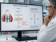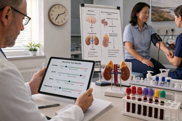
An adrenal hormone panel combines tests that answer different questions about the adrenal cortex and its control systems. Cortisol and ACTH assess the stress-hormone axis. Aldosterone and renin assess sodium balance, potassium handling, and blood-pressure regulation. DHEA-S reflects adrenal androgen production. The pattern can help identify primary adrenal insufficiency, pituitary or glucocorticoid-related suppression, primary aldosteronism, or selected androgen disorders.
There is no single universal panel or one “optimal” set of levels. These hormones follow different rhythms and preparation rules. Cortisol and ACTH are usually paired early in the morning. Aldosterone and renin depend on posture, salt intake, potassium, and many blood-pressure medicines. DHEA-S varies strongly by age and sex. A result may be technically within range yet physiologically inappropriate—for example, normal-range ACTH when cortisol is very low, or normal-range aldosterone when renin is fully suppressed in a person with hypertension. Dynamic tests often confirm a suspected disorder. A panel should be ordered for a defined clinical question, not used as a general stress or fatigue score.
- Low cortisol with high ACTH suggests primary adrenal insufficiency; low cortisol with low or normal ACTH suggests central suppression.
- Low aldosterone with high renin supports mineralocorticoid deficiency, while high or inappropriate aldosterone with suppressed renin raises concern for primary aldosteronism.
- DHEA-S is an age- and sex-specific adrenal androgen marker; it cannot diagnose adrenal insufficiency by itself.
- Morning timing, posture, potassium, salt intake, medicines, and specimen handling can materially change panel results.
- Vomiting, dehydration, confusion, fainting, or shock may indicate adrenal crisis and requires emergency care before complete outpatient testing.
Table of Contents
- What the Panel Measures
- When the Panel Is Useful
- Preparation and Collection
- Cortisol and ACTH Patterns
- Aldosterone and Renin Patterns
- DHEA-S Results
- Follow-Up and Next Steps
What the Panel Measures
The adrenal cortex has three functional zones. The outer zona glomerulosa makes aldosterone, the middle zona fasciculata makes cortisol, and the inner zona reticularis makes adrenal androgens. These zones share cholesterol-based steroid pathways but respond to different signals.
Cortisol
Cortisol helps maintain blood pressure, glucose availability, immune regulation, and the response to illness. The hypothalamus signals the pituitary, the pituitary releases ACTH, and ACTH stimulates adrenal cortisol production. Cortisol peaks around waking for most daytime sleepers and falls toward midnight.
A single total serum cortisol reflects both free and protein-bound hormone at one moment. Estrogen raises cortisol-binding globulin and total cortisol; low albumin or binding globulin can lower it. When baseline results are indeterminate, a cosyntropin stimulation test assesses reserve.
ACTH
ACTH identifies whether abnormal cortisol originates primarily in the adrenal gland or upstream. High ACTH is an appropriate pituitary response to low cortisol. Low ACTH is appropriate when cortisol is high but inappropriate when cortisol is low. The peptide is unstable, so it needs chilled EDTA plasma and rapid processing.
Aldosterone and renin
Aldosterone tells the kidneys to retain sodium and excrete potassium. Renin is released by the kidneys when blood flow, sodium delivery, or effective circulating volume falls. Renin starts a pathway that produces angiotensin II, which stimulates aldosterone. The two values should therefore be interpreted together.
Laboratories measure either plasma renin activity, or PRA, or direct renin concentration, or DRC. These are not interchangeable. The aldosterone-to-renin ratio, or ARR, must use cutoffs matched to the assay and units.
DHEA-S
Dehydroepiandrosterone sulfate is the stable sulfated form of an adrenal androgen. ACTH influences its production. Levels are high in young adulthood and decline with age. Markedly high DHEA-S can point toward an adrenal source of androgen excess; low levels can accompany adrenal insufficiency or ACTH suppression but are common with aging and many illnesses.
An adrenal insufficiency panel emphasizes low hormone production, while a hypertension workup emphasizes aldosterone-renin physiology. Combining markers without a clinical question can produce confusing, unrelated flags.
When the Panel Is Useful
Clinicians choose panel components when symptoms suggest a specific adrenal pathway disorder. Possible reasons include:
- fatigue with weight loss, nausea, low blood pressure, low sodium, high potassium, salt craving, or hyperpigmentation;
- current or recent glucocorticoid exposure with concern for HPA-axis suppression;
- resistant or early-onset hypertension, especially with low potassium;
- an adrenal mass discovered on imaging;
- rapid androgenic changes, severe hirsutism, early puberty, or virilization;
- pituitary disease, surgery, radiation, or head injury;
- follow-up for established adrenal insufficiency or primary aldosteronism; or
- suspected congenital adrenal hyperplasia, together with pathway-specific precursors.
The panel is not a direct screen for every adrenal tumor. Pheochromocytoma requires plasma free or urinary fractionated metanephrines. Cushing syndrome screening uses late-night salivary cortisol, urine free cortisol, or dexamethasone suppression rather than a random morning cortisol. Congenital adrenal hyperplasia requires markers such as 17-hydroxyprogesterone or 11-deoxycortisol.
Symptoms also determine urgency. Gradual hair growth or longstanding hypertension can usually be evaluated through planned outpatient testing. Rapid virilization, severe hypokalemia, a hypertensive emergency, or signs of adrenal crisis need prompt or emergency assessment. A broad panel should never delay treatment of a dangerous electrolyte, blood-pressure, or cortisol problem.
Panels sold directly to consumers may combine valid analytes without a valid indication. The measurements can be accurate while the package interpretation is not. Before ordering, ask whether the result has a recognized decision threshold, whether preparation can be controlled, and whether an abnormal finding has a defined confirmatory path. Testing is valuable when it changes management.
An adrenal incidentaloma does not automatically require every hormone. The imaging appearance, size, blood pressure, potassium, and symptoms guide testing. Cortisol autonomy is commonly assessed with a 1-mg overnight dexamethasone suppression test. Aldosterone and renin are relevant when hypertension or unexplained hypokalemia is present. Metanephrines depend on imaging characteristics and current guideline context.
Fatigue alone rarely justifies a large panel. Sleep disorders, anemia, thyroid disease, medication effects, depression, diabetes, infection, and many other conditions are more common. “Adrenal fatigue” is not a recognized diagnosis and cannot be confirmed by a panel.
Preparation and Collection
One appointment can collect several analytes, but their requirements must be coordinated. The ordering clinician should state whether the goal is low cortisol, aldosterone excess, androgen excess, or monitoring.
For cortisol and ACTH, collection is usually between 7 and 10 a.m. Record wake time, draw time, and the last glucocorticoid dose. Report all steroid routes: tablets, injections, inhalers, creams, nasal sprays, eye drops, and rectal products. Never hold a prescribed steroid without a safe plan.
ACTH requires a prechilled EDTA tube, transport on ice, and prompt centrifugation or freezing according to the laboratory protocol. Delay can falsely lower ACTH. Cortisol usually goes into a separate serum tube.
Aldosterone and renin preparation is more complex. Results change with:
- upright versus seated or supine posture;
- time awake and time of day;
- recent sodium intake;
- potassium level;
- dehydration or volume expansion;
- kidney function;
- menstrual phase and pregnancy; and
- antihypertensive and other medicines.
Low potassium can suppress aldosterone and create a false-negative screen for primary aldosteronism, so potassium should be corrected when safe. Sodium should not be severely restricted before ARR testing unless the clinician instructs otherwise. The laboratory protocol may require the patient to be awake and upright for at least two hours and seated for 5–15 minutes before collection.
Mineralocorticoid receptor antagonists, potassium-wasting diuretics, ACE inhibitors, angiotensin receptor blockers, beta blockers, central alpha-2 agonists, and other drugs can shift renin or aldosterone. Stopping them can be unsafe. The clinician weighs the risk, may substitute less-interfering medicines, or interprets results on treatment. A suppressed renin despite a medicine expected to raise it can still be informative.
Beta blockers and central alpha-2 agonists can suppress renin and create a falsely high ratio. ACE inhibitors, angiotensin receptor blockers, and many diuretics tend to raise renin and can lower the ratio, potentially hiding primary aldosteronism. Mineralocorticoid receptor antagonists often have the largest effect. These tendencies are not rigid correction factors; the clinician cannot simply add or subtract a number. Documenting the drug and dose is essential.
Posture has a physiologic purpose. Standing reduces central blood volume and activates renin-angiotensin signaling, while lying down suppresses it. A sample collected immediately after rushing into the clinic is not equivalent to one collected after a standardized seated rest. The request should specify the intended posture, and the collection team should record what actually happened.
DHEA-S does not fluctuate as sharply as DHEA and generally does not require cycle-specific timing. Report DHEA supplements, androgen therapy, oral contraceptives, pregnancy, and age. High-dose biotin can interfere with some immunoassays; follow the laboratory’s withholding instruction.
Cortisol and ACTH Patterns
Morning cortisol is interpreted with the assay’s reference and decision limits. Very low results support adrenal insufficiency; clearly adequate results make it less likely; intermediate values need stimulation testing. Random afternoon cortisol is harder to interpret because normal levels decline across the day.
| Cortisol | ACTH | Likely direction |
|---|---|---|
| Low | High | Primary adrenal insufficiency or cortisol-synthesis defect |
| Low | Low or inappropriately normal | Pituitary, hypothalamic, or glucocorticoid-induced suppression |
| High after hypercortisolism is confirmed | Low | ACTH-independent adrenal cortisol production |
| High after hypercortisolism is confirmed | Measurable or high | Pituitary or ectopic ACTH-dependent cortisol excess |
Primary adrenal insufficiency often adds high renin, low or inappropriately normal aldosterone, low sodium, high potassium, and low DHEA-S. Central insufficiency usually preserves aldosterone because the renin-angiotensin system, not ACTH, provides its main control. Potassium therefore often remains normal.
A normal-range ACTH does not exclude central disease when cortisol is low; it is physiologically inadequate. Similarly, one normal cortisol does not prove stress reserve. The ACTH stimulation test measures the peak cortisol response, using an assay-specific cutoff.
Glucocorticoid exposure can produce a mixed-looking panel. External steroid suppresses ACTH, which can lower endogenous cortisol and DHEA-S while leaving aldosterone relatively preserved. Hydrocortisone is measured as cortisol, and prednisolone cross-reacts with some assays, so the same exposure can also make total cortisol appear adequate. Exact medication timing resolves more uncertainty than adding another hormone.
In early primary adrenal insufficiency, ACTH and renin may rise before basal cortisol or aldosterone falls clearly below range. That evolving pattern requires specialist follow-up rather than waiting for every marker to become abnormal. Conversely, dehydration alone can raise renin. A trend matters only when posture, medicines, intake, and laboratory methods are comparable.
If Cushing syndrome is suspected, establish cortisol excess first. Morning cortisol may be normal in Cushing syndrome and high during ordinary stress. After confirmation, ACTH classifies the source and guides imaging.
Aldosterone and Renin Patterns
The ARR is a screening tool for primary aldosteronism, not a stand-alone diagnosis. A high ratio is meaningful when renin is suppressed and aldosterone is inappropriate for sodium and potassium status. Dividing by a very low renin value can create a large ratio even when aldosterone is low, so all three—aldosterone, renin, and ratio—must be reviewed.
The 2025 Endocrine Society guideline provides assay-specific guidance. As one example, an aldosterone-to-PRA ratio above 20 when aldosterone is in ng/dL and PRA in ng/mL/h can support a positive screen if aldosterone is sufficiently high and renin suppressed. Direct-renin cutoffs and LC-MS/MS aldosterone thresholds differ. Local laboratory criteria take priority.
| Aldosterone | Renin | Possible explanation |
|---|---|---|
| High or inappropriate | Suppressed | Primary aldosteronism |
| High | High | Secondary aldosterone drive from low volume, diuretics, renal artery disease, or heart failure |
| Low | High | Primary adrenal mineralocorticoid deficiency |
| Low | Low | Hyporeninemic hypoaldosteronism, medicine effect, or another low-renin state |
Primary aldosteronism may occur with normal potassium. Hypertension severity and renin suppression are important even without hypokalemia. A positive screen may be repeated or followed by confirmatory suppression testing, although strongly expressed biochemical patterns can sometimes proceed directly according to guideline criteria.
Confirmatory tests examine whether aldosterone suppresses when sodium or volume increases. Options include saline infusion, oral sodium loading, captopril challenge, and fludrocortisone suppression. Each has contraindications and local protocols. Sodium loading may be unsafe with severe hypertension, heart failure, kidney disease, or marked hypokalemia. The clinician selects the test rather than treating them as interchangeable.
Renin is also useful in replacement therapy for primary adrenal insufficiency. Persistently high renin with salt craving, low pressure, or high potassium may suggest insufficient mineralocorticoid effect, while suppressed renin with hypertension or low potassium may suggest excess. Dose changes use symptoms, pressure, electrolytes, and standardized renin—not a single target in isolation.
If unilateral treatment is considered, adrenal CT shows anatomy but cannot reliably identify which gland is producing aldosterone. Adrenal vein sampling is often used to distinguish unilateral from bilateral production, except in selected patients. An ARR-focused evaluation covers medication and posture effects in more detail.
DHEA-S Results
DHEA-S provides a stable view of adrenal androgen output because its half-life is longer and daily fluctuation smaller than DHEA. Reference intervals decline substantially with age and differ by sex and laboratory. A value that is normal for a 25-year-old may be high for an older adult, or vice versa.
Markedly high DHEA-S, especially with rapid virilization, raises concern for an adrenal source such as an androgen-secreting tumor or congenital adrenal hyperplasia. PCOS can cause mild to moderate elevation, but rapid voice change, clitoral enlargement, sudden severe hirsutism, or a very high androgen level warrants prompt evaluation. Companion tests include total and free testosterone, androstenedione, and 17-hydroxyprogesterone.
Low DHEA-S can occur in primary or central adrenal insufficiency because ACTH-dependent adrenal androgen production falls. It can also reflect normal aging, chronic illness, glucocorticoid use, or other nonspecific factors. A low DHEA-S cannot diagnose cortisol deficiency or justify DHEA treatment by itself.
In people with ovaries, adrenal and ovarian androgen production overlap. DHEA-S is mainly adrenal, while testosterone and androstenedione can arise from both sources. In people with testes, testicular testosterone dominates circulating androgen function, so low DHEA-S does not equal androgen deficiency. The symptom context and companion hormones prevent overinterpretation.
DHEA supplements can raise DHEA-S and downstream androgens. Potential effects include acne, oily skin, unwanted hair growth, scalp hair loss, menstrual change, and interactions with hormone-sensitive conditions. Supplement use should be documented before interpreting the result.
Follow-Up and Next Steps
Abnormal panels require pathway-specific confirmation. For suspected adrenal insufficiency, repeat a properly timed paired sample or perform cosyntropin testing. Primary disease may lead to 21-hydroxylase antibodies, renin and aldosterone confirmation, and cause-directed imaging or genetic testing. Central disease prompts review of other pituitary hormones and MRI.
For suspected primary aldosteronism, correct potassium, review medicines and sodium intake, and repeat the ARR under a documented protocol when needed. Confirmation, adrenal imaging, and adrenal vein sampling depend on the strength of the biochemical result and treatment goals.
For androgen excess, confirm the result with a reliable assay and assess onset and progression. Rapid virilization or a very high DHEA-S deserves prompt imaging and endocrine or gynecologic evaluation; mild stable elevation has a broader differential.
Ask the clinician:
- What exact diagnosis was each panel component intended to assess?
- Were cortisol and ACTH drawn together in the morning and was ACTH handled on ice?
- Which renin method and ARR units did the laboratory use?
- Were posture, potassium, sodium intake, and medicines suitable for ARR interpretation?
- Is DHEA-S compared with the correct age- and sex-specific interval?
- Which abnormality needs confirmation before imaging or treatment?
Do not try to normalize every flagged value with supplements. Treatment targets the disorder: glucocorticoid and sometimes mineralocorticoid replacement for adrenal insufficiency, aldosterone-directed medication or surgery for primary aldosteronism, and cause-specific care for androgen excess. Hormone replacement without a diagnosis can distort future testing and cause harm.
Adrenal crisis requires immediate hydrocortisone and fluids when suspected. Blood for cortisol, ACTH, renin, and aldosterone can be drawn before treatment if it causes no delay. Repeated vomiting, severe dehydration, confusion, fainting, low blood pressure, low glucose, or shock is an emergency.
The panel is most useful as a set of relationships. Cortisol must fit ACTH, aldosterone must fit renin, and DHEA-S must fit age, sex, and symptoms. Preparation and confirmation turn those relationships into a reliable diagnosis.
Keep the original laboratory report rather than only a portal flag. It preserves units, reference intervals, posture notes, collection time, and assay method. If repeat testing occurs, aim for similar conditions. A true change in adrenal physiology is easier to see when preanalytical variables have not changed at the same time.
Results also deserve a time horizon. Some abnormalities require action the same day, while others are best confirmed before treatment. Ask when the clinician expects the repeat or dynamic test, whether current medicines should remain unchanged, and which symptoms should trigger earlier contact. A documented plan prevents a mildly abnormal panel from being ignored or, at the other extreme, treated as an emergency without evidence.
If several components are abnormal, prioritize the result linked to immediate risk—cortisol deficiency, severe potassium disturbance, or dangerous blood pressure—before pursuing less urgent androgen questions.
How Clinicians Reconcile a Mixed Panel
An adrenal panel often contains results that seem to point in different directions. That does not necessarily mean several diseases are present. Cortisol and ACTH vary by time of day and stress, while aldosterone and renin respond strongly to posture, salt intake, potassium, blood volume, and blood-pressure medicines. DHEA-S changes substantially with age and sex. Before forming a conclusion, the clinician checks whether every value used the correct reference interval and whether the samples were collected under conditions appropriate for that specific hormone.
Medication reconciliation is part of interpretation, not an administrative detail. Glucocorticoids can suppress ACTH and endogenous cortisol. Diuretics, mineralocorticoid receptor antagonists, beta blockers, ACE inhibitors, angiotensin receptor blockers, and some calcium-channel blockers can shift renin or aldosterone. Estrogen may change cortisol-binding proteins, and androgen or DHEA supplements can alter androgen measurements. Patients should provide doses, routes, and the time of the last dose rather than stopping medicine on their own.
Next steps should address the dominant clinical pattern. Low morning cortisol with an ACTH value may lead to dynamic adrenal testing. Suppressed renin with inappropriate aldosterone in a patient with hypertension may justify a standardized aldosterone-renin ratio and confirmatory evaluation. Markedly high DHEA-S with rapid-onset virilization can prompt targeted androgen testing and imaging after biochemical confirmation. Mild isolated abnormalities are often repeated under controlled conditions. A concise plan should name the suspected condition, the confounders being corrected, the repeat or confirmatory test, and the symptoms that warrant urgent care. This approach prevents a broad panel from becoming a collection of unrelated flags.
References
- Primary Aldosteronism 2025 (Guideline)
- Physiology, Adrenocorticotropic Hormone (ACTH) 2025 (Review)
- Diagnosis and management of adrenal insufficiency 2024 (Review)
- Adrenocorticotropic Hormone (Cosyntropin) Stimulation Test 2023 (Review)
- Physiology, Aldosterone 2023 (Review)
Disclaimer
This article is educational and cannot diagnose an adrenal or pituitary disorder. Hormone ranges and ratios depend on assay, units, timing, posture, medicines, and clinical context. Suspected adrenal crisis requires immediate emergency care.