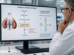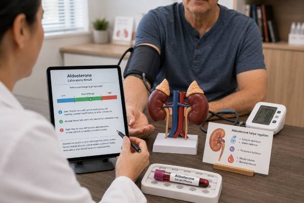
An aldosterone blood test measures the adrenal hormone that helps the kidneys retain sodium and excrete potassium. Clinicians use it with renin to investigate hormonal causes of high or low blood pressure, abnormal potassium, and suspected adrenal insufficiency. The aldosterone number alone is rarely diagnostic because the appropriate level changes with posture, salt intake, potassium, fluid balance, kidney function, and medicines.
For high blood pressure, the central pattern is aldosterone that remains inappropriately high while renin is suppressed. This raises concern for primary aldosteronism, in which one or both adrenal glands produce aldosterone without normal renin control. For low blood pressure or high potassium, low aldosterone may reflect adrenal failure or inadequate renin production. Both aldosterone and renin can be high when the body is responding normally to low circulating volume, diuretics, heart failure, or reduced kidney blood flow. Testing therefore requires standardized preparation and paired interpretation. A result inside the reference range can still be inappropriate for the physiologic setting, and an abnormal screen usually needs confirmation before imaging or long-term treatment.
- Aldosterone should be measured with renin; the relationship is more informative than either value alone.
- High or inappropriately normal aldosterone with suppressed renin suggests primary aldosteronism.
- Low aldosterone with high renin supports primary mineralocorticoid deficiency, including primary adrenal insufficiency.
- Potassium, salt intake, posture, time of day, and many blood-pressure medicines can change the result.
- Severe weakness, paralysis, palpitations, fainting, or a blood-pressure emergency requires urgent care rather than routine repeat testing.
Table of Contents
- What Aldosterone Does
- Why the Test Is Ordered
- Preparation and Collection
- Normal Range and the Aldosterone–Renin Ratio
- High Aldosterone Results
- Low Aldosterone Results
- Confirmation and Follow-Up
What Aldosterone Does
Aldosterone is a mineralocorticoid steroid made in the zona glomerulosa, the outer layer of the adrenal cortex. It acts mainly in the kidney’s distal nephron, increasing sodium reabsorption while promoting potassium and hydrogen-ion excretion. Water follows retained sodium, supporting circulating volume and blood pressure.
The renin-angiotensin-aldosterone system, or RAAS, provides the main control. Specialized kidney cells release renin when they sense reduced renal perfusion, reduced sodium delivery, or sympathetic stimulation. Renin initiates formation of angiotensin II, which constricts blood vessels and tells the adrenal cortex to release aldosterone. Higher potassium can stimulate aldosterone directly. ACTH has a smaller, short-lived effect.
When volume and sodium are adequate, renin and aldosterone should fall. When volume is depleted, both should rise. Disease becomes visible as a mismatch:
- aldosterone high while renin is suppressed suggests autonomous adrenal production;
- aldosterone low while renin is high suggests the adrenal gland cannot respond;
- both high suggests secondary RAAS activation; and
- both low suggests reduced renin drive, medicine effect, or another low-renin state.
Serum or plasma aldosterone is reported in units such as ng/dL or pmol/L. Renin is reported as plasma renin activity in ng/mL/h or as direct renin concentration in mU/L. The methods cannot be mixed in one ratio. The laboratory’s units and assay-specific cutoff are essential.
An aldosterone blood test is different from a 24-hour urine aldosterone test, which assesses daily excretion during a defined sodium intake. It is also different from adrenal vein sampling, an invasive procedure that compares aldosterone secretion from each adrenal gland after primary aldosteronism is established.
Why the Test Is Ordered
Primary aldosteronism is a common, underrecognized cause of hypertension. The 2025 Endocrine Society guideline supports broad screening among people with hypertension, depending on local resources and patient preferences, because targeted treatment can reduce aldosterone-related cardiovascular and kidney risk.
Traditional high-priority reasons include:
- resistant hypertension despite three suitable medicines, or controlled pressure requiring four or more;
- hypertension with spontaneous or diuretic-induced low potassium;
- severe or early-onset hypertension;
- hypertension plus an adrenal mass;
- hypertension plus obstructive sleep apnea;
- hypertension with atrial fibrillation out of proportion to other risk factors;
- a family history of early hypertension or stroke; or
- a first-degree relative with primary aldosteronism.
Normal potassium does not exclude primary aldosteronism. Many affected people never develop hypokalemia. Aldosterone can damage the heart, blood vessels, and kidneys beyond the effect of blood pressure alone, so finding the cause matters even when ordinary antihypertensive treatment lowers the reading.
Blood pressure should be confirmed with accurate technique. Use a correctly sized cuff, rest before measurement, and consider home or ambulatory readings when white-coat or masked hypertension is possible. ARR screening is not a replacement for establishing that hypertension is present. At the same time, a normal office reading on several medicines does not remove the reason to screen if the treatment burden or prior severity is high.
Primary aldosteronism is a spectrum rather than only the classic combination of extreme hypertension and profound hypokalemia. Milder autonomous secretion may still suppress renin and increase long-term risk. The degree of aldosterone excess, duration of hypertension, kidney function, and age all influence presentation.
Testing is also used for high potassium, low sodium, low blood pressure, dehydration, salt craving, or suspected adrenal insufficiency. In primary adrenal failure, cortisol and aldosterone may both be low, while ACTH and renin rise. Central adrenal insufficiency usually preserves aldosterone.
Other uses include investigation of salt-losing kidney disorders, congenital adrenal enzyme defects, pseudohypoaldosteronism, and low-renin hypertension. The same aldosterone concentration can mean different things across these settings.
Preparation and Collection
Preparation should match the local protocol. A typical primary-aldosteronism screen is collected in the morning after the patient has been awake and upright for at least two hours and seated quietly for 5–15 minutes. Other laboratories use supine collection for particular reference intervals. The order and report should state posture.
Maintain usual, unrestricted sodium intake unless the clinician gives a specific plan. Severe sodium restriction activates renin and can hide autonomous aldosterone; very high salt loading is not appropriate before an ordinary screening draw. Dehydration also raises RAAS activity.
Potassium should be measured and low potassium corrected when safe. Hypokalemia can suppress aldosterone and create a false-negative screen. A tourniquet or fist clenching can artifactually raise potassium, so careful phlebotomy matters.
Medicines have predictable directions of effect but no simple correction formula:
- mineralocorticoid receptor antagonists and epithelial sodium-channel blockers raise renin and directly alter the pathway;
- diuretics often raise renin;
- ACE inhibitors and angiotensin receptor blockers raise renin and may lower aldosterone;
- beta blockers and central alpha-2 agonists suppress renin and can raise the ratio;
- nonsteroidal anti-inflammatory drugs may suppress renin; and
- estrogen-containing medicines can affect some direct-renin measurements.
Never stop blood-pressure medication without supervision. A clinician may test on current treatment, substitute medicines with less ARR interference, or arrange a safe washout. Severe hypertension, heart failure, or hypokalemia may make washout inappropriate. Document every drug, dose, and time taken.
Also report pregnancy, menstrual phase when requested, kidney disease, licorice use, vomiting or diarrhea, recent intense exercise, and salt supplements. Pregnancy strongly activates RAAS, so standard nonpregnant ranges may not apply.
Hormonal contraception and sex-hormone therapy deserve method-specific review. Estrogen can raise angiotensinogen and alter PRA or DRC differently, changing the calculated ratio. This does not mean contraception should be stopped automatically. The laboratory and clinician can choose the renin method, interpret the exposure, or repeat under a safe plan if the initial result is unclear.
Age and kidney function also matter. Renin often falls with age and chronic kidney disease, which can inflate the ratio. Reduced filtration and potassium-handling problems broaden the causes of abnormal aldosterone. The test should be interpreted alongside creatinine and estimated glomerular filtration rate.
The blood draw itself is brief. Aldosterone is collected as serum or plasma according to the laboratory, and renin uses its specified tube and handling. Some renin samples have strict temperature requirements because cold can activate prorenin in vitro. Follow the laboratory instructions rather than assuming every component should be placed on ice.
Normal Range and the Aldosterone–Renin Ratio
There is no single normal aldosterone range. Upright values are usually higher than supine values. Sodium intake, age, time of day, pregnancy, and assay method shift the interval. Use the posture-specific range printed on the report.
For hypertension screening, calculate or review the aldosterone-to-renin ratio, or ARR. The 2025 Endocrine Society guideline gives illustrative positive criteria that vary by method. When aldosterone is measured by immunoassay, suppressed PRA at or below 1 ng/mL/h or DRC at or below 8.2 mU/L together with aldosterone at or above 10 ng/dL can support a positive screen. With LC-MS/MS aldosterone, a lower threshold around 7.5 ng/dL may apply. The ratio threshold also differs by renin method and units.
These numbers are guidance, not universal lab rules. A useful screen requires:
- renin that is genuinely suppressed;
- aldosterone that is inappropriate for that suppressed renin;
- a ratio above the local threshold; and
- acceptable potassium, sodium intake, posture, and medicine context.
Unit errors are a common source of apparent disagreement. Aldosterone may be reported in ng/dL, pg/mL, or pmol/L. Renin may be activity or concentration. An ARR of 20 in one unit system does not equal 20 in another. Never recalculate using a cutoff from a different assay without confirming the conversion and denominator.
Suppressed renin is the physiologic hallmark. If renin is not suppressed, primary aldosteronism is less likely, but a diuretic, ACE inhibitor, ARB, or mineralocorticoid antagonist may be forcing it upward. A negative result under strongly interfering medication can merit repeat testing if clinical suspicion remains high.
| Aldosterone | Renin | Possible interpretation |
|---|---|---|
| High or inappropriate | Low | Primary aldosteronism |
| High | High | Secondary aldosteronism |
| Low | High | Primary hypoaldosteronism or adrenal failure |
| Low | Low | Hyporeninemic hypoaldosteronism or low-renin mineralocorticoid-like state |
A high ratio caused only by dividing a low aldosterone by nearly zero renin is not automatically positive. Review the absolute aldosterone. Conversely, a person with very suppressed renin and aldosterone below a generic high cutoff may still have a clinically relevant renin-independent aldosterone phenotype; guideline and specialist interpretation are evolving.
Screening thresholds trade sensitivity for specificity. A lower threshold finds more cases but produces more false positives; a higher threshold misses milder disease. Pretest probability matters. Resistant hypertension and hypokalemia make a given borderline ratio more concerning than the same ratio in a low-risk patient with an uncertain office blood pressure.
High Aldosterone Results
Primary aldosteronism
In primary aldosteronism, one adrenal adenoma or unilateral hyperplasia may overproduce aldosterone, or both glands may produce excess. Sodium retention expands volume and suppresses renin. Potassium can be normal or low; bicarbonate may rise from metabolic alkalosis.
Symptoms, when present, often reflect hypertension or hypokalemia: headache, muscle weakness, cramps, tingling, excessive urination, thirst, palpitations, or arrhythmia. Many people have no specific symptoms. The biochemical pattern, not symptoms alone, identifies disease.
Aldosterone-producing adrenocortical carcinoma is rare. Familial forms can present young or cluster in families. Genetic evaluation is considered according to age, family history, and subtype.
Secondary aldosteronism
When both aldosterone and renin rise, the adrenal gland may be responding appropriately to perceived low volume. Causes include diuretics, vomiting, diarrhea, severe sodium restriction, heart failure, cirrhosis with ascites, nephrotic syndrome, renovascular disease, and renin-producing tumors. Pregnancy also raises both.
Secondary aldosteronism is not treated by removing an adrenal gland. Care targets the trigger, volume state, kidney blood flow, heart or liver disease, and medicines.
High aldosterone with high renin in a dehydrated person is physiologically different from high aldosterone with suppressed renin in a person with hypertension. This is why ordering aldosterone without renin can lead to the wrong conclusion.
Renovascular hypertension deserves separate attention. Narrowing of a renal artery can reduce perfusion to one kidney, raise renin, and drive secondary aldosterone. The person may have abrupt or worsening hypertension, asymmetric kidney size, a rise in creatinine after ACE inhibition, or vascular disease. Imaging is selected from the clinical context; an aldosterone value does not locate a renal-artery lesion.
Heart failure and cirrhosis can activate RAAS even when total body fluid is excessive because effective arterial volume is low. Edema therefore does not guarantee that renin and aldosterone will be suppressed. Treating the underlying hemodynamic disorder differs from treating autonomous adrenal production.
Low Aldosterone Results
Low aldosterone becomes clinically important when potassium is high, sodium or blood pressure is low, or renin shows that the adrenal response is inadequate.
Low aldosterone with high renin suggests primary adrenal mineralocorticoid failure. Causes include autoimmune Addison disease, congenital adrenal hyperplasia, bilateral adrenal damage, infection, hemorrhage, or medicines that impair aldosterone synthesis. Cortisol and ACTH help determine whether the broader adrenal cortex is affected.
Low aldosterone with low renin is common in hyporeninemic hypoaldosteronism, often associated with diabetic kidney disease, chronic tubulointerstitial kidney disease, older age, or medicines. ACE inhibitors, angiotensin receptor blockers, direct renin inhibitors, NSAIDs, heparin, calcineurin inhibitors, trimethoprim, and potassium-sparing drugs can contribute through different mechanisms.
Another low-renin, low-aldosterone pattern occurs when something else acts like a mineralocorticoid. Examples include Liddle syndrome, apparent mineralocorticoid excess, large licorice exposure, 11β-hydroxylase deficiency, 17α-hydroxylase deficiency, and some cortisol-excess states. These conditions often cause hypertension and low potassium rather than the high potassium of hypoaldosteronism.
Pseudohypoaldosteronism produces salt wasting and high potassium despite high aldosterone because the kidney or other tissues resist the hormone. A low aldosterone result would argue against that mechanism.
Low values without symptoms or electrolyte abnormalities may reflect high sodium intake, supine posture, medicine effects, or normal physiology. They should not be treated in isolation.
Hyperkalemia requires confirmation because hemolysis, fist clenching, and delayed sample processing can falsely raise potassium. A genuine high potassium with ECG changes, weakness, or arrhythmia is urgent. Aldosterone testing can identify the chronic mechanism later, but immediate management stabilizes the heart and lowers potassium safely.
Type 4 renal tubular acidosis is a common setting for hypoaldosteronism or aldosterone resistance. It typically combines hyperkalemia with a mild normal-anion-gap metabolic acidosis. Diabetes and kidney disease are frequent contexts. Bicarbonate, kidney function, glucose, and medicine review help identify it.
Confirmation and Follow-Up
After a positive aldosterone–renin ratio, the clinician reviews collection quality and may repeat testing. Depending on the strength of the pattern and guideline criteria, confirmation may use saline infusion, oral sodium loading with urine aldosterone, captopril challenge, or fludrocortisone suppression. Severe hypertension, kidney disease, heart failure, or marked hypokalemia can make sodium-loading tests unsafe.
Once primary aldosteronism is established, adrenal CT evaluates anatomy. CT alone cannot reliably tell which gland is overproducing hormone because nonfunctioning nodules are common and small functioning lesions may be missed. Adrenal vein sampling usually determines unilateral versus bilateral secretion when surgery is considered, with selected exceptions.
Unilateral disease may be treated with adrenalectomy. Bilateral disease is usually treated with a mineralocorticoid receptor antagonist such as spironolactone or eplerenone. Treatment aims to control blood pressure and potassium and to reverse renin suppression when feasible, while monitoring kidney function.
After targeted treatment, potassium may rise and estimated filtration may fall because the prior aldosterone excess had increased renal filtration and potassium excretion. This can reveal underlying kidney disease rather than represent treatment failure. Clinicians monitor electrolytes and creatinine soon after surgery or medication changes and adjust other antihypertensives as blood pressure improves.
For low aldosterone, repeat electrolytes and review medicines first. Cortisol, ACTH, renin, glucose, kidney function, and acid-base status help identify adrenal failure versus hyporeninemic disease. Primary adrenal insufficiency requires glucocorticoid replacement and, when aldosterone is deficient, fludrocortisone. A full adrenal insufficiency evaluation confirms the pattern.
Ask the clinician:
- Was renin measured as PRA or DRC, and which ARR cutoff applies?
- What posture and rest period were used?
- Was potassium corrected and sodium intake adequate?
- Which medicines could cause a false positive or false negative?
- Is the aldosterone appropriate for the renin, rather than merely inside range?
- Does the result need repeat screening, suppression testing, imaging, or adrenal vein sampling?
Seek urgent care for severe weakness, paralysis, a dangerous heartbeat, chest pain, neurologic symptoms, fainting, or extreme blood pressure. Do not self-treat potassium or salt abnormalities; both under- and overcorrection can be dangerous.
An aldosterone result becomes clinically meaningful only when paired with renin and the person’s volume, potassium, posture, and medication state. Standardized collection and pathway-specific confirmation prevent a normal physiologic response from being mistaken for adrenal disease.
Keep the full report with the collection time, posture, potassium, drug list, aldosterone units, renin method, and calculated ratio. A portal flag that shows only “high” or “normal” cannot be reinterpreted accurately later. If the screen is repeated, matching the preparation conditions makes change over time meaningful. Ask for a written next step—repeat testing, suppression testing, treatment, or no further action—so a borderline result does not remain unresolved.
Home blood-pressure readings can strengthen follow-up when they use a validated cuff and a consistent schedule. Record readings rather than changing salt, potassium, or medication in response to one high value. Bring the log and device to a visit so technique and calibration can be checked. Hormone-directed treatment should be judged by both biochemical response and real-world pressure control.
Why Renin and Potassium Complete the Picture
Aldosterone cannot be classified as simply high or low without considering renin. A seemingly normal aldosterone may be inappropriate when renin is strongly suppressed, especially in a person with resistant hypertension or unexplained low potassium. In contrast, both hormones may rise when the kidneys sense reduced blood flow, dehydration, diuretic use, or narrowing of a renal artery. Low aldosterone with high renin suggests a different mechanism from low values of both hormones. The paired pattern therefore provides more information than either result alone.
Potassium influences the test in both directions. Low potassium can reduce aldosterone secretion and potentially hide primary aldosteronism, while high potassium normally stimulates aldosterone. Clinicians commonly correct significant hypokalemia before standardized screening when it is safe to do so. Sodium intake, posture, collection time, menstrual status, and kidney function can also shift results. The report’s units and assay-specific reference range must be used because laboratories measure and calculate these hormones differently.
If the first result conflicts with the clinical picture, repeating aldosterone and renin under documented conditions is often more useful than acting on one number. The clinician may adjust interfering blood-pressure medicine only when the risks are acceptable and substitute agents that have less effect on the system. A suspicious pattern may lead to an aldosterone-renin ratio, confirmatory suppression testing, adrenal imaging, and sometimes adrenal vein sampling. Imaging alone cannot establish hormone production because nonfunctioning adrenal nodules become more common with age. Severe hypertension with chest pain, neurologic symptoms, breathing difficulty, or acute vision change requires emergency evaluation rather than outpatient hormone interpretation.
References
- Primary Aldosteronism 2025 (Guideline)
- Primary Aldosteronism: An Endocrine Society Clinical Practice Guideline 2025 (Guideline)
- Endocrine Hypertension in Children and Adolescence 2025 (Review)
- Hyperaldosteronism 2024 (Review)
- Hypoaldosteronism 2023 (Review)
Disclaimer
This information is educational and cannot diagnose or treat an aldosterone disorder. Reference ranges and ARR thresholds depend on assay, units, posture, medicines, sodium, and potassium. Severe blood-pressure or potassium-related symptoms require urgent medical care.