
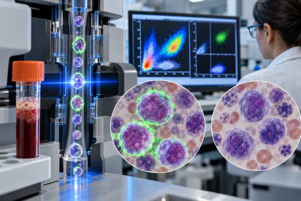
A CD33 test checks whether blood or bone marrow cells express CD33, a protein associated mainly with myeloid development. It is commonly included in flow cytometry panels used to investigate acute myeloid leukemia (AML), myelodysplastic syndromes, and other myeloid disorders. Many AML blasts are CD33-positive, but CD33 is not specific for leukemia: normal myeloid precursors and monocytes can also express it. A positive result therefore supports myeloid differentiation only when it fits the cell population, morphology, and the rest of the immunophenotype. CD33 can also matter after diagnosis because it is the target of gemtuzumab ozogamicin and is being studied in other antibody and cellular therapies. There is no universal “normal CD33 level” for leukemia testing. Reports usually describe the percentage of abnormal cells that are positive, the intensity of expression, and whether staining is uniform or heterogeneous. The most useful interpretation combines CD33 with markers such as myeloperoxidase, CD13, CD34, CD117, HLA-DR, monocytic markers, and molecular and chromosome findings.
- CD33 positivity is common in AML but does not diagnose AML by itself. Normal myeloid cells also express CD33.
- CD33 is usually measured by flow cytometry. Reports may include percentage positive, dim/bright intensity, and whether expression is uniform or variable.
- There is no single diagnostic CD33 cutoff for all AML cases. The full marker pattern and genetic classification are more important than one percentage.
- CD33 can be a treatment target. Gemtuzumab ozogamicin binds CD33, so expression can have therapeutic relevance in selected AML settings.
- CD33 should not be confused with a specific myeloid-lineage proof test. Myeloperoxidase and other findings may carry greater lineage specificity in difficult acute leukemia classification.
Table of Contents
- What CD33 Is and Why It Is Tested
- How CD33 Testing Is Done
- What a CD33-Positive Result Means
- CD33 in Acute Myeloid Leukemia
- CD33 and Targeted Treatment
- CD33 in MRD and Follow-Up Testing
- Limitations and Next Steps
What CD33 Is and Why It Is Tested
CD33 is a myeloid-associated cell-surface protein that helps laboratories recognize cells developing along granulocytic and monocytic pathways. It is also called SIGLEC-3. The protein is found on many myeloid progenitors and mature monocytes and on a large proportion of AML blasts.
Because acute leukemia can arise from different blood-cell lineages, one of the first laboratory questions is whether the abnormal blasts are myeloid, B-lymphoid, T-lymphoid, or show features of more than one lineage. CD33 contributes to that assessment, but it is not used alone. A leukemia flow cytometry panel evaluates multiple antigens at the same time and identifies the pattern carried by the abnormal population.
CD33 testing can be useful for:
- supporting myeloid differentiation in suspected AML;
- describing the immunophenotype of leukemic blasts at diagnosis;
- identifying aberrant antigen combinations useful for follow-up;
- helping characterize acute promyelocytic leukemia and other AML phenotypes;
- assessing whether a CD33-directed treatment may be relevant; and
- studying antigen persistence or change after therapy.
CD33 is not restricted to malignant cells. A normal marrow contains CD33-positive myeloid cells, and reactive changes can alter their proportions. For this reason, “CD33 positive” only becomes meaningful after the laboratory has identified which cells are positive.
It is also important to separate a lineage-associated marker from modern disease classification. Current AML classification relies heavily on recurrent genetic abnormalities. A patient can have AML even if CD33 is weak or absent, and strong CD33 does not identify a specific AML genetic subtype by itself.
CD33 outside AML
CD33 can appear in several settings that are not acute myeloid leukemia. Normal monocytes and developing myeloid cells carry the antigen, so marrow recovery after chemotherapy or growth-factor treatment may produce many CD33-positive cells without indicating relapse. Myelodysplastic neoplasms and chronic myeloid disorders can also contain CD33-positive myeloid populations. In some hematologic cancers outside the myeloid lineage, aberrant CD33 expression can occur as part of an unusual immunophenotype.
This broad distribution explains why a CD33-positive blood test is not used as a population screening test for leukemia. The laboratory first has to identify a suspicious population by cell size, CD45 intensity, immaturity markers, maturation pattern, morphology, or another abnormal feature. CD33 then helps characterize that population. If the cells show normal maturation across multiple markers, their CD33 expression is usually a normal biological finding rather than a tumor marker result.
How CD33 Testing Is Done
CD33 is most often measured by multiparameter flow cytometry on bone marrow or peripheral blood. Fluorescent antibodies bind CD33 and other markers, allowing the instrument to analyze individual cells rapidly.
A flow cytometry report may state that a blast population is, for example, “CD33 positive, CD13 positive, CD117 positive, CD34 partial, HLA-DR positive, and myeloperoxidase positive.” The importance lies in the combination. Flow software also compares the abnormal cells with expected maturation patterns in normal marrow.
The test can report several dimensions of expression:
- percentage positive: the estimated proportion of the gated abnormal population that exceeds the laboratory’s threshold;
- intensity: often described as dim, moderate, or bright relative to controls or normal populations;
- uniformity: whether nearly all blasts show similar expression;
- heterogeneity: whether distinct CD33-high and CD33-low/negative subpopulations exist; and
- coexpression: which other antigens appear on the same cells.
Immunohistochemistry can detect CD33 in fixed tissue, but flow cytometry is usually more informative for acute leukemia because it evaluates many markers simultaneously. Bone marrow morphology, cytochemistry, chromosome studies, FISH, and sequencing complete the diagnostic workup.
No fasting is required for CD33 analysis itself. If bone marrow is needed, the patient undergoes the usual marrow aspiration and biopsy procedure. When circulating blasts are abundant, peripheral blood may provide useful flow and molecular material, although marrow assessment is often still needed for complete classification and baseline evaluation.
A sample can be affected by hemodilution, low cell viability, or prior therapy. These issues matter because they can alter the apparent blast percentage or antigen intensity. Laboratories therefore interpret flow findings alongside the aspirate smear and core biopsy rather than assuming every tube is equally representative.
The denominator is also important when reading percentages. “Eighty percent CD33-positive” may refer to 80% of gated blasts, not 80% of all marrow cells. If blasts themselves represent 20% of nucleated cells, those are very different quantities. Good reports identify the abnormal population clearly so the reader can tell whether a percentage refers to blasts, monocytes, total leukocytes, or another gate. Asking the laboratory or hematologist to clarify the denominator can prevent major misinterpretation of a flow result. This is especially important when a patient compares reports from different time points, because the gating strategy and specimen quality may not be identical between separate clinical laboratories.
What a CD33-Positive Result Means
CD33 positivity means that the analyzed cells carry a myeloid-associated antigen; it does not by itself establish malignancy or prove AML. Normal myeloid cells can be strongly positive, so the first step is deciding whether the CD33-positive population is abnormal.
| Finding | Typical interpretation | Key limitation |
|---|---|---|
| CD33 on an abnormal blast population | Supports myeloid differentiation and is common in AML | Needs other lineage and genetic evidence |
| Bright, uniform CD33 | Seen in some AML phenotypes, including many APL cases | Not specific for one AML subtype |
| Dim or partial CD33 | Still compatible with AML | May complicate targeted-treatment or MRD interpretation |
| CD33 on mature monocytes/myeloid cells | Can be normal | Must not be mistaken for leukemic blasts |
| CD33 absent on blasts | Does not exclude AML | Other myeloid markers and genetics may establish the diagnosis |
CD33 is less lineage-specific than myeloperoxidase in difficult cases of acute leukemia. For example, when laboratories assess possible mixed-phenotype acute leukemia, the criteria for proving myeloid lineage rely on more specific evidence than CD33 alone. CD33 is therefore best thought of as supportive, not definitive, lineage evidence.
There is also no universal percentage that separates “CD33-positive AML” from “CD33-negative AML” for every purpose. Research studies may use different thresholds, and treatment trials can define expression differently. Antigen density—the amount of CD33 per cell—may sometimes matter more biologically than a simple percentage positive.
CD33 percentage should also not be treated as a stand-alone prognosis score. A person with 90% CD33-positive blasts does not automatically have a worse outlook than someone with 40% positive blasts. AML prognosis is driven much more strongly by age and fitness, cytogenetic and molecular risk, response to therapy, and MRD status. Antigen expression can influence a targeted-treatment question without functioning as a general survival scale.
If a report includes both CD33 and CD34, the two markers answer different questions. CD33 supports myeloid differentiation, while CD34 is associated with immature hematopoietic cells and is present on only a subset of AML cases. AML can be CD34-positive or CD34-negative.
CD33 in Acute Myeloid Leukemia
Most AML cases express CD33 on at least part of the leukemic blast population, but the pattern varies by patient and subtype. AML is now classified using an integrated approach that prioritizes defining genetic abnormalities when present and uses morphology and immunophenotyping to support classification.
A typical AML flow phenotype may include CD13, CD33, CD117, myeloperoxidase, and variable CD34 and HLA-DR. Monocytic differentiation can add CD64, CD14, CD11c, CD36, or lysozyme-related findings. Megakaryocytic and erythroid leukemias require different lineage markers.
Some patterns can raise suspicion for specific AML types. Acute promyelocytic leukemia (APL), for example, often shows strong myeloid markers including CD33, frequently with absent or weak HLA-DR and CD34. This pattern can prompt urgent testing for the PML::RARA fusion, but flow cytometry cannot replace the molecular diagnosis. APL is a medical emergency because early coagulopathy can be life-threatening, so suspected cases are handled rapidly.
Other AML genotypes can be associated with recognizable immunophenotypes, but none should be diagnosed from CD33 expression alone. Molecular tests such as FLT3 mutation testing, NPM1, IDH1/2, TP53, and broader sequencing can affect prognosis and treatment selection.
CD33 expression may also be heterogeneous within the leukemic clone. A sample can contain CD33-bright and CD33-dim blasts, and the distribution can change during treatment. This heterogeneity matters because it can affect how well a CD33-targeted therapy reaches all leukemic cells and how confidently CD33 can be used in follow-up gating.
The most clinically important conclusion from a diagnostic flow report is therefore not “CD33 is positive,” but “an abnormal myeloid blast population has been identified and characterized,” followed by the genetic and clinical classification.
CD33 and Targeted Treatment
CD33 is a treatment target in AML because it is present on many leukemic blasts and can internalize antibody-bound drug complexes. Gemtuzumab ozogamicin is an antibody-drug conjugate that binds CD33 and delivers a calicheamicin derivative into the cell.
Gemtuzumab is used in selected CD33-positive AML settings, often as part of a combination regimen. Its role depends on age, disease genetics, treatment phase, regimen, and patient-specific risk. Certain favorable-risk AML groups have shown particular benefit in established treatment strategies, while toxicity and disease biology affect the risk-benefit balance.
CD33 expression is necessary for the biological target to be present, but more staining does not guarantee a better response. Response can also depend on:
- antigen density per leukemic cell;
- drug internalization and intracellular processing;
- efflux pumps and resistance mechanisms;
- the fraction of blasts with low or absent CD33;
- genetic subtype and cytogenetic risk;
- treatment combination and dose schedule; and
- liver-related toxicity risk and prior transplant exposure.
Researchers have also studied CD33 gene variants and alternative splicing that may alter antibody binding or expression, but these are not a universal routine decision tool in all AML patients.
Other CD33-directed strategies include bispecific antibodies, antibody constructs, and CAR-T approaches. A major challenge is that CD33 is also present on normal myeloid progenitors. A therapy that efficiently eliminates CD33-positive leukemia can therefore damage healthy myelopoiesis, causing prolonged cytopenias. This “on-target, off-leukemia” effect is one of the central obstacles in developing cellular therapies against CD33.
Treatment decisions should therefore use the exact approved indication or clinical-trial criteria rather than a generic CD33 percentage found online.
CD33 in MRD and Follow-Up Testing
CD33 can contribute to flow-cytometric measurable residual disease assessment, but it should never be the only marker used to track AML. MRD flow cytometry looks for rare cells that retain a leukemia-associated immunophenotype or occupy an abnormal “different-from-normal” maturation space.
At diagnosis, a laboratory may document an abnormal combination such as CD34-positive blasts with unusually bright CD33, asynchronous marker expression, or aberrant lymphoid antigens. During follow-up, the laboratory searches for cells matching or evolving from that phenotype.
Several factors make single-marker tracking unsafe:
- AML clones can change phenotype under treatment pressure;
- CD33 intensity can increase or decrease;
- normal regenerating marrow contains CD33-positive precursors;
- targeted therapy can alter antigen availability; and
- a relapse can arise from a subclone with a different surface pattern.
Modern MRD strategies therefore combine multiple antibodies and often integrate molecular testing. The MRD test for leukemia may use flow cytometry, PCR, or next-generation sequencing depending on the detectable target and AML subtype.
A result reported as MRD-negative does not mean zero leukemic cells exist. It means disease was not detected above the assay’s validated sensitivity in that specimen. Sample quality is especially important in marrow MRD because hemodilution can lower sensitivity. Clinicians interpret the result with timing, treatment phase, blood count recovery, and molecular findings.
Limitations and Next Steps
CD33 is useful because it is common on AML blasts, but its lack of complete specificity and its variable expression limit stand-alone interpretation. A technically correct positive result can still be clinically misleading if the wrong cell population is being evaluated.
Important limitations include:
- normal monocytes and myeloid precursors are CD33-positive;
- some AML cases are weak, partial, or negative for CD33;
- different laboratories may use different antibodies and positivity thresholds;
- prior therapy can alter antigen expression;
- small or hemodiluted samples can misrepresent the disease; and
- expression may differ between diagnosis and relapse.
After an abnormal CD33 result in suspected AML, the next steps commonly include confirmation of blast morphology, myeloperoxidase and broader flow findings, chromosome analysis, FISH, and rapid molecular testing for actionable or defining abnormalities. The urgency depends on the clinical picture. Suspected APL requires particularly rapid action because of bleeding risk. Very high white-cell counts, severe infection, bleeding, tumor lysis, respiratory symptoms, or neurologic changes can also require emergency management.
When reviewing a report, useful questions include: What percentage of cells are blasts? Which cells are CD33-positive? Is expression dim or bright? Are myeloperoxidase and CD117 present? Is CD34 present? What genetic AML category was identified? Does CD33 affect a treatment choice? Which marker or molecular target will be used for response monitoring?
CD33 is best understood as one coordinate in a multidimensional leukemia map. It becomes clinically powerful when the cell population, genetic subtype, treatment target, and follow-up strategy all point in the same direction.
References
- Flow Cytometry in Acute Myeloid Leukemia (AML): A Critical Tool for Accurate Diagnosis, Classification, and Monitoring 2026 (Review)
- Current status and research directions in acute myeloid leukemia 2024 (Review)
- Recent advances in CAR-T therapy for the treatment of acute myeloid leukemia. 2024 (Review)
- Flow cytometry in acute myeloid leukemia and detection of minimal residual disease 2025 (Review)
- Advances in Acute Myeloid Leukemia Classification, Prognostication and Monitoring by Flow Cytometry 2023 (Review)
Disclaimer
This article is for general education and cannot diagnose AML or determine whether a person should receive CD33-directed therapy. CD33 results require interpretation with morphology, complete flow cytometry, cytogenetic and molecular findings, treatment history, and clinical status by qualified professionals. Suspected acute leukemia, severe bleeding, high fever, breathing difficulty, confusion, or rapidly worsening illness requires prompt medical evaluation.





