
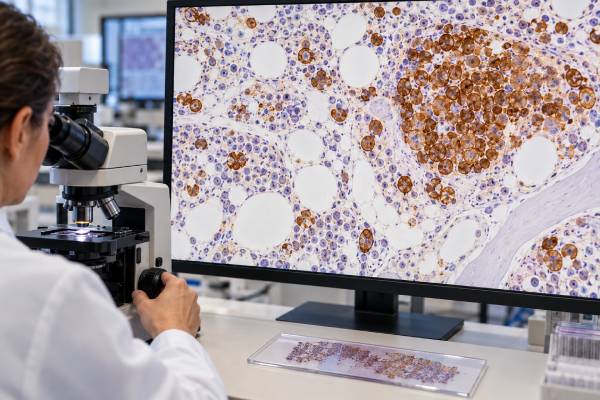
A CD34 test identifies cells that express CD34, a protein found on many early blood-forming stem and progenitor cells and on vascular endothelial cells. In hematology, CD34 is commonly used to help recognize immature cells, including blasts in acute leukemia, but a positive result does not mean cancer. Normal bone marrow contains a small CD34-positive progenitor population, and many leukemias are only partly CD34-positive or completely CD34-negative. CD34 also has a separate practical role in stem cell transplantation, where laboratories count CD34-positive cells to estimate the number of hematopoietic progenitors collected for a graft. In leukemia diagnosis, however, the result is interpreted as part of a larger immunophenotype rather than as a stand-alone number. Pathologists ask which cells are CD34-positive, what other markers they express, whether the cells look and behave like blasts, and what genetic abnormalities are present. Understanding that distinction prevents a common error: equating “CD34 positive” with “leukemia positive.”
- CD34 marks immaturity, not malignancy. Normal hematopoietic stem/progenitor cells can be CD34-positive, as can blasts in many leukemias.
- Acute leukemia can be CD34-positive or CD34-negative. Absence of CD34 does not exclude AML, B-ALL, or another acute leukemia.
- There is no universal leukemia cutoff for CD34 positivity. Flow reports describe percentage, intensity, and coexpression within the abnormal cell population.
- CD34 counts used for stem cell collection are a different test context. Graft doses are quantified as CD34-positive cells, often normalized to patient weight.
- The blast percentage and CD34-positive percentage are not the same thing. Some blasts lack CD34, and some normal progenitors are CD34-positive.
Table of Contents
- What CD34 Is and Why It Is Tested
- How CD34 Testing Is Performed
- What CD34-Positive Staining Means
- CD34 in Acute Leukemia
- CD34 and Stem Cell Collection
- CD34 in MRD and Marrow Recovery
- Limitations and Next Steps
What CD34 Is and Why It Is Tested
CD34 is a cell-surface sialomucin expressed on many early hematopoietic stem and progenitor cells. As blood cells mature, most lose CD34. This makes the marker useful for identifying immature populations in bone marrow and blood.
CD34 is not exclusive to blood-forming cells. Endothelial cells lining blood vessels also express it, and CD34 can be found in several nonhematopoietic stromal or progenitor cell populations. In a bone marrow or tumor biopsy, the pathologist therefore interprets CD34 based on cell shape and location, not color alone.
In hematology, CD34 testing serves several distinct purposes:
- identifying an immature blast population in suspected acute leukemia;
- helping distinguish blasts from more mature myeloid or lymphoid cells;
- describing the leukemia-associated immunophenotype at diagnosis;
- supporting measurable residual disease analysis with other markers;
- quantifying hematopoietic progenitor cells for stem cell collection and transplantation; and
- highlighting vascular structures in some tissue pathology settings.
Because these purposes are different, a CD34 result must be read in context. A “2% CD34-positive cells” marrow result in a person recovering after chemotherapy is not interpreted the same way as a “90% of abnormal blasts are CD34-positive” leukemia flow result. Likewise, a stem cell collection report giving a dose in millions of CD34-positive cells per kilogram is not a leukemia marker result.
The related CD33 test can help characterize myeloid differentiation, while CD34 mainly marks cellular immaturity. Using them together is more informative than either marker alone.
How CD34 Testing Is Performed
CD34 can be measured by flow cytometry or immunohistochemistry, with flow cytometry being especially important in leukemia and stem cell enumeration. The method chosen depends on whether the question is diagnostic classification, tissue localization, MRD assessment, or graft collection.
In leukemia flow cytometry, fluorescent antibodies bind CD34 and many other antigens. The instrument measures expression on individual cells and allows the laboratory to isolate a population based on size, complexity, CD45 intensity, and marker combinations. The report may describe CD34 as bright, dim, partial, or negative and may provide the percentage of abnormal cells that express it.
In bone marrow core biopsies, CD34 immunohistochemistry can highlight immature cells in their tissue setting. It may help estimate blast distribution, but pathologists do not count every CD34-positive cell as a leukemic blast. Normal precursors and endothelial cells can stain, and some leukemic blasts do not.
For stem cell collection, flow cytometry uses standardized counting methods to determine the absolute number of viable CD34-positive cells in peripheral blood or an apheresis product. Clinicians can use a pre-collection peripheral blood CD34 count to judge whether mobilization has produced enough circulating progenitors and can use the product count to determine whether the collection goal has been reached.
A leukemia flow cytometry panel typically includes CD34 with markers that establish lineage and maturation. In suspected AML, these may include CD117, CD13, CD33, myeloperoxidase, HLA-DR, monocytic markers, and selected lymphoid antigens. In B-ALL or T-ALL, the panel shifts toward lineage-specific markers and TdT.
No fasting is required for CD34 itself. Preparation depends on whether the sample is blood, bone marrow, tissue, or an apheresis product.
What CD34-Positive Staining Means
CD34 positivity means a cell expresses an immaturity-associated marker; it does not tell whether that cell is normal or malignant without additional evidence. This is the most important principle for interpreting the test.
| Finding | Possible meaning | What must be checked |
|---|---|---|
| Small CD34-positive progenitor population in marrow | Can represent normal hematopoietic precursors | Maturation pattern and overall marrow findings |
| Large abnormal CD34-positive blast population | Supports an immature leukemia phenotype | Lineage markers, morphology, genetics, blast percentage |
| CD34-negative abnormal blasts | Still compatible with acute leukemia | Other blast markers and defining genetic findings |
| CD34-positive vascular structures in tissue | Normal endothelial staining | Cell location and morphology |
| High CD34 count in an apheresis product | Usually indicates a progenitor-rich stem cell collection | Collection target, viability, and dose per kilogram |
The denominator matters. If a report says “CD34 is expressed in 70%,” determine whether that means 70% of blasts, 70% of a gated abnormal population, or 70% of all nucleated cells. These statements are not equivalent.
CD34 intensity also has context. A leukemic population may show abnormally uniform or altered CD34 compared with normal progenitors, but there is no single intensity value that proves malignancy across all laboratories. Instruments, antibody clones, fluorochromes, calibration, and gating strategies differ.
Another common mistake is assuming CD34 percentage equals blast percentage. Morphologic blast counts include blasts whether they express CD34 or not. Conversely, a small normal CD34-positive progenitor population should not be added automatically to a leukemic blast count. The final interpretation integrates morphology and immunophenotyping.
CD34 and the blast count are separate measurements
This distinction is especially important because acute leukemia classification may use a blast-percentage threshold in some settings, while genetically defined leukemias can have disease-specific rules. The blast count is based on identifying immature malignant cells by morphology and integrated laboratory evidence. CD34 is only one possible feature of those cells.
For example, a marrow can contain 25% blasts even if only half of those blasts express CD34. The CD34-positive fraction would then be smaller than the total blast burden. The reverse misunderstanding can occur in regenerating marrow: a small wave of normal CD34-positive precursors may be visible after therapy even when there is no abnormal blast population.
Pathologists resolve this by combining the aspirate differential, core biopsy, flow cytometry, and genetic results. If the numbers do not agree, sample quality and patchy disease are considered. A hemodiluted aspirate can underestimate blasts, while a core biopsy may better show a focal infiltrate. The final report should explain which estimate is being used for diagnosis and why.
CD34 in Acute Leukemia
CD34 is frequently expressed in acute leukemia, but its presence is variable and depends on the biological subtype. It can appear on blasts in AML, B-lymphoblastic leukemia/lymphoma, and some T-lymphoblastic leukemias.
In AML, CD34 often accompanies CD117 and other immaturity-associated features, but many AML cases lack it. Some genetically defined AML subtypes are commonly CD34-negative, although phenotype alone cannot establish the genotype. Acute promyelocytic leukemia, for example, often lacks CD34 and HLA-DR, while showing strong myeloid antigen expression. NPM1-mutated AML is also frequently CD34-negative. These are patterns that can raise suspicion, not diagnostic rules.
In B-ALL, CD34 is common but not universal. Blasts are characterized with B-lineage markers such as CD19, CD22, cytoplasmic CD79a, and often CD10 and TdT. In T-ALL, immaturity is shown with markers such as cytoplasmic CD3 and TdT, with variable CD34.
CD34 can also help identify leukemia stem/progenitor-like compartments in research and specialized flow approaches. A CD34-positive/CD38-low or negative phenotype resembles primitive hematopoietic stem cells, but leukemia stem cells are biologically heterogeneous and cannot be defined in every patient by CD34/CD38 alone. Some AML is entirely CD34-negative yet still contains leukemia-propagating cells.
Modern AML diagnosis increasingly depends on genetic classification. Tests for NPM1, FLT3, IDH1/2, TP53, PML::RARA, core-binding factor fusions, and other abnormalities can define disease entities or influence risk and therapy. CD34 provides useful phenotypic context but does not replace those tests.
CD34 has also been studied as a prognostic marker, particularly in AML, but it should not be used as an independent modern risk score. Older studies sometimes associated CD34 expression with lower remission rates or adverse outcomes, yet today prognosis is shaped by a much richer set of variables. Cytogenetic and molecular risk, age, treatment intensity, transplant strategy, measurable residual disease, and response kinetics are generally more actionable than a simple CD34-positive/negative label. A clinician may still note CD34 as part of the leukemia phenotype, but treatment should not be inferred from that marker alone.
CD34 and Stem Cell Collection
In stem cell transplantation, CD34 is used as a practical counting marker for hematopoietic progenitor cells rather than as a leukemia marker. This is one of the most important alternative meanings of a “CD34 test.”
Before autologous or allogeneic transplantation, stem cells can be collected from peripheral blood after mobilization, from bone marrow, or in some settings from cord blood. Peripheral blood collection is performed by apheresis. The laboratory measures CD34-positive cells to estimate the progenitor content of the product.
Results may be reported as an absolute CD34-positive cell concentration and as a total dose normalized to recipient body weight, commonly in units of ×10^6 CD34-positive cells/kg. Collection targets depend on transplant type, disease, institutional practice, whether one or multiple transplants are planned, and product quality. A clinician may therefore say a collection “reached the CD34 target,” which has nothing to do with whether leukemia blasts are CD34-positive.
Flow cytometric stem cell enumeration uses dedicated standardized gating and viability assessment. It is not simply the same panel used to diagnose acute leukemia. Mobilized blood contains normal progenitor cells whose CD34 positivity is expected and desired.
This distinction also matters after transplantation. The presence of normal CD34-positive precursors as marrow regenerates can be a sign of hematopoietic recovery. It should not be called relapse unless the cells show the patient’s abnormal leukemia phenotype or molecular disease evidence.
CD34 in MRD and Marrow Recovery
CD34 can help find residual leukemic blasts only when it is combined with other abnormal markers and a different-from-normal analysis. After chemotherapy, the marrow can contain many regenerating precursors. These cells may be CD34-positive and can mimic immature leukemia if the panel is interpreted too narrowly.
At diagnosis, the laboratory documents the leukemia-associated phenotype. A CD34-positive AML clone might also show abnormal CD13/CD33 intensity, asynchronous maturation, CD117, or aberrant lymphoid markers. At follow-up, the laboratory looks for rare cells that reproduce that multidimensional pattern or occupy an abnormal maturation space.
The challenge is that leukemia changes. Treatment can select subclones, and relapse may show different CD34 expression from the original sample. A CD34-positive leukemia can become less positive, and a CD34-negative leukemia can relapse without ever gaining the marker. This is why modern leukemia MRD testing uses multiple antigens and, when possible, molecular targets.
Regenerating marrow has its own recognizable patterns. Normal myeloid precursors mature through coordinated changes in CD34, CD117, HLA-DR, CD13, CD33, CD15, CD11b, and other markers. Experienced flow laboratories use these maturation trajectories to distinguish normal recovery from aberrant blasts.
A result of “no abnormal CD34-positive blast population identified” is not identical to “no leukemia detected” unless the assay is specifically validated for MRD and the patient’s leukemia is known to be trackable by that approach. Molecular MRD may detect disease that flow does not, and the reverse can occur depending on the target and specimen.
Limitations and Next Steps
CD34 has high practical value but low stand-alone specificity for leukemia. Its interpretation is especially vulnerable to errors when readers confuse immaturity with malignancy or compare results from different clinical contexts.
Important limitations include:
- normal stem and progenitor cells are CD34-positive;
- some leukemias are CD34-negative;
- endothelial cells stain with CD34 in tissue;
- marrow recovery can temporarily increase normal immature cells;
- sample hemodilution can alter apparent blast proportions;
- treatment can change antigen expression; and
- laboratory methods and reporting thresholds differ.
If CD34-positive blasts are found in a new acute leukemia workup, the next steps are usually lineage assignment, genetic classification, cytogenetic risk assessment, and baseline MRD planning. If a small CD34-positive population appears after therapy, the laboratory compares its full phenotype with normal precursors and the diagnostic leukemia clone rather than labeling it relapse from CD34 alone.
In a tissue report, it is also worth checking whether the pathologist is using CD34 to highlight blood vessels rather than hematopoietic blasts. CD34 is a common vascular marker, so endothelial staining can act as an internal positive control. In some soft-tissue tumors, CD34 may be part of a completely different diagnostic panel. The same marker name can therefore have very different meanings depending on whether the specimen is marrow, blood, lymph node, skin, or a soft-tissue mass. The report’s microscopic description and final diagnosis determine which interpretation applies. For that reason, a CD34 result copied out of context from a pathology report can be misleading. The specimen type, gated population, and purpose of the test should always travel with the number or positive/negative label when results are discussed carefully with patients, clinicians, or another laboratory team.
For patients reviewing a report, useful questions include: Are the CD34-positive cells normal progenitors or abnormal blasts? What percentage of total marrow cells are blasts? Which lineage markers are present? Does the phenotype suggest a specific genetic subtype that was confirmed molecularly? Is the result from a diagnostic panel, an MRD assay, or a stem cell collection count?
Urgency comes from the underlying condition, not CD34 positivity itself. Suspected acute leukemia with severe bleeding, infection, very high white-cell counts, respiratory symptoms, or neurologic changes requires rapid medical assessment. In transplantation, collection adequacy and timing are managed by the transplant team according to the treatment plan.
References
- Human CD34 as a biomarker and functional tool in progenitor cell isolation: A comprehensive review 2026 (Review)
- Immunophenotypic features of early haematopoietic and leukaemia stem cells. 2024 (Review)
- CD34-Structure, Functions and Relationship with Cancer Stem Cells 2023 (Review)
- Advances in Acute Myeloid Leukemia Classification, Prognostication and Monitoring by Flow Cytometry 2023 (Review)
- Flow Cytometric Identification of Hematopoietic and Leukemic Blast Cells for Tailored Clinical Follow-Up of Acute Myeloid Leukemia. 2022 (Review)
Disclaimer
This article is for general education and cannot diagnose leukemia or determine stem cell transplant decisions. CD34 results must be interpreted according to the specimen, testing method, full immunophenotype, morphology, genetics, treatment history, and clinical setting by qualified professionals. Seek prompt medical attention for severe bleeding, fever with immune suppression, breathing difficulty, confusion, or other rapidly worsening symptoms.





