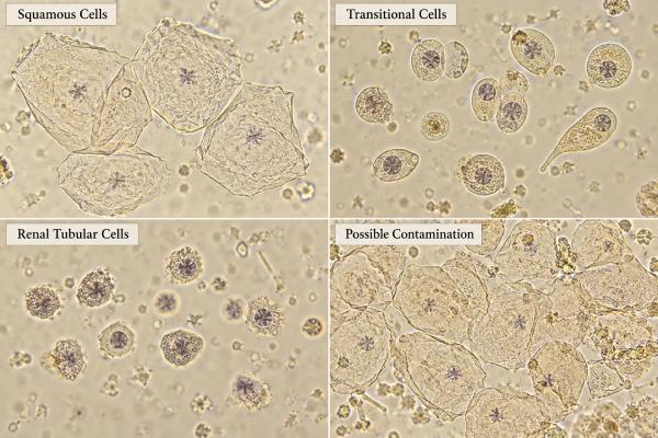
Epithelial cells in urine are cells shed from surfaces that line the urinary and genital tracts. A small number is common, but the type of epithelial cell matters more than the word “epithelial” alone. Squamous epithelial cells usually come from the skin, vulva, or distal urethra and can suggest sample contamination when numerous. Transitional, or urothelial, cells come from the bladder, ureters, and renal pelvis and may increase with irritation, instrumentation, stones, or infection. Renal tubular epithelial cells come from the kidney tubules and are more clinically important because increased numbers can signal tubular injury. Automated urinalysis systems may report epithelial cells as a total count, while manual microscopy can better distinguish subtypes. Interpretation should include symptoms, collection method, bacteria, white and red blood cells, casts, protein, and kidney function. A “high epithelial cell” result does not automatically mean infection, cancer, or kidney damage.
- Squamous epithelial cells most often reflect collection quality: many squamous cells can mean skin or genital contamination, especially in a voided sample.
- Transitional cells come from the urinary lining: small numbers can be normal, while increases may occur after catheterization, irritation, stones, or inflammation.
- Renal tubular epithelial cells are different: increased RTECs can indicate kidney-tubule injury and deserve correlation with creatinine and urine output.
- There is no single universal normal cutoff: laboratories report cells per high-power field or per microliter, with method-specific reference ranges.
- Repeat collection can be useful: when squamous cells are abundant and the urine culture or urinalysis is hard to interpret, a properly collected repeat sample may clarify the result.
Table of Contents
- What Epithelial Cells in Urine Are
- Squamous Epithelial Cells and Contamination
- Transitional or Urothelial Cells
- Renal Tubular Epithelial Cells
- Normal, High, and Abnormal Results
- Collection, Microscopy, and Automated Testing
- How Results Are Interpreted With Other Urine Findings
- Common Scenarios and Practical Next Steps
- When Repeat Testing or Medical Follow-Up Is Needed
What Epithelial Cells in Urine Are
Epithelial cells form protective linings throughout the urinary system and nearby genital surfaces. They naturally shed as cells age and are replaced. That means an epithelial-cell result is not automatically abnormal.
The most useful distinction is where the cells came from. Urine microscopy can show three broad groups:
- squamous epithelial cells from external genital or distal urethral surfaces;
- transitional, or urothelial, cells from the bladder, ureters, and renal pelvis;
- renal tubular epithelial cells from the nephron itself.
These categories have very different implications. A urine sample with many squamous cells may mainly tell you that the specimen was contaminated during collection. In contrast, increased renal tubular cells can be a clue to intrinsic kidney injury.
A routine urine microscopy test evaluates epithelial cells together with RBCs, WBCs, casts, crystals, bacteria, yeast, and other particles. The entire sediment pattern is usually more informative than any one cell count.
Some laboratories report only “epithelial cells” or “epithelial cells, non-renal.” Others separate squamous, transitional, and renal tubular cells. Automated flow cytometry may provide counts per microliter, while manual microscopy often uses cells per high-power field. Because methods differ, the reference range printed by the performing laboratory should be used rather than a universal internet cutoff.
Squamous Epithelial Cells and Contamination
Squamous epithelial cells are large, flat cells with abundant cytoplasm and a relatively small nucleus. They commonly enter a urine cup from skin, the vulva, vagina, or distal urethra during voiding. Finding a few is common even when collection is careful.
When numerous, squamous cells can raise suspicion that the specimen contains material from outside the urinary tract. This matters most when clinicians are trying to decide whether bacteria, white cells, or a urine culture represent a true urinary infection.
However, squamous cells are not a perfect contamination test. Older studies showed poor predictive value when squamous cells were used alone. More recent studies using automated particle counts have found that higher squamous-cell numbers are associated with mixed or contaminated cultures, but the relationship still depends on sex, collection method, bacterial count, and laboratory threshold.
So a report of “many squamous epithelial cells” should not be read as “this urine is definitely contaminated.” A better interpretation is: collection quality may be reduced, and infection markers may be less reliable.
If the patient has urinary symptoms and the sample also contains pyuria, nitrites, or substantial bacteriuria, clinicians may still have useful evidence for infection. If symptoms are absent and the culture grows mixed organisms in a sample full of squamous cells, repeating a clean-catch specimen may be more informative than treating the first result.
A related article on urine culture contamination and mixed flora helps explain why sample quality can change the meaning of a culture report.
Transitional or Urothelial Cells
Transitional epithelial cells, also called urothelial cells, line the renal pelvis, ureters, bladder, and much of the urethra. They can change shape as the urinary tract stretches, which is why they are called “transitional.”
Small numbers may be present in otherwise normal urine. Increased shedding can occur when the urothelium is irritated or mechanically disturbed. Common situations include:
- recent urinary catheter placement or removal;
- cystoscopy or other urinary instrumentation;
- urinary tract infection or inflammation;
- kidney or bladder stones;
- urinary obstruction or trauma.
A cluster of transitional cells after catheterization is very different from persistent atypical cells in a person with unexplained hematuria. Routine urinalysis microscopy is not the same as urine cytology, which is designed to examine cellular atypia for possible urothelial malignancy.
If a laboratory flags “atypical urothelial cells,” the next step depends on the exact report, symptoms, age, hematuria pattern, and risk factors. That finding should not be equated with cancer, but it may prompt cytology, imaging, or urologic evaluation when clinically appropriate.
The distinction between transitional cells and renal tubular cells can be difficult on automated systems or in degenerated specimens. Size, nuclear features, cell clustering, and clinical context help a trained observer classify them.
Renal Tubular Epithelial Cells
Renal tubular epithelial cells, or RTECs, line the nephron and are the epithelial subtype most directly linked to kidney parenchymal injury. Healthy tubules continually renew cells, so an occasional RTEC may be seen. Increased numbers, especially in the right clinical setting, suggest tubular stress, injury, or repair.
RTECs can appear in acute tubular injury caused by ischemia, sepsis, nephrotoxins, severe systemic illness, pigment injury, or other insults. They may also be present in tubulointerstitial inflammation and in glomerular disorders with secondary tubular damage.
The finding becomes more convincing when RTECs occur with epithelial cell casts or granular casts. Casts form within the kidney tubules, so they provide stronger localization than free cells alone.
RTECs are not simply a “high epithelial cell count.” If a report does not specify the subtype, ask whether the laboratory distinguishes squamous cells from renal tubular epithelial cells. The difference can completely change the clinical meaning.
Research also shows that urinary tubular cells can appear during recovery from severe AKI, including viable epithelial clusters with regenerative features. That reinforces an important point: tubular-cell shedding is a marker of kidney epithelial activity and injury, but the count does not map neatly onto one stage or one prognosis without the creatinine trend and clinical course.
Normal, High, and Abnormal Results
There is no single normal epithelial-cell range that applies to every laboratory. Reports may use “none,” “rare,” “few,” “moderate,” or “many,” a number per high-power field, or an automated count per microliter.
| Cell type | Small amount | Higher amount may suggest |
|---|---|---|
| Squamous | Common in voided urine | Reduced collection quality or external contamination |
| Transitional/urothelial | Can be normal | Irritation, infection, stones, catheterization, instrumentation |
| Renal tubular | Occasional cells may occur | Tubular injury, inflammation, severe illness, or recovery from AKI |
A “high epithelial cells” flag is therefore incomplete without the subtype. Ten squamous cells and ten renal tubular cells do not mean the same thing.
Sex and anatomy also influence expected contamination. Voided samples from women often contain more squamous cells because of contact with vulvovaginal surfaces. Catheterized samples generally reduce external contamination but can increase urothelial shedding because the catheter itself contacts the urinary lining.
The number of epithelial cells should also be interpreted relative to other sediment findings. A sample with many squamous cells but no pyuria, no bacteria, and no protein may simply be a poor collection. A sample with renal tubular cells, granular casts, rising creatinine, and reduced urine output is a much more kidney-specific abnormal pattern.
Collection, Microscopy, and Automated Testing
Proper collection improves the usefulness of every urine result. For a routine clean-catch sample, the goal is to collect midstream urine while minimizing contact with external skin and genital secretions. Instructions vary by laboratory, so follow the collection kit rather than improvising aggressive cleansing that may irritate tissue.
A contaminated specimen is not dangerous by itself; it is a data-quality problem. It can lead to unnecessary cultures, false concern for infection, or repeated testing. When accurate diagnosis matters, a repeat voided sample or, in selected situations, a catheterized specimen may be used.
Sample handling after collection is equally important. Cells change with time. Dilute or alkaline urine can cause cell lysis, while delays can alter morphology. Modern urinalysis guidance recommends prompt particle examination or appropriate refrigeration when delays are unavoidable.
Automated analyzers are fast and consistent for high-volume testing. They are especially good at counting RBCs, WBCs, and bacteria. Their limitation is classification of less common particles such as renal tubular cells, pathological casts, lipids, and some crystals. When the analyzer flags abnormal cells or the result conflicts with the clinical picture, manual microscopy can add important detail.
A 2023 biopsy-correlation study emphasized that manual sediment examination can identify dysmorphic RBCs, renal tubular epithelial cells, lipids, casts, and other particles that automated systems may miss or misclassify. This is particularly relevant in suspected intrinsic kidney disease.
How Results Are Interpreted With Other Urine Findings
Epithelial cells rarely answer a clinical question by themselves. Their meaning comes from combinations.
Squamous cells + mixed bacterial growth: contamination becomes more likely, especially when symptoms are absent. A repeat specimen may be preferable before labeling someone with a UTI.
Squamous cells + strong UTI pattern: many squamous cells reduce confidence, but leukocyte esterase, nitrites, pyuria, symptoms, and a dominant uropathogen can still support infection. The clinician decides whether the evidence is strong enough to treat or whether a repeat culture is needed.
Transitional cells + recent catheterization: increased shedding may be expected from instrumentation and is usually interpreted cautiously if there are no other concerning findings.
Renal tubular cells + rising creatinine: this pattern raises concern for tubular injury. Urine output, granular casts, medication exposure, blood pressure, and systemic illness help determine the cause.
Renal tubular cells + RBC casts or dysmorphic RBCs: a glomerular process may be causing secondary tubular injury. The overall sediment should not be forced into a single “tubular” category.
Renal tubular cells + crystals: crystal-related nephropathy, medication precipitation, uric acid injury, oxalate injury, or another crystalline process may be relevant depending on crystal type and clinical history.
This pattern-based approach is more reliable than trying to interpret every line of a urinalysis separately.
Common Scenarios and Practical Next Steps
The wording on an epithelial-cell report can sound alarming, but common scenarios are easier to interpret when the cell type and the clinical question are clear.
“Many squamous epithelial cells” on a routine check with no symptoms: this most often raises a specimen-quality issue. If the rest of the urinalysis is unremarkable, no treatment is usually directed at the squamous cells themselves. A repeat sample may be requested only if the clinician still needs an accurate answer about blood, infection, or protein.
“Many squamous cells, bacteria, and leukocytes” in a person with burning and frequency: contamination is possible, but so is a real UTI. Symptoms, nitrites, pyuria, culture growth pattern, and bacterial quantity determine whether the sample is interpretable enough to act on. A culture dominated by one recognized uropathogen is more convincing than mixed flora from several organisms.
“Transitional epithelial cells” after a catheter or cystoscopy: recent instrumentation is a common explanation. The urothelial lining can shed cells after mechanical contact. Persistent hematuria or atypical cellular features may still need follow-up, but the procedure changes the baseline expectation.
“Renal tubular epithelial cells present” after a severe illness: this deserves more attention. Clinicians often review creatinine, urine output, blood pressure, fluid status, medications, and other casts. If tubular cells are numerous or accompanied by granular or epithelial casts, intrinsic kidney injury becomes more likely.
“Epithelial cells” without a subtype: the report may not be specific enough for clinical conclusions. If kidney injury is suspected, a manual sediment review can be more useful than assuming a total epithelial count represents contamination.
The practical rule is to match the finding to the question being asked. If the question is “Is this urine culture reliable?”, squamous cells matter most. If the question is “Is there tubular kidney injury?”, renal tubular cells and casts matter most. If the question is “Could there be a urothelial lesion?”, persistent atypical urothelial cells and hematuria need a different pathway.
When Repeat Testing or Medical Follow-Up Is Needed
A repeat urine sample is commonly useful when abundant squamous epithelial cells make the original test difficult to interpret, especially if a culture shows mixed flora or if the result does not fit the symptoms. Collecting a new midstream specimen with careful technique often resolves the uncertainty.
Medical follow-up is more important when the report specifically identifies renal tubular epithelial cells, epithelial casts, or other pathologic casts. Serum creatinine, estimated GFR, electrolytes, and urine output may need review, particularly after severe illness or a new medication exposure.
Prompt assessment is appropriate for markedly reduced urine, swelling, shortness of breath, persistent vomiting or dehydration, new gross hematuria, severe flank pain, high fever with urinary symptoms, or a rapidly rising creatinine. These symptoms are not caused by “epithelial cells” themselves; they indicate a potentially important underlying urinary or kidney problem.
The practical takeaway is simple: epithelial-cell subtype determines meaning. Squamous cells mostly speak to sample quality, transitional cells reflect the urothelial lining, and renal tubular cells can signal kidney-tubule injury. Read the result with the rest of the urinalysis rather than treating one flagged number as a diagnosis.
If a repeat sample is requested, try to use the collection method specified by the laboratory and deliver the specimen promptly. Changing collection quality can be more useful than repeatedly interpreting the same contaminated pattern. For renal tubular cells, however, follow-up should focus on kidney-function trends and the underlying illness rather than on obtaining a “cleaner” specimen alone.
References
- The EFLM European Urinalysis Guideline 2023 2024 (Guideline)
- Quality indicators for urine sample contamination: can squamous epithelial cells and bacteria count be used to identify properly collected samples? 2025
- Urinary Sediment Microscopy and Correlations with Kidney Biopsy: Red Flags Not To Be Missed 2023
- Diagnostic Utility of Urine Microscopy in Kidney Diseases 2024 (Review)
- Urinary renal tubular epithelial cells and casts as predictors of renal outcomes in patients with biopsy-proven diabetic nephropathy 2024
- Tubular epithelial progenitors are excreted in urine during recovery from severe acute kidney injury and are able to expand and differentiate in vitro 2022
Disclaimer
Epithelial cells in urine are interpreted differently depending on whether they are squamous, transitional, or renal tubular cells. A flagged count does not by itself diagnose contamination, infection, cancer, or kidney injury. Discuss persistent abnormalities, renal tubular cells, pathologic casts, or kidney-function changes with a clinician, and seek prompt care for severe urinary symptoms, markedly reduced urine, or signs of acute illness.