
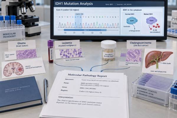
An IDH1 mutation test looks for abnormal changes in the IDH1 gene, most commonly at amino acid R132. These mutations alter cell metabolism and cause production of the oncometabolite D-2-hydroxyglutarate, or 2-HG, which can disrupt normal cell differentiation and gene regulation. IDH1 status has important but different roles in glioma, acute myeloid leukemia (AML), and cholangiocarcinoma.
In adult diffuse glioma, IDH mutation status is central to modern tumor classification and can now also identify patients eligible for mutant-IDH targeted therapy in defined grade 2 disease. In AML, a susceptible IDH1 mutation can guide use of IDH1 inhibitors in specific treatment settings. In advanced cholangiocarcinoma, especially intrahepatic disease, IDH1 testing is part of molecular profiling because a qualifying mutation can lead to targeted treatment after prior therapy. A result therefore should never be interpreted without the cancer type, exact variant, specimen, and testing method.
- A positive IDH1 result usually identifies an activating mutation at R132 that produces the abnormal metabolite 2-HG.
- In adult diffuse glioma, IDH status helps define the tumor type; oligodendroglioma also requires 1p/19q codeletion.
- In AML, susceptible IDH1 mutations can qualify patients for approved IDH1-targeted therapies in defined newly diagnosed or relapsed/refractory settings.
- In advanced cholangiocarcinoma, an IDH1 mutation can be an actionable biomarker, particularly in intrahepatic tumors.
- There is no normal numeric IDH1 range; results are interpreted by the exact mutation, classification, tumor type, and assay quality.
Table of Contents
- What an IDH1 mutation test detects
- IDH1 in glioma
- IDH1 in acute myeloid leukemia
- IDH1 in cholangiocarcinoma
- How IDH1 testing is performed
- How to interpret positive, negative, and VUS results
- Treatment, follow-up, and questions to ask
What an IDH1 mutation test detects
IDH1 encodes isocitrate dehydrogenase 1, an enzyme involved in normal cellular metabolism. Cancer-associated IDH1 mutations give the enzyme a new activity: instead of performing its usual reaction, mutant IDH1 converts alpha-ketoglutarate into D-2-hydroxyglutarate (2-HG).
High 2-HG levels interfere with enzymes that regulate DNA and histone methylation. The result is a broad epigenetic change that can block normal cell maturation and contribute to tumor development.
Most clinically important IDH1 mutations affect codon R132. The most common glioma variant is IDH1 R132H. AML and cholangiocarcinoma can contain R132C, R132G, R132S, R132L, R132H, and other susceptible variants depending on the disease and assay.
A report may classify the finding as:
- Pathogenic or oncogenic.
- Likely pathogenic.
- Variant of uncertain significance, or VUS.
- Not detected.
- Indeterminate or failed because of specimen limitations.
IDH1 is often tested on a multigene cancer mutation panel. This is useful because the interpretation commonly depends on other molecular findings. In glioma, 1p/19q status, ATRX, TP53, CDKN2A/B, and other markers may affect classification or grade. In AML, co-mutations influence risk and treatment planning. In cholangiocarcinoma, other actionable targets such as FGFR2 fusions, BRAF, HER2, NTRK, and RET may be assessed at the same time.
An IDH1 mutation is generally a somatic tumor alteration. Its presence in a tumor is not usually interpreted as an inherited cancer-risk finding.
IDH1 in glioma
IDH status is one of the defining molecular features of adult-type diffuse gliomas. Modern classification does not rely on microscopic appearance alone.
Two major IDH-mutant adult diffuse glioma categories are:
- Astrocytoma, IDH-mutant, graded according to additional histologic and molecular features.
- Oligodendroglioma, IDH-mutant and 1p/19q-codeleted, which requires both an IDH mutation and whole-arm codeletion of chromosomes 1p and 19q.
This means an IDH1-positive result can be essential to the final integrated diagnosis, but it does not by itself distinguish astrocytoma from oligodendroglioma.
IDH1 R132H immunohistochemistry
Because IDH1 R132H is so common in diffuse glioma, pathology laboratories often begin with an immunohistochemistry antibody specific for the R132H mutant protein. Strong tumor-cell staining supports that mutation and can provide a rapid, tissue-efficient answer.
A negative R132H stain does not exclude every IDH mutation. Noncanonical IDH1 mutations and IDH2 mutations require sequencing or another molecular method when the patient’s age, tumor appearance, or diagnostic setting makes an IDH-mutant glioma plausible.
An IDH2 mutation test can therefore be relevant when sequencing is used to evaluate the broader IDH1/2 status.
Targeted treatment in grade 2 IDH-mutant glioma
The treatment meaning of IDH testing changed substantially after the phase III INDIGO trial. Vorasidenib, a brain-penetrant inhibitor of mutant IDH1 and IDH2, prolonged progression-free survival and delayed the need for the next anticancer intervention in selected patients with grade 2 IDH-mutant glioma after surgery.
In August 2024, the FDA approved vorasidenib for adults and children age 12 years and older with grade 2 astrocytoma or oligodendroglioma harboring a susceptible IDH1 or IDH2 mutation after surgery, including biopsy, subtotal resection, or gross total resection.
The mutation result therefore can now have both classification and predictive treatment value in glioma. Whether vorasidenib is appropriate depends on grade, prior therapy, timing, tumor characteristics, and the current treatment plan.
IDH1 in acute myeloid leukemia
IDH1 mutations occur in a subset of AML and create the same 2-HG-producing abnormal enzyme. In leukemia, however, the test is not primarily used to name the disease. It is a molecular biomarker that can affect treatment selection and should be interpreted alongside the broader AML genomic profile.
Testing is commonly performed at diagnosis because treatment decisions can depend on actionable mutations. It may also be repeated at relapse if the molecular profile needs to be reassessed.
IDH1-targeted therapies in AML
Several IDH1-directed options are established for susceptible mutations in defined settings. Ivosidenib has approvals that include newly diagnosed IDH1-mutated AML in older adults or people with comorbidities that preclude intensive induction, including use with azacitidine. The randomized trial of ivosidenib plus azacitidine showed improved clinical outcomes compared with azacitidine alone in this population.
Olutasidenib is approved for adults with relapsed or refractory AML with a susceptible IDH1 mutation detected by an FDA-approved test.
These treatments illustrate why the phrase “IDH1-positive” is not enough for prescribing. The report must show a susceptible pathogenic mutation, and the patient must fit the clinical indication for the particular drug.
IDH inhibitors can cause differentiation syndrome, a potentially serious treatment complication caused by rapid maturation of malignant cells. This is a drug-safety issue rather than a feature of the mutation test itself, but it underscores why targeted therapy requires specialist monitoring.
An IDH1 mutation also does not replace other AML risk markers. Mutations such as NPM1, FLT3, TP53, RUNX1, and others can change prognosis or treatment strategy. The complete molecular profile and cytogenetic findings remain important.
IDH1 in cholangiocarcinoma
IDH1 mutations are most strongly associated with intrahepatic cholangiocarcinoma, a bile duct cancer arising within the liver. They are less characteristic of extrahepatic cholangiocarcinoma.
For patients with advanced biliary tract cancer, current molecular guidance supports genomic profiling early enough that actionable results are available when systemic treatment choices are needed. IDH1 is one of the established genes included in this workup for intrahepatic cholangiocarcinoma.
Why the result can change treatment
Ivosidenib is approved for adults with previously treated, locally advanced or metastatic cholangiocarcinoma with an IDH1 mutation detected by an appropriate test. The ClarIDHy trial showed improved progression-free survival compared with placebo and established mutant IDH1 as a clinically actionable target in this disease.
This treatment setting is different from glioma or AML even though the same gene is involved. The relevant cancer type, prior treatment, stage, and exact mutation determine whether the biomarker is actionable.
Broad profiling can be efficient because intrahepatic cholangiocarcinoma may contain other targetable alterations. A gene fusion panel or combined DNA/RNA NGS approach can help identify FGFR2 and other rearrangements while DNA sequencing assesses IDH1 and additional mutations.
A negative IDH1 result does not mean there are no targeted options. It simply redirects attention to other molecular alterations and the standard treatment framework for the disease.
How IDH1 testing is performed
The specimen and method differ by disease.
| Disease | Common specimen | Common approach |
|---|---|---|
| Glioma | Brain tumor biopsy or resection | IDH1 R132H immunohistochemistry plus sequencing when needed |
| AML | Bone marrow or peripheral blood | PCR-based companion assay or NGS panel |
| Cholangiocarcinoma | Tumor biopsy/resection; sometimes plasma ctDNA | Targeted NGS or validated mutation assay |
Glioma testing workflow
A neuropathologist may use IDH1 R132H immunohistochemistry first because it is rapid and preserves tissue. If the stain is negative but an IDH mutation remains clinically plausible, sequencing can detect non-R132H IDH1 mutations and IDH2 mutations.
The result is then integrated with morphology and markers such as 1p/19q codeletion and ATRX to reach the final WHO-aligned diagnosis.
AML testing workflow
AML testing uses marrow or blood containing leukemic cells. Rapid targeted tests may be used when an IDH1-directed treatment decision is urgent, while larger NGS panels provide broader risk and target information.
A companion diagnostic may be required for a particular drug indication. The laboratory report should identify the exact R132 variant and whether it is considered susceptible to the therapy under consideration.
Cholangiocarcinoma testing workflow
Tumor NGS is commonly used because multiple actionable biomarkers need evaluation. Limited biopsy tissue can be a challenge, so laboratories may coordinate DNA and RNA testing to conserve material.
Plasma circulating tumor DNA can sometimes identify an IDH1 mutation when tissue is limited, but a negative liquid-biopsy result is less definitive when little tumor DNA is circulating. Tissue testing may still be needed if the result would affect treatment.
How to interpret positive, negative, and VUS results
Positive pathogenic IDH1 mutation
A pathogenic R132 mutation confirms mutant-IDH biology. Its clinical meaning then depends on the disease:
- In glioma, it contributes directly to integrated tumor classification and may determine eligibility for mutant-IDH therapy in a defined grade 2 setting.
- In AML, it identifies a treatment-relevant molecular subgroup and may qualify the patient for an IDH1 inhibitor.
- In cholangiocarcinoma, it can provide a targeted treatment option after prior therapy in advanced disease.
No IDH1 mutation detected
A negative result means the assay did not find a reportable IDH1 mutation within its validated scope. It does not mean the cancer is benign or low risk.
In glioma, an IDH1 R132H IHC-negative result may need sequencing before a tumor is considered IDH-wildtype, especially when noncanonical mutations remain possible. In AML or cholangiocarcinoma, a negative IDH1 result directs treatment planning toward other molecular and clinical features.
Variant of uncertain significance
A VUS is not the same as a susceptible activating R132 mutation. It should not automatically be used to classify a glioma as IDH-mutant or to qualify a patient for targeted treatment unless additional evidence supports pathogenicity.
The laboratory may update variant classifications over time. Keeping the complete report, including the exact DNA and protein change, allows later reinterpretation.
Variant allele frequency
NGS reports may list the percentage of sequencing reads carrying IDH1. This value can reflect tumor purity, copy number, and clonal burden, but it is not a universal “IDH1 level.” There is no normal, optimal, or toxic VAF range.
In AML, serial molecular measurements can sometimes be clinically informative in broader disease monitoring, but routine measurable residual disease strategies are disease- and assay-specific. A single VAF should not be interpreted without hematologic context.
Treatment, follow-up, and questions to ask
The strongest way to use an IDH1 result is to connect it to the exact clinical decision it can change.
For glioma, ask whether the mutation establishes an IDH-mutant diffuse glioma and what additional testing is needed to distinguish astrocytoma from oligodendroglioma. If the tumor is grade 2 after surgery, ask whether the current clinical situation matches the criteria in which vorasidenib is considered.
For AML, ask which IDH1 variant was detected, whether an FDA-approved companion test is required for the planned therapy, and how the IDH1 finding interacts with other AML risk markers. If an IDH inhibitor is prescribed, patients should understand symptoms that require urgent evaluation for differentiation syndrome and other treatment toxicities.
For cholangiocarcinoma, ask whether the tumor is intrahepatic, whether broad molecular profiling has been completed, and whether the IDH1 result qualifies for ivosidenib based on prior treatment and current disease status.
Other useful questions include:
- What exact IDH1 mutation was found? R132 variants have the strongest established cancer relevance.
- Was the result found by IHC, sequencing, PCR, or more than one method? The method determines what variants can be excluded.
- If glioma R132H IHC was negative, was sequencing performed? This helps avoid missing noncanonical IDH1 or IDH2 mutations.
- Is the finding pathogenic or a VUS? Targeted therapy should not be based on an uncertain variant without supporting evidence.
- Does the result change the formal tumor diagnosis? This is especially important in glioma.
- Does the result qualify for a current targeted therapy in this disease and treatment setting? The answer differs across glioma, AML, and cholangiocarcinoma.
- Were other actionable mutations or fusions tested? Broad profiling can reveal alternatives when IDH1 is negative.
- Would repeat molecular testing be useful at relapse or progression? Tumors can evolve, and the answer depends on disease type and prior therapy.
Does IDH1 mutation status predict prognosis?
There is no single prognosis attached to an IDH1-positive result. In adult diffuse glioma, IDH-mutant tumors generally belong to biologically different categories from IDH-wildtype glioblastoma, but outcome still depends on tumor type, grade, 1p/19q status, age, extent of resection, treatment, and other molecular features. In AML, IDH1 is interpreted within a larger genomic and cytogenetic risk framework rather than used alone as a favorable or adverse label. In cholangiocarcinoma, the mutation is most clinically useful as a treatment biomarker; stage, resectability, response to systemic therapy, and other disease features remain central to prognosis.
This is why a molecular report should not convert “IDH1 mutation detected” into a survival estimate without disease-specific evidence. The mutation can be highly important while still being only one part of the overall risk picture. When comparing reports over time, keep the tumor diagnosis, specimen date, and treatment status with the molecular result so later clinicians do not interpret IDH1 outside its original context.
IDH1 is a good example of precision medicine that cannot be reduced to one universal interpretation. The same molecular alteration can be a classification marker in brain cancer, a therapeutic biomarker in leukemia, and an actionable target in biliary tract cancer. The exact disease context determines what “IDH1 mutation positive” means.
The specimen source should stay attached to the IDH1 result as well. A mutation detected in a resected glioma, a bone-marrow AML sample, and a cholangiocarcinoma biopsy may share the same gene name but come from completely different disease processes. If a person has more than one malignancy, clinicians should not automatically carry an IDH1 result from one cancer into treatment decisions for another. The pathology diagnosis, collection date, and exact variant identify which disease the biomarker belongs to.
References
- IDH inhibition in gliomas: from preclinical models to clinical trials 2024 (Review)
- Vorasidenib in IDH1- or IDH2-Mutant Low-Grade Glioma 2023 (RCT)
- FDA approves vorasidenib for Grade 2 astrocytoma or oligodendroglioma with a susceptible IDH1 or IDH2 mutation 2024
- Ivosidenib and Azacitidine in IDH1-Mutated Acute Myeloid Leukemia 2022 (RCT)
- FDA Approval Summary: Ivosidenib for the Treatment of Patients with Advanced Unresectable or Metastatic, Chemotherapy Refractory Cholangiocarcinoma with an IDH1 Mutation 2022
- ESMO Clinical Practice Guideline interim update on the management of biliary tract cancer 2025 (Guideline)
Disclaimer
This article is for general educational purposes and does not replace interpretation by a neuropathologist, hematologist, oncologist, or molecular pathologist. IDH1 results have different diagnostic and treatment implications in glioma, AML, and cholangiocarcinoma, and eligibility for targeted therapy depends on the exact mutation, disease setting, testing method, and current drug label or guideline. Treatment decisions should be based on the complete clinical and molecular picture.





