
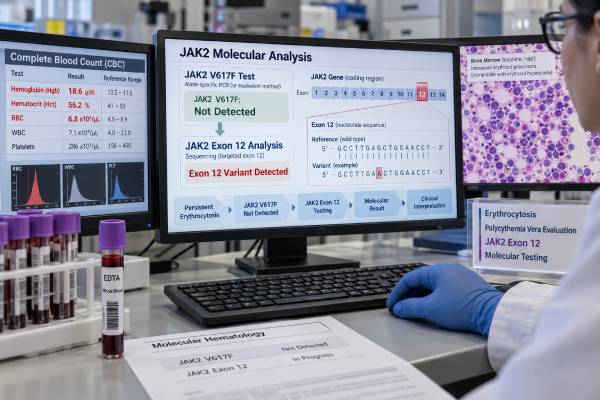
A JAK2 exon 12 mutation test is most often used when polycythemia vera (PV) is suspected but the common JAK2 V617F mutation is not detected. PV is a clonal myeloproliferative neoplasm that causes excessive red blood cell production. Almost all patients with PV have an activating JAK2 mutation: most have V617F in exon 14, while a smaller group has an exon 12 mutation. Exon 12 variants are diverse and include deletions, insertions, substitutions, and complex changes clustered around a short region of the gene. A positive result strongly supports PV when persistent erythrocytosis and the rest of the clinical picture fit. A negative result makes PV less likely but does not end the evaluation. Current erythrocytosis workups may examine JAK2 exons 12–15 and then investigate low erythropoietin, hypoxia, medications, kidney disease, inherited causes, and other sources of elevated hemoglobin or hematocrit. The mutation result should always be interpreted with blood counts, serum erythropoietin, and often bone marrow morphology.
- JAK2 exon 12 testing is a key follow-up when PV is suspected and JAK2 V617F is negative.
- JAK2 V617F accounts for about 95% of PV in many cohorts; exon 12 mutations explain much of the remaining JAK2-mutated PV.
- A positive exon 12 mutation is a major molecular clue for PV but should be integrated with hemoglobin/hematocrit, marrow findings, and clinical context.
- Exon 12-mutated PV can present with marked erythrocytosis while white blood cell and platelet counts are less elevated than in typical V617F-positive PV.
- If JAK2 testing is negative, the next step is a structured erythrocytosis workup rather than assuming the high hemoglobin is benign.
Table of Contents
- What a JAK2 Exon 12 Mutation Is
- When JAK2 Exon 12 Testing Is Ordered
- How the Test Is Performed and Reported
- What a Positive Exon 12 Result Means
- How Exon 12 Fits Polycythemia Vera Diagnosis
- What a Negative Result Means
- Allele Frequency, Limitations, and Next Steps
What a JAK2 Exon 12 Mutation Is
JAK2 exon 12 mutations are acquired activating changes in the JAK2 gene that drive abnormal blood-cell signaling. JAK2 encodes a tyrosine kinase that transmits growth signals from blood-cell receptors. When JAK2 is abnormally activated, hematopoietic stem and progenitor cells can grow without normal regulatory control.
The best-known JAK2 mutation is V617F, located in exon 14. Exon 12 mutations affect a different part of the gene and are more genetically diverse. They include small deletions, insertions, substitutions, and combinations of these changes. Because there is no single exon 12 variant comparable to V617F, the laboratory method must be able to recognize multiple mutation patterns.
These mutations are closely associated with PV and erythrocytosis. In a 2025 study of 532 patients with PV, JAK2 V617F was found in 94.9% and exon 12 mutations in 5.1%. Other cohorts have reported somewhat different percentages, but the general pattern is consistent: exon 12 disease is uncommon compared with V617F-positive PV and important precisely because it can explain a V617F-negative case.
Exon 12-mutated PV often has a strong red-cell phenotype. Some patients present with relatively isolated erythrocytosis, meaning hemoglobin and hematocrit are high while white blood cell and platelet counts are normal or only mildly increased. That pattern can make the molecular test especially helpful.
Most JAK2 exon 12 mutations in PV are somatic, meaning they were acquired in the blood-forming clone. They are not usually inherited from a parent. The mutation identifies clonal hematopoiesis in the appropriate setting, but a complete diagnosis still requires integration with clinical and pathologic data.
When JAK2 Exon 12 Testing Is Ordered
The classic reason to order the test is persistent erythrocytosis with a negative JAK2 V617F result. Erythrocytosis means the red-cell mass is increased or, in routine practice, that hemoglobin or hematocrit is repeatedly above the expected range.
PV should be considered when hemoglobin and hematocrit are persistently elevated without a clear secondary explanation. Contemporary diagnostic frameworks commonly use thresholds around hemoglobin greater than 16.5 g/dL or hematocrit greater than 49% in men and hemoglobin greater than 16.0 g/dL or hematocrit greater than 48% in women, although interpretation also depends on the laboratory, altitude, hydration, and the rest of the case.
A typical molecular sequence is:
- test for JAK2 V617F when PV is clinically suspected;
- if V617F is not detected, evaluate JAK2 exon 12 or use a broader assay covering JAK2 exons 12–15;
- review serum erythropoietin and bone marrow findings when appropriate; and
- if JAK2 remains negative, evaluate non-PV causes of erythrocytosis.
Some modern laboratories skip the two-step approach and use a single JAK2 assay that covers multiple exons. This can reduce the chance of missing an unusual JAK2 variant and aligns with recent recommendations to exclude PV using JAK2 screening that includes exons 12–15.
Serum erythropoietin is useful but not decisive by itself. A low level supports autonomous red-cell production and strengthens concern for PV, whereas a normal or high level makes secondary causes more likely. However, EPO values can overlap, so the molecular and marrow findings remain important.
Before interpreting the molecular test, clinicians also ask whether the patient has absolute erythrocytosis or only a high measured concentration caused by reduced plasma volume. Dehydration, diuretic use, burns, and other causes of volume contraction can raise hemoglobin and hematocrit without a true increase in red-cell mass. Repeating the complete blood count after a transient cause resolves and reviewing older results can prevent an unnecessary clonal workup. Persistent elevation across months is more persuasive than one isolated value.
The time course helps separate acquired from hereditary erythrocytosis as well. A new rise in an older adult suggests a different differential diagnosis from lifelong high hemoglobin documented since adolescence. Family history, oxygen saturation, medications, kidney imaging when indicated, and EPO all help direct testing once PV has been excluded. These steps are complementary to JAK2 testing rather than alternatives to it.
How the Test Is Performed and Reported
JAK2 exon 12 testing is usually performed on peripheral blood, although bone marrow can also be tested. Blood is convenient because the mutation is present in the circulating myeloid clone and does not require special fasting or preparation.
Testing methods include targeted sequencing, allele-specific approaches, next-generation sequencing, and specialized techniques such as pyrosequencing. The diversity of exon 12 mutations creates a technical challenge. An assay that is excellent for one specific point mutation may be less suitable for a region containing many small insertions and deletions.
A 2026 pyrosequencing study illustrated this issue: the method detected multiple exon 12 variants, but a complex insertion/deletion required manual interpretation and Sanger confirmation. This is why a negative result should always be interpreted in light of the assay’s covered region and sensitivity.
Reports may include:
- the exact DNA and protein change;
- “detected” or “not detected” wording;
- a pathogenicity classification;
- variant allele frequency or allele burden; and
- the assay’s limit of detection.
There is no universal normal numeric range. A negative mutation result means no covered exon 12 mutation was detected above that laboratory’s threshold. If the clinical suspicion for PV remains high, clinicians may consider a more sensitive method or a broader JAK2 assay rather than assuming that every possible exon 12 change has been excluded.
A myeloproliferative neoplasm molecular panel can be useful when the blood-count pattern is not purely erythrocytosis or when the differential diagnosis includes another MPN. However, CALR and MPL are primarily associated with essential thrombocythemia and myelofibrosis, not with classic PV, so they do not replace adequate JAK2 testing in a PV workup.
What a Positive Exon 12 Result Means
A positive JAK2 exon 12 result provides strong evidence for a clonal JAK2-driven myeloproliferative process and, in the setting of erythrocytosis, strongly supports PV. It is especially informative when JAK2 V617F was already negative.
The result should be interpreted alongside the blood counts. Exon 12-mutated PV often emphasizes red-cell overproduction. In the 2025 comparative study, patients with exon 12 mutations had higher hemoglobin and hematocrit and lower white blood cell and platelet counts than patients with V617F-positive PV. Another multi-institutional study described a substantial subset presenting with isolated erythrocytosis.
A positive result does not indicate how advanced the disease is. It does not tell whether a patient has had thrombosis, how large the spleen is, or whether myelofibrosis has developed. Those are separate clinical and pathologic questions.
It also does not make the exact mutant allele percentage a treatment target by itself. The primary goals in PV are reducing thrombosis risk and controlling hematocrit, symptoms, and other blood counts. Molecular burden is biologically interesting, but routine management is not based on driving the exon 12 VAF to zero.
Because JAK2 exon 12 variants are strongly linked to PV, a confirmed positive result in a patient with persistent erythrocytosis usually shifts the workup away from common secondary causes and toward formal PV classification. Bone marrow morphology can then document panmyelosis and help establish baseline disease features.
How Exon 12 Fits Polycythemia Vera Diagnosis
PV is not diagnosed by hemoglobin alone. Modern WHO and ICC approaches integrate erythrocytosis, bone marrow morphology, and a JAK2 mutation, with serum erythropoietin providing supportive information.
Bone marrow in PV typically shows age-adjusted hypercellularity with proliferation of multiple myeloid lineages, especially erythroid, granulocytic, and megakaryocytic cells. This pattern is called panmyelosis. The megakaryocytes have characteristic variable sizes and maturation patterns that help separate PV from reactive erythrocytosis.
Marrow interpretation can be particularly useful in exon 12 disease because the peripheral blood may not show the broad panmyelosis pattern people associate with V617F-positive PV. A patient can have striking erythrocytosis with relatively ordinary platelet and white blood cell counts. In that setting, the combination of an exon 12 mutation, low EPO, and PV-compatible marrow carries more diagnostic weight than any one feature alone. Conversely, a molecular result detected at very low level with marrow that does not fit should be reviewed carefully before a lifelong MPN label is assigned.
Iron deficiency can further blur the picture. Microcytosis lowers the amount of hemoglobin carried per red cell, so a patient with an expanded red-cell clone may have a hemoglobin value that looks less dramatic than expected. Ferritin, transferrin saturation, mean corpuscular volume, red-cell count, and prior phlebotomy history can reveal this “masked” presentation. Iron replacement decisions in established PV should be individualized because increasing available iron can increase erythropoiesis and phlebotomy needs.
A JAK2 exon 12 mutation can satisfy the molecular component just as JAK2 V617F can. That is an important point for a V617F-negative patient: “JAK2 negative” should not be used as a final label until exon 12 and, in appropriate algorithms, the broader exon 12–15 region have been adequately assessed.
Hemoglobin and hematocrit can also be affected by iron deficiency. PV patients sometimes become iron deficient because of repeated phlebotomy or because iron is consumed by increased erythropoiesis. In severe iron deficiency, the hemoglobin may appear less elevated than expected even though the underlying clone is driving red-cell production. Red-cell count, marrow findings, iron studies, and prior blood counts can clarify that situation.
Clinical features supporting PV can include headache, dizziness, aquagenic itching, erythromelalgia, splenomegaly, thrombosis, or unusual venous clots such as splanchnic thrombosis. None is specific. The molecular result gains meaning when these findings align with the hematologic pattern.
What a Negative Result Means
A negative exon 12 test after a negative V617F test makes classic PV less likely, but the next step is not simply to label the erythrocytosis “idiopathic.” Recent erythrocytosis guidance recommends first making sure PV has truly been excluded, preferably with JAK2 testing that covers exons 12–15 when suspicion remains.
The workup then asks whether the erythrocytosis is hereditary or acquired. Useful clues include how long the hemoglobin has been elevated, family history, oxygen saturation, smoking history, medication exposure, and serum EPO.
Acquired causes can include:
- chronic lung or heart disease causing low oxygen;
- sleep-disordered breathing;
- residence at high altitude;
- smoking or carbon monoxide exposure;
- testosterone or other androgen use;
- erythropoiesis-stimulating drugs;
- certain kidney conditions;
- EPO-producing tumors; and
- relative erythrocytosis from reduced plasma volume.
Longstanding erythrocytosis, especially from youth or with affected relatives, can raise concern for inherited causes. These include high-oxygen-affinity hemoglobin variants and mutations involving EPOR or oxygen-sensing pathway genes. Specialized testing is guided by EPO level and clinical history.
A negative mutation test is therefore useful because it changes the diagnostic pathway. It does not mean the elevated hematocrit should be ignored, and it does not mean that every person with erythrocytosis needs a bone marrow biopsy. The next test should be chosen based on how strongly the rest of the case still suggests PV.
Allele Frequency, Limitations, and Next Steps
Some laboratories provide an exon 12 variant allele frequency. A higher VAF generally indicates that a larger fraction of sampled blood cells carries the mutation, but the number is not the same as red-cell mass or disease stage. Different exon 12 variants may also behave differently in an assay, making direct comparison across laboratories difficult.
A 2025 PV cohort found an association between higher mutant allele frequency and worse overall survival in both exon 12- and V617F-mutated groups, but a single study-derived cutoff should not be turned into a universal clinical threshold. Prognosis and treatment decisions still rely on validated clinical factors, especially age, thrombosis history, blood counts, symptoms, disease progression, and additional mutations when relevant.
False-negative results can occur when the clone is small, the assay has limited sensitivity, or a complex variant is difficult to detect. False interpretation can also occur if a report is read without knowing whether it tested only V617F, only exon 12, or a wider region. The phrase “JAK2 negative” should always be tied to the exact assay performed.
Practical questions after testing include:
- Was JAK2 V617F already tested, and by what method?
- Did the exon 12 assay cover insertions, deletions, and complex variants?
- What were the hemoglobin, hematocrit, EPO, iron studies, and prior blood counts?
- Does marrow morphology support PV?
- If all JAK2 testing is negative, what secondary or hereditary causes remain?
- Would broader JAK2 exons 12–15 testing or repeat testing change management?
The central interpretation is that JAK2 exon 12 testing closes an important gap in the V617F-negative PV workup. A positive result can strongly support PV; a negative result redirects the evaluation toward comprehensive JAK2 exclusion and other causes of erythrocytosis.
For follow-up after PV is established, clinicians usually track hematocrit, leukocytes, platelets, symptoms, treatment tolerance, and thrombotic events rather than repeating exon 12 testing at every visit. Molecular burden may be measured in selected situations, but routine care is driven mainly by clinical disease control.
References
- The Pyrosequencing-Based Method for JAK2 Exon 12 Somatic Mutation Detection 2026
- JAK2 Unmutated Erythrocytosis: 2026 Update on Diagnosis and Management 2025 (Review)
- Features and Allele Frequency of JAK2 Exon 12-Mutated Polycythemia Vera in Comparison with JAK2V617F-Mutated Disease 2025
- Clinicopathologic characteristics of myeloproliferative neoplasms with JAK2 exon 12 mutation 2023
- International Consensus Classification of myeloid and lymphoid neoplasms: myeloproliferative neoplasms 2023 (Review)
- The 5th edition of the World Health Organization Classification of Haematolymphoid Tumours: Myeloid and Histiocytic/Dendritic Neoplasms 2022 (Review)
Disclaimer
JAK2 exon 12 testing should be interpreted as part of a complete erythrocytosis and polycythemia vera evaluation. A positive result strongly supports a clonal JAK2-driven process but does not replace assessment of blood counts, marrow morphology, thrombosis risk, and other clinical findings. A negative test may require broader JAK2 testing or evaluation for secondary and hereditary erythrocytosis.





