
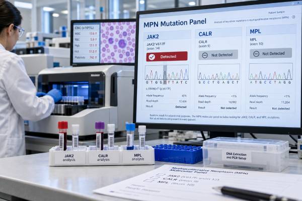
A myeloproliferative neoplasm (MPN) panel tests for acquired gene mutations that drive abnormal blood-cell production, most importantly JAK2, CALR, and MPL. These mutations are central to the molecular diagnosis of the classic Philadelphia chromosome–negative MPNs: polycythemia vera (PV), essential thrombocythemia (ET), and primary myelofibrosis (PMF). The panel is commonly ordered for persistent erythrocytosis, unexplained thrombocytosis, marrow findings suspicious for an MPN, or clinical features such as splenomegaly that suggest a clonal myeloid disorder.
A positive driver mutation strongly supports clonality, but it does not identify the exact MPN on its own. JAK2 can occur in PV, ET, and PMF; CALR and MPL are mainly associated with ET and PMF. Diagnosis still depends on the complete blood count, bone marrow morphology, erythropoietin level when relevant, exclusion of BCR::ABL1-positive chronic myeloid leukemia, and other clinical findings. A negative JAK2/CALR/MPL panel also does not rule out an MPN, because some patients are triple-negative or carry uncommon variants detectable only by broader or more sensitive testing.
- What it measures: The panel looks for major MPN driver mutations, usually JAK2 V617F, CALR exon 9 variants, and MPL exon 10 variants; many assays also evaluate JAK2 exon 12.
- What a positive result means: A pathogenic driver mutation supports a clonal MPN but must be matched with marrow and blood findings to distinguish PV, ET, and PMF.
- What a negative result means: A negative panel does not exclude MPN; assay coverage, low-level clones, noncanonical variants, and triple-negative disease must be considered.
- Typical disease pattern: JAK2 dominates in PV, while ET and PMF can be driven by JAK2, CALR, or MPL.
- Preparation: No fasting is usually needed. Peripheral blood is often adequate for molecular testing, although a bone marrow examination may still be required for formal classification.
Table of Contents
- What an MPN panel tests
- When an MPN panel is ordered
- How JAK2, CALR, and MPL results differ
- Using molecular results to diagnose PV, ET, and PMF
- Negative and triple-negative MPN results
- How MPN molecular testing is performed and reported
- Prognosis, limitations, and next steps
What an MPN panel tests
The core MPN panel evaluates mutations that activate growth signaling in hematopoietic stem cells. JAK2, CALR, and MPL converge on the JAK-STAT pathway, which helps regulate blood-cell production. An acquired driver mutation can make this signaling persist when it should be tightly controlled, giving the mutant clone a growth advantage.
The most common targets are:
- JAK2 V617F, a hotspot mutation in exon 14;
- JAK2 exon 12 mutations, especially important in JAK2 V617F-negative erythrocytosis when PV is suspected;
- CALR exon 9 insertions or deletions, often classified as type 1/type 1-like or type 2/type 2-like patterns; and
- MPL exon 10 mutations, including classic codon 515 variants such as W515L and W515K.
Many laboratories run these tests sequentially. For example, they may test JAK2 V617F first, then CALR and MPL if JAK2 is negative. Other laboratories use a multiplex NGS panel from the beginning. A broader assay may also include genes such as ASXL1, TET2, SRSF2, U2AF1, EZH2, IDH1/2, DNMT3A, CBL, and others that can provide additional clonal or prognostic information.
The panel is designed mainly for BCR::ABL1-negative MPNs. Chronic myeloid leukemia is biologically different and is defined by BCR::ABL1. When CML is a realistic alternative diagnosis, a BCR::ABL1 test is needed rather than assuming a negative MPN driver panel excludes CML.
A dedicated JAK2 V617F mutation test may be used as the first molecular step, while a panel is convenient when ET or PMF is also under consideration.
When an MPN panel is ordered
The panel is most useful when blood counts remain abnormal and common reactive causes do not fully explain the pattern.
Persistent erythrocytosis
Erythrocytosis means an increased red-cell mass reflected by elevated hemoglobin or hematocrit. PV is strongly associated with JAK2 mutation, but secondary erythrocytosis is much more common and can result from chronic hypoxia, smoking, sleep apnea, testosterone or other medications, kidney disease, or tumors that increase erythropoietin.
A PV evaluation therefore usually combines JAK2 testing with clinical assessment, serum erythropoietin, and often bone marrow morphology. Current diagnostic frameworks use hemoglobin/hematocrit thresholds and molecular evidence together rather than relying on one test.
Persistent thrombocytosis
A platelet count of 450 × 10⁹/L or higher is part of the diagnostic framework for ET, but reactive thrombocytosis can occur with iron deficiency, infection, inflammation, recent surgery, malignancy, or splenectomy. JAK2, CALR, or MPL positivity supports a clonal explanation.
The driver does not by itself distinguish ET from prefibrotic PMF. Both can present with high platelets, and the decisive distinction often comes from marrow megakaryocyte morphology and cellularity.
A CALR mutation test is particularly useful in JAK2-negative thrombocytosis, while an MPL mutation test evaluates another less common ET/PMF driver.
Suspected myelofibrosis
PMF may present with anemia, splenomegaly, constitutional symptoms, leukoerythroblastosis, elevated lactate dehydrogenase, abnormal platelets, or marrow fibrosis. Driver testing supports clonality, while broader sequencing and cytogenetics can add prognostic information.
How JAK2, CALR, and MPL results differ
JAK2, CALR, and MPL are often grouped together, but they do not have identical disease associations.
| Driver | Common MPN associations | Key interpretation point |
|---|---|---|
| JAK2 V617F | PV, ET, PMF | Very common in PV and also frequent in ET/PMF; not specific for one MPN |
| JAK2 exon 12 | PV | Important in JAK2 V617F-negative patients with convincing erythrocytosis |
| CALR exon 9 | ET, PMF | Strongly supports a clonal MPN in the right setting; not typical of PV |
| MPL exon 10 | ET, PMF | Less common driver; classic variants affect codon 515 |
JAK2
JAK2 mutation is the hallmark molecular finding in PV. JAK2 V617F accounts for the great majority of cases, while exon 12 variants explain many of the remaining JAK2 V617F-negative cases. In ET and PMF, JAK2 V617F is also common, so the mutation must be interpreted with marrow morphology and the dominant blood-count pattern.
The presence of JAK2 V617F in a patient with thrombocytosis does not automatically mean ET. Prefibrotic PMF and, in some settings, masked PV can enter the differential diagnosis.
CALR
CALR mutations are concentrated in ET and PMF and are usually found when JAK2 is absent. Most are frameshift insertions or deletions in exon 9. In PMF, CALR subtype can carry prognostic information; type 1/type 1-like CALR has generally been associated with more favorable survival than some alternative driver states in validated models.
In ET, CALR-mutated disease often has a different clinical phenotype from JAK2-mutated disease, including younger age and different thrombosis patterns at the population level. Those patterns do not replace individualized risk assessment.
MPL
MPL encodes the thrombopoietin receptor. Activating MPL variants drive JAK-STAT signaling and occur in a smaller proportion of ET and PMF. A pathogenic MPL variant supports clonality but cannot distinguish ET from PMF without marrow and clinical correlation.
The three major driver groups are often mutually exclusive, although highly sensitive sequencing can occasionally detect more than one mutation or a small additional clone. Such results deserve careful laboratory and clinical review rather than a simplified “double positive” interpretation.
Using molecular results to diagnose PV, ET, and PMF
A positive MPN panel is strongest when it explains a matching disease phenotype. The molecular finding is integrated with formal diagnostic criteria rather than used as a stand-alone cancer test.
Polycythemia vera
PV is a JAK2-mutated MPN characterized by clonal erythrocytosis. JAK2 mutation plus sustained elevation of hemoglobin or hematocrit strongly raises the probability of PV, but clinicians still assess bone marrow morphology and serum erythropoietin and exclude secondary causes of erythrocytosis.
A low serum erythropoietin concentration can support PV, while a normal or high level may point toward other causes, although no single EPO value settles the diagnosis by itself. JAK2 exon 12 testing becomes important when V617F is negative but the clinical pattern remains convincing.
Essential thrombocythemia
ET requires persistent thrombocytosis, characteristic marrow megakaryocyte morphology, exclusion of other myeloid neoplasms, and evidence of clonality or absence of a reactive cause. JAK2, CALR, or MPL mutations are found in the large majority of patients and provide strong clonal evidence.
The main diagnostic trap is confusing ET with prefibrotic PMF. Both can show high platelets and the same driver mutations, but prefibrotic PMF has different marrow morphology and can have a different clinical course. The mutation cannot resolve that distinction alone.
Primary myelofibrosis
PMF can be prefibrotic or overtly fibrotic. JAK2, CALR, or MPL is present in most cases, but a driver mutation is not mandatory if other clonal evidence and diagnostic criteria are met. Bone marrow examination is central because megakaryocyte atypia, cellularity, and fibrosis grade establish the morphologic framework.
In overt PMF, anemia, splenomegaly, elevated LDH, leukoerythroblastosis, and constitutional symptoms may provide additional clinical support. A blood-cancer LDH result can reflect increased cell turnover but is nonspecific and cannot diagnose PMF.
Negative and triple-negative MPN results
A negative JAK2/CALR/MPL panel means no covered driver mutation was detected above the assay’s limit of detection. It does not automatically mean the blood-count abnormality is reactive.
Several explanations are possible:
- the patient truly has a non-clonal/reactive blood-count abnormality;
- the MPN is triple-negative for the three major drivers;
- the driver is present at a level below the test’s analytic sensitivity;
- the assay did not cover a noncanonical JAK2, CALR, or MPL variant;
- another clonal mutation provides evidence of a myeloid neoplasm; or
- the correct diagnosis is a different myeloid disorder.
Triple-negative status is most relevant in ET and PMF. It requires extra care because the molecular panel no longer provides the usual major-driver evidence of clonality. Bone marrow morphology, chromosome studies, and broader NGS become more important.
In PMF, additional mutations such as ASXL1, SRSF2, U2AF1, EZH2, IDH1/2, or others may help establish clonality and contribute prognostic information. In ET, clinicians must be especially careful to exclude reactive thrombocytosis and other myeloid neoplasms.
A negative panel in suspected PV requires a different approach because essentially all well-established PV is JAK2-mutated. If JAK2 V617F is negative, testing for JAK2 exon 12 or other appropriately sensitive JAK2 variants is important before accepting a JAK2-negative PV label.
How MPN molecular testing is performed and reported
Peripheral blood is often sufficient because the mutated myeloid clone circulates. Bone marrow DNA can also be tested, particularly when a marrow biopsy is already being performed. Fasting is not generally required.
Laboratories may use several methods:
- allele-specific PCR or quantitative PCR for highly sensitive detection of known hotspots such as JAK2 V617F;
- fragment analysis for CALR exon 9 insertions and deletions;
- targeted sequencing for defined regions of JAK2, CALR, and MPL; or
- next-generation sequencing for the major drivers plus a wider group of myeloid genes.
Current classification guidance emphasizes adequate assay sensitivity because low variant allele frequencies can occur, particularly early in disease. A result should therefore be interpreted with the laboratory’s stated limit of detection.
Variant allele frequency
NGS and some quantitative assays report variant allele frequency (VAF), the percentage of sequence reads containing the mutation. VAF can provide information about clone representation but is not a direct measurement of the percentage of malignant cells.
For JAK2 V617F, a higher allele burden may correlate with certain disease features at a population level, but the number should not be used alone to diagnose an MPN, classify PV versus ET versus PMF, or decide that treatment is working. Copy-number changes and clonal architecture can change VAF independently of simple cell counts.
Pathogenic variants versus VUS
A pathogenic or likely pathogenic driver supports an MPN in the right setting. A variant of uncertain significance (VUS) does not carry the same weight. If a report identifies an unusual JAK2, CALR, or MPL alteration, the classification and laboratory interpretation are essential before it is treated as a true driver.
Prognosis, limitations, and next steps
The major driver mutation can influence phenotype and prognosis, but it is only one part of risk assessment. In ET, thrombosis risk depends strongly on age, previous thrombosis, JAK2 status, and other clinical factors. In PV, management focuses heavily on thrombosis prevention and hematocrit control. In PMF, modern prognostic systems can incorporate clinical variables, karyotype, and mutations beyond JAK2/CALR/MPL.
Broader molecular testing becomes particularly valuable in PMF because mutations such as SRSF2, ASXL1, U2AF1-Q157, and others can carry adverse prognostic information. Cytogenetic abnormalities also matter. A basic driver panel therefore answers the question “Is a classic MPN driver present?” more directly than “What is this patient’s complete prognosis?”
After an MPN panel, common next steps include:
- review of repeat complete blood counts and the peripheral smear;
- ferritin and iron studies when thrombocytosis or masked erythrocytosis is possible;
- serum erythropoietin when PV is suspected;
- BCR::ABL1 testing when CML is part of the differential diagnosis;
- bone marrow aspirate and biopsy for definitive ET/PMF classification or when PV morphology is needed;
- cytogenetic testing and expanded NGS for suspected PMF or atypical disease; and
- clinical risk assessment for thrombosis, bleeding, progression, symptoms, and transplant eligibility.
The most important interpretation principle is that driver positivity establishes molecular evidence of a clone, not the complete disease identity. A JAK2-positive result may fit PV, ET, or PMF. CALR and MPL narrow the pattern toward ET or PMF, but marrow morphology remains essential to separate them.
Similarly, a negative result should trigger review of assay coverage and the differential diagnosis rather than an automatic conclusion that no MPN exists. This is especially important when the patient has persistent unexplained counts, splenomegaly, marrow abnormalities, or progressive symptoms.
Ask the hematology team which exact mutations were tested, whether JAK2 exon 12 was included, what the assay sensitivity was, whether a bone marrow examination is needed, and whether broader NGS would add diagnostic or prognostic value. Those details turn a molecular panel from a list of gene names into a clinically useful diagnosis.
Why mutation burden is usually not a treatment target by itself
Some therapies can reduce JAK2 V617F allele burden, but a falling VAF is not automatically equivalent to clinical remission or proven disease modification. Blood counts, symptoms, spleen size, marrow findings when relevant, thrombosis prevention, and progression risk remain more direct clinical endpoints. A molecular change is most informative when it is interpreted alongside those outcomes rather than used alone.
This distinction is especially important when comparing serial results from different laboratories. Assay design and sensitivity can shift the reported VAF. If molecular monitoring is clinically important, consistent methodology and attention to the laboratory’s analytic range make trends more reliable.
Small clones require clinical context
Highly sensitive assays can occasionally detect a very small JAK2-mutated clone in a person who does not yet meet criteria for PV, ET, or PMF. The result may represent early clonal hematopoiesis rather than established MPN. Persistent blood-count abnormalities, marrow morphology, allele burden, and follow-up determine whether that molecular finding is clinically diagnostic.
References
- Molecular diagnostic criteria of myeloproliferative neoplasms 2023 (Review)
- Advances in Molecular Understanding of Polycythemia Vera, Essential Thrombocythemia, and Primary Myelofibrosis: Towards Precision Medicine 2024 (Review)
- Essential thrombocythemia: 2024 update on diagnosis, risk stratification, and management 2024 (Review)
- Polycythemia vera: 2024 update on diagnosis, risk-stratification, and management 2023 (Review)
- Primary myelofibrosis: 2023 update on diagnosis, risk-stratification, and management 2023 (Review)
- International Consensus Classification of myeloid and lymphoid neoplasms: myeloproliferative neoplasms 2023 (Review)
Disclaimer
This article is for general education and does not diagnose or exclude a myeloproliferative neoplasm. JAK2, CALR, and MPL results must be interpreted with blood counts, bone marrow findings, other laboratory tests, and clinical features by a qualified hematology team. Seek urgent care for symptoms of a serious clot or bleeding event, including sudden chest pain, shortness of breath, one-sided weakness, severe new headache, or uncontrolled bleeding.





