Kidney injury molecule-1 (KIM-1) is a protein that rises when cells in the kidney’s proximal tubules are injured. A KIM-1 blood test measures the circulating form of this protein and is being studied as a way to detect kidney damage that may not be obvious from creatinine alone. Unlike creatinine and estimated glomerular filtration rate (eGFR), which mainly describe how well the kidneys are filtering, KIM-1 is considered a kidney injury marker. That distinction can matter when tubule cells are damaged before a large change in filtration appears.
Blood KIM-1 is not yet a routine stand-alone kidney test in most clinical settings, and there is no single universal “normal” cutoff that applies across laboratories. Results depend heavily on the assay, units, clinical setting, and whether the test is being used in research, drug-safety monitoring, acute kidney injury (AKI), or chronic kidney disease (CKD). A KIM-1 result is therefore most useful when interpreted beside creatinine, eGFR, urine findings, medications, and the person’s overall clinical picture.
- KIM-1 measures kidney tubule injury, not filtration directly: higher blood levels generally indicate more proximal tubular stress or damage.
- There is no universal blood KIM-1 reference range: values and units vary by assay, so the laboratory’s own method and reference information matter.
- A high KIM-1 result does not identify one specific disease: AKI, CKD, diabetes-related kidney injury, toxins, ischemia, and other conditions can raise it.
- KIM-1 may increase before creatinine clearly changes in some settings, but it has not replaced creatinine, eGFR, urine albumin, or urinalysis in routine kidney care.
- Seek prompt medical assessment for possible AKI symptoms such as sharply reduced urine output, worsening swelling, shortness of breath, persistent vomiting, confusion, or severe weakness regardless of the KIM-1 value.
Table of Contents
- What the KIM-1 Blood Test Measures
- Why a KIM-1 Blood Test May Be Ordered
- How to Interpret KIM-1 Blood Test Results
- Causes of High KIM-1 Levels
- KIM-1 Compared With Other Kidney Tests
- Testing, Preparation, and Important Limitations
- What to Do After a KIM-1 Result
What the KIM-1 Blood Test Measures
KIM-1 is a cell-surface protein produced in very small amounts by healthy proximal tubule cells. The proximal tubules are an early part of the kidney’s microscopic filtering system. After blood is filtered through the glomeruli, these tubules reclaim water, electrolytes, glucose, amino acids, and other useful substances while helping move wastes into the urine.
When proximal tubule cells are injured, expression of KIM-1 rises sharply. Part of the protein can be shed from the injured cell surface and detected in urine, while circulating KIM-1 can be measured in blood. This is why KIM-1 is often described as a damage biomarker rather than a conventional kidney function marker.
That difference is important. A creatinine blood test mainly reflects filtration after creatinine has accumulated in the bloodstream. KIM-1 is tied more directly to an injury response in kidney tubule cells. In theory, this can reveal biological damage earlier or add information when filtration markers are difficult to interpret.
KIM-1 also has a biological role. Injured proximal tubule cells use it to recognize and clear dead or dying cells and debris. Short-term expression may be part of tissue repair. Persistently high expression, however, has been linked in experimental and clinical research with ongoing inflammation, maladaptive repair, and fibrosis.
Blood KIM-1 should not be confused with urinary KIM-1. Both reflect related kidney biology, but they are different measurements. Their concentrations, reference values, and associations with clinical outcomes are not interchangeable.
Why a KIM-1 Blood Test May Be Ordered
A KIM-1 blood test is most often used in specialized or research settings rather than as part of a standard annual kidney panel. Its main appeal is the possibility of detecting or characterizing tubular injury that conventional tests may miss or show only indirectly.
Potential uses include:
- evaluating suspected acute tubular injury or AKI in hospital or research settings;
- studying kidney effects of surgery, sepsis, reduced blood flow, contrast exposure, or nephrotoxic drugs;
- investigating CKD progression and kidney failure risk;
- monitoring possible drug-induced kidney toxicity during medication development;
- studying diabetic kidney disease and other conditions that can damage tubules;
- combining KIM-1 with other biomarkers to improve risk assessment.
KIM-1 is especially attractive because a rise in serum creatinine can lag behind the initial injury. Creatinine may also be influenced by muscle mass, diet, age, and fluid balance. KIM-1 is not immune to confounding, but it measures a different part of the kidney-injury process.
Still, KIM-1 is not a replacement for established evaluation. When clinicians suspect AKI, they continue to rely heavily on serial creatinine measurements, urine output, history, physical examination, medication review, and often urinalysis. A broader acute kidney injury blood test panel may also be needed to assess potassium, bicarbonate, urea nitrogen, and other complications.
For CKD, routine evaluation still centers on eGFR and urine albumin. KIM-1 may add prognostic information in selected populations, but its exact place in everyday CKD care remains under study.
How to Interpret KIM-1 Blood Test Results
The most important rule is simple: interpret KIM-1 using the reporting laboratory’s assay information, not a universal cutoff found online. There is currently no standardized blood KIM-1 range that can be applied across all laboratories, platforms, and patient groups.
Different studies report KIM-1 in units such as pg/mL, ng/L, or ng/mL. Assays may use different antibodies, calibration materials, sample processing methods, and detection technologies. A number that is “high” in one method may not map cleanly to the same number from another method.
| Result pattern | What it can suggest | What is usually needed next |
|---|---|---|
| Low or within the laboratory’s expected range | No strong KIM-1 signal of proximal tubule injury at that time | Interpret with creatinine, eGFR, urine albumin, symptoms, and clinical risk |
| Mildly elevated | Possible tubule stress or injury; may also reflect CKD burden or another clinical factor | Repeat or correlate with established kidney tests and the clinical setting |
| Markedly elevated | Stronger evidence of active or substantial tubular injury, depending on assay and context | Assess promptly for AKI, nephrotoxic exposure, hemodynamic problems, infection, obstruction, or CKD progression |
| Rising on serial tests | Potentially worsening or ongoing tubular injury | Look for a continuing cause and compare with creatinine, urine output, electrolytes, and treatment changes |
| Falling on serial tests | May indicate decreasing injury or recovery | Confirm that kidney function and the clinical condition are also improving |
A high KIM-1 level does not by itself diagnose AKI. AKI is still defined clinically using changes in serum creatinine and/or urine output. Research suggests that blood KIM-1 can help identify AKI and may improve risk prediction, but performance varies across patient groups and study designs.
A useful way to think about KIM-1 is to separate injury from function. For example, imagine two people with the same creatinine of 1.0 mg/dL. One has stable kidney function and a low KIM-1 result. The other has just had major surgery and shows a sharp rise in KIM-1. Their filtration numbers may look similar at that moment, but the second person may have a stronger signal of recent tubular stress. The reverse can also happen: a person with long-standing CKD may have a reduced eGFR but no major new rise in an injury marker, suggesting chronic loss of filtration rather than a large new tubular insult. These patterns are simplified examples, not diagnostic rules, but they show why combining markers can be more informative than treating one number as the whole story.
Serial results can also be more useful than a single isolated measurement. A value that doubles or falls substantially over a short interval may carry different meaning from one stable result, even if both remain above a laboratory threshold. That said, clinicians must compare serial values using the same assay whenever possible. Changing laboratories or platforms can introduce analytical differences that look like biological change.
Similarly, a normal KIM-1 value does not prove that the kidneys are normal. Glomerular diseases can cause major albumin leakage with less prominent tubular injury, and early CKD can be present with little change in an injury biomarker. An eGFR test and urine albumin measurement answer different questions and remain central to kidney assessment.
Causes of High KIM-1 Levels
High blood KIM-1 generally points toward proximal tubular stress or damage, but it does not reveal the cause on its own. The same biomarker response can occur after several types of injury.
Acute kidney injury
AKI is one of the most studied settings for KIM-1. Tubule cells can be injured by low blood flow, severe infection, surgery, toxins, or inflammation. Because KIM-1 expression can rise soon after proximal tubular injury, researchers have studied it as an earlier signal than creatinine.
Common clinical situations include major surgery, shock, severe dehydration, sepsis, and intensive care illness. The timing of the sample matters because a biomarker may rise and fall over hours or days as the injury evolves.
Medication or toxin-related kidney injury
Some medicines and chemicals can damage proximal tubule cells. Examples include certain chemotherapy drugs, some antibiotics, and other nephrotoxic exposures. In drug development, KIM-1 has been studied as a safety biomarker because it may reveal tubular damage before large functional changes occur.
A high result does not prove that a medication caused the injury. Clinicians must consider dose, timing, other drugs, blood pressure, hydration, infection, and baseline kidney function before attributing kidney damage to one agent.
Chronic kidney disease
Blood KIM-1 may also be elevated in CKD. Recent cohort research has linked higher plasma KIM-1 with lower eGFR, more albuminuria, diabetes, CKD progression, and kidney failure risk. This does not mean KIM-1 should be used alone to stage CKD. Standard staging still relies on GFR and albuminuria categories.
A CKD blood test panel provides the broader context needed to interpret a damage marker, including filtration and metabolic complications.
Other sources of elevation
KIM-1 is strongly associated with injured kidney tubules, but circulating concentrations can also be affected by the broader disease context. Kidney cancer, especially renal cell carcinoma, has been studied as another setting in which KIM-1 may be elevated. For this reason, an unexplained result should never be interpreted as proof of a single kidney disease without standard clinical evaluation.
KIM-1 Compared With Other Kidney Tests
KIM-1 is best understood as one piece of a kidney “dashboard.” Different markers describe filtration, structural damage, electrolyte control, or protein leakage. No single test captures every function of the kidneys.
| Test | Main information | Key limitation |
|---|---|---|
| Blood KIM-1 | Proximal tubule injury signal | No universal clinical cutoff; not routinely standardized |
| Creatinine | Indirect marker of filtration | Influenced by muscle mass, diet, fluid status, and delayed rise after acute injury |
| eGFR | Estimated filtering capacity | Calculated estimate, not a direct injury marker |
| Cystatin C | Alternative filtration marker used to estimate GFR | Can be affected by inflammation, thyroid status, glucocorticoids, and other factors |
| Urine albumin-to-creatinine ratio | Albumin leakage and glomerular kidney damage | Can vary with exercise, fever, infection, and temporary physiologic stress |
| NGAL | Another tubular injury/inflammation biomarker | Can rise with systemic inflammation and infection; thresholds vary |
KIM-1 and NGAL are both studied as earlier injury biomarkers, but they respond through different biological pathways and may be affected by different non-kidney factors. Combining biomarkers may eventually improve precision, yet most routine clinical decisions still depend on established tests.
For a broad assessment, a kidney function blood test panel remains more useful than KIM-1 alone because it can identify reduced filtration and electrolyte or acid-base problems that may require immediate treatment.
Testing, Preparation, and Important Limitations
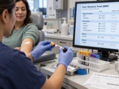
A KIM-1 blood test uses a standard blood sample, usually drawn from a vein in the arm. Fasting is generally not required solely for KIM-1, but preparation can depend on other tests ordered at the same time. Follow the laboratory or clinician’s instructions if the sample is part of a larger research protocol or panel.
The major limitation is lack of standardization. There is no universally accepted decision threshold comparable with familiar clinical cutoffs for potassium or eGFR. Results from different assay platforms may not be directly comparable.
Other important limitations include:
- Clinical context changes test performance. Results in cardiac surgery patients may not perform the same way in sepsis, outpatient CKD, cancer therapy, or community screening.
- Timing matters. A sample taken before injury, during the biomarker rise, or after recovery can produce very different values.
- Kidney disease is heterogeneous. Some conditions primarily affect glomeruli, blood vessels, or the urinary tract rather than proximal tubules.
- KIM-1 can add prognostic information without proving causation. A higher level may identify higher-risk patients but does not explain why their kidneys are injured.
- Research cutoffs should not be self-applied. A threshold from one study may use a different assay and patient population than a person’s own laboratory test.
These limitations explain why a KIM-1 result should not be used to start, stop, or change medication without clinician review. It is especially important not to discontinue kidney-protective medicines solely because an experimental biomarker is above a research threshold.
What to Do After a KIM-1 Result
The next step depends more on the overall kidney picture than on the KIM-1 number alone. A reasonable approach is to confirm what assay was used, compare the result with the laboratory’s expected range, and review established kidney markers from the same period.
If KIM-1 is elevated, clinicians may consider:
- repeating serum creatinine and calculating eGFR;
- checking urine output and hydration status;
- measuring electrolytes, especially potassium and bicarbonate;
- reviewing prescription drugs, over-the-counter pain relievers, supplements, and recent contrast exposure;
- performing urinalysis and urine albumin testing;
- considering imaging when urinary obstruction is possible;
- repeating KIM-1 only if serial testing is part of the relevant clinical or research protocol.
A single elevated result without symptoms or changes in routine kidney tests may lead to repeat testing rather than urgent treatment. By contrast, an elevated KIM-1 result in a person with rapidly rising creatinine, falling urine output, abnormal potassium, sepsis, or low blood pressure fits a much more concerning pattern.
Seek urgent medical care for very low or absent urine output, severe shortness of breath, chest symptoms, confusion, fainting, rapidly increasing swelling, uncontrolled vomiting, or profound weakness. These symptoms can occur with serious AKI or dangerous electrolyte disturbances, and waiting for a biomarker result is not appropriate.
The practical value of KIM-1 is that it can add information about tubular damage to tests that mainly show kidney function. Its promise is real, but so are its current limitations. Until assays and clinical thresholds become more standardized, KIM-1 is best interpreted as a complementary biomarker rather than a stand-alone diagnosis.
References
- Kidney injury molecule 1 in the early detection of acute kidney injury-a systematic review and meta-analysis 2025 (Systematic Review)
- Plasma and Urinary KIM-1 in Chronic Kidney Disease: Prognostic Value, Associations with Albuminuria, and Implications for Kidney Failure and Mortality 2025
- From acute tubular injury to tubular repair and chronic kidney diseases – KIM-1 as a promising biomarker for predicting renal tubular pathology 2025 (Review)
- Plasma Biomarkers and Incident CKD Among Individuals Without Diabetes 2023
- Elevated Serum KIM-1 in Sepsis Correlates with Kidney Dysfunction and the Severity of Multi-Organ Critical Illness 2024
Disclaimer
KIM-1 blood testing is an evolving biomarker test and is not a substitute for a clinical diagnosis of acute or chronic kidney disease. Reference values and clinical significance vary by assay and setting, so results should be interpreted by a qualified healthcare professional together with creatinine, eGFR, urine tests, symptoms, medications, and medical history. Seek urgent medical care for symptoms that could indicate severe kidney injury or dangerous electrolyte problems.