
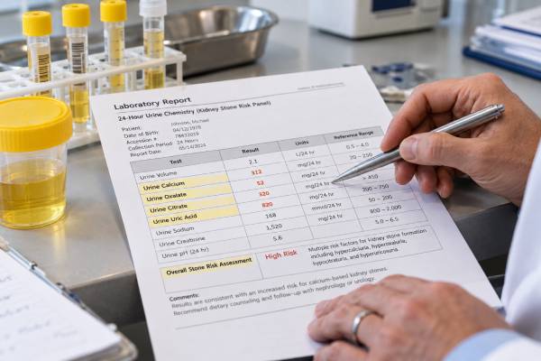
A kidney stone test panel measures the urine chemistry that makes crystals more or less likely to form. The core 24-hour urine markers usually include calcium, oxalate, citrate, uric acid, volume, sodium, pH, and creatinine, with many laboratories also reporting calcium oxalate, calcium phosphate, and uric acid supersaturation. The results are most useful as a pattern rather than as isolated “high” or “low” numbers. High urine calcium may be much more important when urine volume is low, while adequate citrate can reduce calcium crystallization. Acidic urine can drive uric acid stones even when the total uric acid excretion is not dramatically elevated. A detailed panel is especially useful after recurrent, bilateral, multiple, early-onset, or high-risk stones, and it can turn general advice into specific targets that can be checked again after diet or medication changes.
- A standard 24-hour kidney stone panel commonly measures calcium, oxalate, citrate, uric acid, volume, sodium, pH, and creatinine.
- Low urine volume raises the concentration of every stone-forming solute, so many adult prevention plans aim for more than 2.5 L of urine per day.
- High calcium and high oxalate increase calcium oxalate risk, while low citrate removes a natural crystal inhibitor.
- Low urine pH is the main chemical driver of uric acid stones, often more important than the blood uric acid level.
- Supersaturation and repeat testing show whether the overall urine environment improves, not just whether one marker reaches a reference range.
Table of Contents
- What a Kidney Stone Urine Panel Measures
- 24-Hour Collection and Preparation
- Urine Calcium and Oxalate Results
- Citrate, Urine Volume, and Sodium
- Uric Acid and Urine pH
- Supersaturation and Common Result Patterns
- Follow-Up Tests and Next Steps
What a Kidney Stone Urine Panel Measures
A kidney stone urine panel is usually a 24-hour collection designed to quantify stone-forming and stone-protective substances over an entire day. It is different from a routine urinalysis, which is a spot test used for blood, protein, infection clues, crystals, specific gravity, and other screening findings.
The most common analytes are:
- urine volume;
- calcium;
- oxalate;
- citrate;
- uric acid;
- sodium;
- potassium;
- pH;
- creatinine;
- sometimes magnesium, phosphate, sulfate, ammonium, urea nitrogen, and calculated supersaturation.
Each marker answers a different question. Calcium and oxalate are the raw materials for calcium oxalate crystals. Citrate helps keep calcium soluble. Urine volume determines concentration. Sodium often reflects salt intake and can influence calcium excretion. Uric acid and pH help assess uric acid stone risk. Creatinine helps judge whether the collection was reasonably complete.
Many stone laboratories combine these measurements into supersaturation values. A urine supersaturation test estimates whether the urine is chemically favorable for a particular crystal. That can be more informative than looking at calcium, oxalate, or pH alone.
A panel does not directly tell what an existing stone is made of. When stone material is available, a kidney stone analysis provides that information and helps clinicians decide which parts of the urine panel deserve the most attention.
24-Hour Collection and Preparation
Accurate collection is essential because the laboratory calculates daily excretion from everything passed during the timed period. A missed void can make several results falsely low, while collecting longer than 24 hours can make daily excretion look falsely high.
A typical collection starts after discarding the first morning urine and recording the time. Every urine sample after that is collected, including overnight, and the final sample is collected at the same time the next morning. The laboratory’s instructions take priority because some containers require refrigeration, preservatives, or special handling.
For a baseline evaluation, the goal is usually to capture the patient’s usual diet, fluid intake, and routine. Deliberately drinking far more than normal, avoiding all salt for one day, or stopping medication without instruction can create an artificial profile that does not explain everyday stone formation.
Many guidelines favor two 24-hour collections for a detailed initial metabolic assessment because urine chemistry varies from day to day. One collection can still be useful, but two samples can reveal whether an abnormality is consistent.
Day-to-day variation is not just random laboratory noise. Sodium intake, protein intake, sweating, exercise, work schedule, restaurant meals, and bowel function can all change urinary chemistry. If two collections differ greatly, the difference itself can be informative. One may represent a high-sodium workday and the other a lower-sodium home day, or one may have much lower volume because of heat exposure. Reviewing what happened on each collection day can make the panel more useful than simply averaging the numbers.
Testing is best done after the acute stone episode has settled and the patient is back to normal eating and drinking. Vomiting, infection, temporary fasting, or unusually aggressive hydration after an emergency visit can distort volume, pH, sodium, and other markers.
Creatinine is often used as a collection-quality check. Expected excretion depends on muscle mass, age, sex, body size, diet, and kidney function, so an unusual value is a clue rather than absolute proof that the sample is incomplete.
Urine Calcium and Oxalate Results
Urine calcium and oxalate are central to calcium oxalate stone risk, but neither should be interpreted in isolation.
Hypercalciuria means urinary calcium excretion is high. Common clinical thresholds are around 250 mg/day in women and 300 mg/day in men, although laboratories and guidelines vary. Risk is continuous: a value does not suddenly become harmless one milligram below a cutoff.
High urine calcium can reflect inherited tendency, high sodium intake, endocrine disease, medication effects, increased intestinal absorption, or other mineral disorders. It often occurs with normal blood calcium. This is why a urine calcium test for kidney stones answers a different question from a serum calcium test.
If blood calcium is elevated, primary hyperparathyroidism becomes an important possibility. A blood calcium test and PTH testing when indicated can identify a systemic cause that urine testing alone cannot diagnose.
Hyperoxaluria means urine oxalate is elevated. Typical adult reference ranges are often roughly 20–40 mg/day, but methods differ. Even modest oxalate increases can meaningfully raise calcium oxalate supersaturation.
Causes of high oxalate include frequent high-oxalate foods, low calcium intake with meals, high-dose vitamin C, fat malabsorption, inflammatory bowel disease, bariatric surgery, short bowel, and rare primary hyperoxaluria. Urine oxalate measurement is particularly important when stones occur with chronic diarrhea or gastrointestinal disease.
One of the most common misunderstandings is that calcium oxalate stones require a low-calcium diet. In most adults, normal dietary calcium—often about 1,000–1,200 mg/day depending on age and guideline—is preferred. Calcium eaten with meals binds oxalate in the gut and can reduce oxalate absorption. Severe calcium restriction can therefore make urinary oxalate worse.
The timing of calcium matters most when oxalate exposure occurs. Calcium from food eaten with an oxalate-containing meal can bind oxalate before it is absorbed. A calcium supplement taken far away from meals may not provide the same gut-binding effect and may increase urinary calcium in some patients. Whether supplements are appropriate depends on bone health, diet, kidney function, and the 24-hour urine pattern, so supplementation should be individualized rather than added automatically.
When calcium and oxalate are both elevated, the combination can be more important than either result alone because crystal formation depends on their interaction and concentration. Urine volume and citrate can either amplify or partly offset that pattern, which is why the complete panel and supersaturation values are more informative than isolated numbers.
Citrate, Urine Volume, and Sodium
Citrate is a natural inhibitor of calcium stone formation. It binds calcium and reduces the amount available to combine with oxalate or phosphate. It can also interfere with crystal growth and aggregation.
A urine citrate value below roughly 320 mg/day is often described as hypocitraturia in adults, but laboratories and clinicians use different definitions. Some interpret citrate in relation to calcium or urine volume rather than relying on one absolute threshold.
Low citrate can occur with chronic diarrhea, metabolic acidosis, distal renal tubular acidosis, high dietary acid load, low potassium intake, or certain medications. A urine citrate test can identify a treatable protective deficit.
Urine volume is one of the highest-value measurements because it changes concentration without changing how much calcium or oxalate the body produces. A person excreting the same amount of calcium into 3 L of urine is generally at lower supersaturation than someone excreting it into 1.2 L.
Many guidelines advise enough fluid to produce more than about 2.5 L of urine per day for adult stone prevention. The required fluid intake is higher than that because water is also lost through sweat, skin, breathing, and stool. Hot climates, physical jobs, exercise, fever, and diarrhea can raise fluid needs substantially.
Urine sodium often acts as an objective marker of sodium intake. High sodium intake increases urinary calcium in many stone formers and can work against efforts to lower calcium supersaturation. Reducing excess sodium can therefore improve urinary calcium without sacrificing normal dietary calcium.
A single urine sodium result is not a perfect food diary, but it is often more informative than asking someone to estimate salt intake from memory. Hidden sodium in restaurant meals, processed foods, sauces, breads, and packaged foods can contribute substantially.
Uric Acid and Urine pH
Urinary uric acid and pH are interpreted together because uric acid solubility depends strongly on acidity.
Common thresholds for hyperuricosuria are around 800 mg/day in men and 750 mg/day in women, although laboratory definitions vary. High excretion can result from high purine intake, gout-related metabolism, rapid cell turnover, certain genetic disorders, or other causes.
For uric acid stones, however, low urine pH is usually more important than high total uric acid. When urine pH remains below about 5.5, more uric acid exists in its poorly soluble form and crystallizes more easily.
People with obesity, insulin resistance, metabolic syndrome, type 2 diabetes, and gout often have persistently acidic urine. A uric acid stone risk test therefore emphasizes pH and volume as well as uric acid excretion.
A high blood uric acid level does not prove that a kidney stone is made of uric acid, and a normal blood level does not rule it out. Blood and urine urate measure different parts of uric acid physiology.
Urine pH also matters for calcium phosphate. More alkaline urine can raise calcium phosphate supersaturation, especially when urinary calcium is high. That creates an important treatment tradeoff: alkali can help low citrate and uric acid stones, but excessive alkalinization may increase calcium phosphate risk.
For that reason, patients using potassium citrate or another alkali often need follow-up urine pH and supersaturation rather than assuming that a higher pH is always better.
Supersaturation and Common Result Patterns
Supersaturation brings the panel together. It estimates whether the urine contains enough dissolved material, under its current pH and volume conditions, to favor a particular crystal.
A report may include calcium oxalate supersaturation, calcium phosphate supersaturation, and uric acid supersaturation. Lowering the relevant supersaturation is often a more useful treatment goal than forcing every individual analyte into a textbook range.
Common patterns include:
| Urine pattern | What it suggests | Common next step |
|---|---|---|
| Low volume + high calcium | Concentrated calcium stone risk | Increase fluids, reduce excess sodium, assess hypercalciuria |
| High oxalate + low calcium intake | Increased intestinal oxalate absorption | Normalize calcium with meals and review oxalate sources |
| Low citrate + high calcium | Reduced protection against calcium crystallization | Address acid load and consider citrate therapy |
| Low pH + low volume | Strong uric acid stone environment | Increase volume and use supervised alkalinization when indicated |
| High pH + high calcium | Possible calcium phosphate tendency | Review stone composition, citrate, bicarbonate, and causes of alkaline urine |
| High sodium + high calcium | Salt intake may be driving calciuria | Reduce dietary sodium and recheck |
The same abnormality can have different significance depending on the stone. Low citrate is important in calcium oxalate disease, but a patient with a pure uric acid stone may have a different primary target. A very alkaline urine is concerning in some calcium phosphate stone formers but desirable up to a controlled target in cystinuria.
Mixed stones require especially careful interpretation. A stone that is mostly calcium oxalate with a meaningful calcium phosphate fraction may call for citrate therapy while also requiring close attention to urine pH. A patient with uric acid and calcium oxalate components may need alkalinization plus evaluation of calcium, oxalate, and citrate. The panel helps avoid a one-diagnosis, one-treatment approach when more than one crystal pathway is active, and it provides measurable targets that can be reassessed after each major prevention change.
This is why stone composition and the urine panel should be read together. The laboratory numbers become clinically useful only when connected to the actual crystal type and the patient’s history.
Follow-Up Tests and Next Steps
After the initial panel, follow-up depends on what was abnormal and which treatment was started. Dietary measures may include more fluid, lower sodium, normal calcium with meals, moderation of excessive animal protein, targeted reduction of high-oxalate foods, or more fruits and vegetables.
Medication may be used when the urine pattern and clinical history justify it. Thiazide or thiazide-like drugs can reduce urinary calcium. Potassium citrate can raise citrate and pH. Allopurinol may be useful for selected hyperuricosuric stone formers. Cystinuria requires separate high-volume, alkalinization, and sometimes thiol-drug therapy.
The panel can objectively show whether treatment worked over time and whether a practical change produced the intended biochemical effect rather than only a subjective sense of improvement. If a patient increases fluid intake, urine volume should rise. If sodium intake falls, urine sodium should fall and urinary calcium may improve. If potassium citrate is effective, urine citrate and pH should change in the expected direction.
Current European guidance suggests repeating 24-hour urine testing around 8–12 weeks after starting pharmacologic prevention. Once urine risk factors are controlled, testing may be spaced to about yearly for many stable high-risk patients, although individual follow-up varies.
Imaging remains important because laboratory improvement is not the final outcome. Ultrasound or CT can show whether existing stones are stable and whether new stones are forming. A patient can have a better panel but still need treatment for an existing obstructing stone.
Blood tests are often paired with the urine panel. Creatinine, electrolytes, calcium, bicarbonate, and uric acid can reveal kidney, endocrine, or acid-base disorders and can monitor the safety of prevention medication.
If the results look inconsistent, check collection accuracy before making major changes. Missing urine, overcollection, illness, unusual diet, or intentional overhydration can distort the profile. Repeating a questionable collection is often more useful than treating an unreliable number.
Seek prompt medical care for suspected stones with fever or chills, inability to urinate, persistent vomiting, uncontrolled pain, pregnancy, or a single functioning kidney. A metabolic panel is designed to prevent future stones, not to rule out an acute infected obstruction.
The strongest use of a kidney stone test panel is as a feedback system: measure the urine environment, identify the dominant risks, change the factors that are modifiable, and then remeasure to confirm that the chemistry actually moved in a safer direction.
References
- Metabolic Evaluation and Recurrence Prevention – EAU Guidelines on Urolithiasis – Uroweb 2026 (Guideline)
- Summary of the clinical practice guideline for the management of urinary stones, third edition 2025 (Guideline)
- Metabolic Evaluation and Recurrence Prevention for Urinary Stone Patients: An EAU Guidelines Update 2024 (Review)
- Urological Guidelines for Kidney Stones: Overview and Comprehensive Update 2024 (Review)
- Kidney Stone Pathophysiology, Evaluation and Management: Core Curriculum 2023 2023 (Review)
- UPDATE – Canadian Urological Association guideline: Evaluation and medical management of kidney stones 2022 (Guideline)
Disclaimer
This article is for general education and does not replace individualized medical care. Kidney stone urine ranges, supersaturation targets, medication choices, and follow-up schedules vary with stone type, kidney function, laboratory methods, and other health conditions. Review abnormal results and treatment changes with a qualified clinician.