
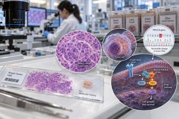
A PDGFRA mutation test looks for activating changes in the platelet-derived growth factor receptor alpha gene, most often as part of the molecular evaluation of a gastrointestinal stromal tumor (GIST). The exact mutation matters because PDGFRA variants do not all respond to the same tyrosine kinase inhibitors. The best-known example is PDGFRA D842V in exon 18, which is strongly resistant to imatinib but highly sensitive to avapritinib in advanced GIST. Other PDGFRA mutations, including some exon 12 and non-D842V exon 18 variants, may remain sensitive to imatinib. A report therefore should not stop at “PDGFRA positive”; it should identify the exon and exact protein change. PDGFRA mutations occur in a minority of GISTs and are usually mutually exclusive with a primary KIT driver mutation. The result helps confirm molecular subtype, select therapy, avoid ineffective treatment, and sometimes clarify why a tumor has a particular gastric or epithelioid pattern.
- A positive PDGFRA result means a PDGFRA alteration was detected, but treatment meaning depends on the exact variant.
- PDGFRA D842V is an exon 18 activation-loop mutation that is resistant to imatinib and is a key indication for avapritinib in advanced GIST.
- Some non-D842V PDGFRA mutations can be imatinib-sensitive, so all PDGFRA-positive GISTs should not be treated as D842V.
- A negative PDGFRA result does not rule out GIST; many GISTs instead carry KIT mutations or belong to other molecular subgroups.
- PDGFRA testing is usually performed on tumor tissue and typically reflects a somatic cancer driver rather than an inherited mutation.
Table of Contents
- What PDGFRA Testing Measures in GIST
- Common PDGFRA Exon Variants
- What a PDGFRA D842V Result Means
- Positive, Negative, and Wild-Type Results
- Testing Methods and Pathology Context
- How Mutation Status Guides GIST Treatment
- Follow-Up, Resistance, and Questions to Ask
What PDGFRA Testing Measures in GIST
PDGFRA encodes a receptor tyrosine kinase on the cell surface. Under normal conditions, binding of platelet-derived growth factors activates the receptor in a controlled way and sends growth and survival signals into the cell. Activating mutations can hold the receptor in an active conformation even without its normal ligand, creating a persistent growth signal.
GIST is the cancer in which PDGFRA testing has its clearest routine role. Most GISTs are driven by an activating mutation in either KIT or PDGFRA, with KIT being more common. The two primary drivers are usually mutually exclusive. Molecular testing therefore often evaluates both genes together, sometimes within a broader GIST biomarker panel.
PDGFRA-mutant GISTs often arise in the stomach and can have epithelioid or mixed cell morphology. Many have a relatively indolent natural history compared with some KIT-mutant GISTs, but behavior still varies with tumor size, mitotic rate, rupture, location, and other clinical factors. Mutation status does not replace standard pathologic risk assessment.
The test is a DNA test, not a measurement of PDGFRA protein concentration. Results are reported as specific sequence changes such as p.D842V or p.V561D, often with the exon number. Exon location matters because different parts of the receptor control different functions and drug-binding conformations.
A KIT mutation test in GIST is the closest companion assay. If neither KIT nor PDGFRA has a pathogenic driver mutation, the tumor may be called KIT/PDGFRA wild-type and may require evaluation for SDH deficiency, NF1-associated disease, BRAF or RAS alterations, or rare gene fusions depending on the clinicopathologic setting.
Common PDGFRA Exon Variants
PDGFRA mutations cluster in functional regions of the receptor. The major clinically relevant sites in GIST are exons 12, 14, and 18.
| PDGFRA region | Example | General treatment relevance |
|---|---|---|
| Exon 12, juxtamembrane domain | V561D and related variants | Many are sensitive to imatinib |
| Exon 14, ATP-binding region | Rare substitutions | Drug sensitivity depends on the exact variant |
| Exon 18, activation loop | D842V | Primary resistance to imatinib; strong sensitivity to avapritinib |
| Exon 18, non-D842V | Other substitutions or indels around the activation loop | Some remain imatinib-sensitive; interpretation must be variant-specific |
Exon 18 mutations are the most common PDGFRA alterations in GIST, and D842V is the dominant individual variant. D842V changes aspartic acid (D) to valine (V) at amino acid 842. This stabilizes an active kinase conformation that type II inhibitors such as imatinib bind poorly.
Exon 12 mutations affect the juxtamembrane domain, a regulatory region that normally restrains kinase activity. V561D is a recognized example. Many exon 12-mutant tumors can respond to imatinib, which is why assuming that every PDGFRA mutation is imatinib-resistant can deny a patient an effective and well-established treatment.
Exon 14 mutations are uncommon. They affect the ATP-binding region and need individual interpretation. A laboratory report or molecular tumor board may cite preclinical and clinical data for a rare variant when evidence is limited.
Not every sequence change in PDGFRA is a proven driver. A variant of uncertain significance should not be handled as though it were D842V simply because it appears in the same gene. The classification, location, allele fraction, tumor histology, and evidence from GIST databases or literature all contribute to interpretation.
What a PDGFRA D842V Result Means
A PDGFRA D842V-positive GIST represents a distinct molecular subgroup with major treatment implications. Historically, this mutation was one of the clearest examples of primary resistance to imatinib in GIST. Patients with advanced D842V-mutant disease had very low response rates to imatinib and several other earlier tyrosine kinase inhibitors.
Avapritinib changed that treatment landscape. It is a type I inhibitor designed to bind the active conformation of KIT and PDGFRA and has shown very high response rates in PDGFRA D842V-mutant GIST. Current GIST guidelines recommend avapritinib for advanced disease with this genotype, subject to local regulatory labeling and individual clinical factors.
This distinction is important because the phrase “PDGFRA-mutant” is too broad for treatment selection. Two tumors can both have exon 18 changes yet differ in imatinib sensitivity. D842V is the canonical resistant variant, while some non-D842V exon 18 variants can respond to imatinib. Newer research continues to refine which rare exon 18 mutations behave more like D842V and which may be treated effectively with older inhibitors.
D842V status also affects adjuvant treatment after surgery. Imatinib is used after resection for selected GISTs with a significant recurrence risk when the mutation is sensitive. It should not be prescribed as adjuvant therapy for a D842V-driven tumor simply because the conventional pathologic risk category is high; the molecular resistance makes the drug biologically inappropriate.
Similarly, mutation testing should be available before prolonged neoadjuvant therapy when treatment response is needed to shrink a tumor before surgery. Starting imatinib without knowing that a tumor carries D842V can waste time while the mass remains resistant.
D842V should not be interpreted as a hereditary diagnosis. Most PDGFRA mutations in sporadic GIST are somatic. Rare familial GIST syndromes involving germline PDGFRA variants exist, but they have different clinical patterns and require dedicated germline evaluation if family history or multifocal disease raises concern.
Positive, Negative, and Wild-Type Results
A positive PDGFRA mutation result means a reportable PDGFRA variant was detected in the tested tumor. The next questions are: Is it pathogenic? Which exon is involved? What is the exact amino-acid change? What therapy is known to work against that variant?
A negative result means the assay did not detect a qualifying PDGFRA mutation in the regions it tested. It does not mean the tumor is not a GIST. KIT mutations are more common, and a complete molecular evaluation may identify a different driver.
The term KIT/PDGFRA wild-type GIST is used when neither gene contains a typical activating driver mutation. This is not one single disease. It is a collection of molecular subgroups that can include SDH-deficient GIST, NF1-associated GIST, BRAF-mutant disease, and rare kinase fusions. These tumors may respond differently to standard KIT/PDGFRA inhibitors, so further classification can be clinically useful.
A laboratory may also report no mutation detected because of insufficient tumor or an indeterminate result. That is different from a technically adequate negative test. Small biopsies, low tumor percentage, degraded DNA, or decalcified tissue can reduce sensitivity. If mutation status will determine systemic therapy, repeat testing on another tissue block or specimen may be justified.
Variant allele fraction should be interpreted cautiously. A PDGFRA variant found in 30% of sequencing reads does not mean 30% of the tumor is “positive.” Normal-cell contamination, tumor purity, copy number, and clonality influence the number. In routine GIST care, the exact variant and its drug sensitivity are generally more important than the VAF itself.
Testing Methods and Pathology Context
PDGFRA mutations can be detected by Sanger sequencing, targeted PCR methods, or next-generation sequencing. Many centers now use NGS because it can assess KIT, PDGFRA, and other GIST-relevant genes in one assay. A solid-tumor NGS panel can be especially helpful when initial KIT/PDGFRA testing is negative or when a rare alternative driver is suspected.
Testing is usually performed on formalin-fixed tumor tissue from biopsy or surgery. The pathologist selects an area with sufficient viable tumor and may enrich that area before DNA extraction. The methods section of the report should state which PDGFRA exons are covered and the assay’s sensitivity.
Molecular testing complements, rather than replaces, pathology. GIST diagnosis commonly uses morphology plus immunohistochemical markers such as KIT (CD117) and DOG1. PDGFRA-mutant GIST can sometimes show weaker KIT expression, but many still stain positively. A mutation result should therefore be interpreted with the microscopic and immunophenotypic findings.
Testing has particular value when systemic therapy is planned. Guidelines encourage mutational analysis for most patients with advanced GIST and for localized disease in which adjuvant therapy is being considered. The reason is practical: the genotype predicts sensitivity more directly than the fact that a tumor is simply labeled “GIST.”
A common misconception is that immunohistochemistry can reveal the exact PDGFRA mutation. It cannot. Protein staining may support the diagnosis of GIST, but D842V versus another exon 18 change requires molecular analysis.
The timing of molecular testing also matters. Testing a newly diagnosed resectable GIST that clearly will not receive systemic therapy may not be as urgent as testing a tumor for which neoadjuvant, adjuvant, or metastatic treatment is planned. Even so, retaining adequate tumor material is important because mutation status can become clinically relevant later. For a small biopsy, the pathology team may need to balance diagnostic stains against the amount of tissue reserved for DNA testing.
Another point is that PDGFRA mutation status does not determine recurrence risk on its own. A small gastric PDGFRA-mutant GIST with a low mitotic rate can have a very different prognosis from a large ruptured tumor with high mitotic activity, even if both carry the same driver. Molecular subtype informs therapy, while conventional pathologic features remain essential for estimating relapse risk and planning imaging follow-up. This separation is clinically useful: mutation testing answers which kinase inhibitor is likely to work, while pathology and stage help answer whether systemic treatment is needed at all and how closely the patient should be monitored.
Another limitation is tumor heterogeneity after years of treatment. Primary PDGFRA mutations initiate the tumor, but additional resistance alterations can emerge during tyrosine kinase inhibitor therapy. A biopsy from one progressing lesion may not capture every resistant clone elsewhere in the body. This becomes more relevant in heavily treated advanced disease than at initial diagnosis.
How Mutation Status Guides GIST Treatment
For advanced or unresectable GIST, mutation status can change the first treatment choice. Imatinib remains a standard first-line drug for many KIT- or PDGFRA-mutant GISTs that are sensitive to it, but D842V is the major exception. For D842V-mutant disease, avapritinib is the targeted standard in contemporary guidelines.
For localized GIST after complete surgery, the need for adjuvant treatment is based on recurrence risk and mutation sensitivity. Tumor size, site, mitotic count, and rupture are major risk factors. If adjuvant imatinib is otherwise indicated, the molecular result confirms whether the driver is likely to respond. A D842V result argues against adjuvant imatinib.
For neoadjuvant treatment, the stakes are similar. The goal may be to shrink a large or anatomically difficult tumor before surgery. Because early tumor control matters, mutation testing should ideally precede therapy when feasible so the chosen inhibitor matches the driver.
Avapritinib has important adverse-effect considerations. These include nausea, edema, fatigue, anemia, cognitive effects, and a risk of intracranial bleeding in certain settings. Dose reduction or interruption may be needed, particularly for cognitive symptoms. The treating team balances these risks against the substantial benefit seen in D842V disease.
Patients with a non-D842V PDGFRA variant need variant-specific interpretation. Some exon 12 and exon 18 changes are imatinib-sensitive. Rare mutations may have limited clinical data, and recommendations can rely on structural biology, laboratory sensitivity data, case reports, and expert review. This is one setting where a sarcoma center or molecular tumor board can add value.
Later-line treatment after progression also depends on the original driver, acquired mutations, prior drugs, and regulatory options. Sunitinib, regorafenib, and ripretinib are established agents in advanced GIST treatment sequences, but their activity varies across molecular subtypes. The best sequence for an individual PDGFRA-mutant tumor should be chosen from current GIST guidance rather than from a generic “line of therapy” rule.
Follow-Up, Resistance, and Questions to Ask
PDGFRA is not usually re-measured as a serial blood tumor marker. After diagnosis, response and recurrence are followed with imaging, symptoms, examination, and GIST-specific clinical assessment. CT is commonly used; PET can be useful in selected situations where very early metabolic response information is needed.
When advanced disease progresses on targeted therapy, resistance can arise from secondary kinase mutations or from more complex tumor evolution. Re-biopsy or repeat sequencing may be considered if the result could affect access to another inhibitor or a clinical trial. Plasma ctDNA is being studied in GIST, but low tumor DNA shedding and clonal heterogeneity can limit a negative result.
Useful questions for a pathology or oncology visit include:
- What exact PDGFRA mutation was detected, including exon and protein change?
- Is the variant D842V or a different exon 18 alteration?
- Is there evidence that this specific mutation is sensitive or resistant to imatinib?
- Was KIT tested at the same time?
- If both KIT and PDGFRA are negative, should the tumor be evaluated for SDH deficiency or another driver?
- Is adjuvant or neoadjuvant treatment being considered, and does the mutation support the proposed drug?
- If D842V is present, is avapritinib appropriate under current local guidelines and drug labeling?
- Was the sample adequate, or could a negative result reflect limited tumor or test coverage?
- If the tumor has progressed on therapy, would repeat molecular testing change the next treatment choice?
A PDGFRA report is most useful when it is read at the variant level. “PDGFRA positive” is only the starting point. The exact exon and amino-acid change can determine whether imatinib is a good option, whether D842V-specific management is needed, and whether another molecular explanation should be sought.
References
- 2023 GEIS Guidelines for gastrointestinal stromal tumors 2023 (Guideline)
- SEOM-GEIS clinical guideline for gastrointestinal stromal tumors (2022) 2023 (Guideline)
- NCCN Guidelines® Insights: Gastrointestinal Stromal Tumors, Version 2.2022 2022 (Guideline)
- KIT/PDGFRA inhibitors for the treatment of gastrointestinal stromal tumors: getting to the gist of the problem 2024 (Review)
- Predicting Clinical Sensitivities of PDGFRA Exon 18 Mutations to Imatinib and Avapritinib to Optimize Gastrointestinal Stromal Tumor Treatment 2026
Disclaimer
PDGFRA mutation results in GIST should be interpreted by a sarcoma oncology and pathology team using the exact variant, disease stage, recurrence risk, prior therapy, and current drug guidance. D842V and other PDGFRA variants can have very different drug sensitivity, so treatment should not be chosen from the gene name alone. This article is educational and does not replace individualized medical care.





