
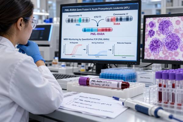
A PML-RARA fusion test looks for the defining genetic abnormality of acute promyelocytic leukemia (APL). In most APL, parts of the PML gene on chromosome 15 and the RARA gene on chromosome 17 join together, creating the PML::RARA fusion. Detecting that fusion confirms the molecular diagnosis and has immediate treatment importance because APL responds to therapies that directly target the abnormal fusion protein, especially all-trans retinoic acid (ATRA) and arsenic trioxide (ATO). The test is also valuable after treatment: sensitive reverse-transcription PCR can measure tiny amounts of PML-RARA RNA as measurable residual disease (MRD) and can help identify molecular persistence or relapse. APL is a medical emergency because it can cause severe bleeding and clotting problems early in the disease. If APL is strongly suspected from blood and marrow findings, treatment with ATRA is often started before molecular confirmation is complete. The final test result then establishes whether the suspected leukemia truly carries PML-RARA.
- A positive PML-RARA result confirms the characteristic molecular lesion of APL in the appropriate clinical and pathologic setting.
- APL treatment should not be delayed while waiting for final molecular results when the disease is strongly suspected, because early bleeding risk can be life-threatening.
- RT-qPCR is commonly used for MRD monitoring because it can detect PML-RARA transcripts far below the level visible by routine microscopy.
- A negative PML-RARA test makes classic APL unlikely but does not exclude every RARA-rearranged leukemia; rare alternative RARA fusion partners require additional testing.
- The key post-treatment milestone is molecular remission after consolidation. Persistent or recurrent PML-RARA generally requires prompt hematology review.
Table of Contents
- What the PML-RARA Fusion Means
- When PML-RARA Testing Is Ordered
- How the Test Is Performed
- How to Interpret PML-RARA Results
- PML-RARA MRD Monitoring
- How the Result Guides APL Treatment
- Limitations, Rare Variants, and Follow-Up
What the PML-RARA Fusion Means
The PML-RARA fusion is created by a balanced chromosome translocation, classically written as t(15;17)(q24;q21). The rearrangement joins the promyelocytic leukemia gene, PML, with the retinoic acid receptor alpha gene, RARA. The resulting PML-RARA protein interferes with normal gene regulation and prevents immature myeloid cells from completing their normal differentiation into mature granulocytes.
That block produces the abnormal promyelocytes that define APL. The same fusion protein also explains why APL is uniquely sensitive to differentiation therapy. ATRA binds the RARA portion of the fusion protein, while arsenic trioxide acts mainly through the PML portion. Together, these drugs promote degradation of the abnormal protein and restore normal maturation and tumor-suppressive pathways.
APL belongs to the acute myeloid leukemia family but differs from most AML in several important ways. It has a specific molecular driver, distinctive coagulopathy, and highly effective targeted therapy. For this reason, rapid molecular confirmation is not a minor classification step; it directly changes treatment.
The fusion is present in the great majority of APL cases. Most patients have one of several recurrent transcript forms depending on the PML breakpoint. Laboratories may describe these as bcr1, bcr2, or bcr3 isoforms. The exact transcript type usually matters more for assay design and later MRD tracking than for the basic diagnosis.
A broad hematologic cancer biomarker panel can identify many AML-associated mutations, but PML-RARA requires dedicated attention because a rapid answer can affect same-day treatment.
When PML-RARA Testing Is Ordered
PML-RARA testing is ordered whenever APL is suspected. Clues may come from the complete blood count, blood smear, bone marrow morphology, flow cytometry, or a bleeding pattern that seems disproportionate to the platelet count.
Typical findings can include abnormal promyelocytes with heavy granulation, bundles of Auer rods, low platelets, anemia, and variable white blood cell counts. APL often produces disseminated intravascular coagulation and excessive fibrinolysis, which can lead to bruising, nosebleeds, gastrointestinal bleeding, pulmonary bleeding, or intracranial hemorrhage. Because those complications can occur before full diagnostic workup is complete, clinicians treat suspected APL as an emergency.
Testing is used in three main situations:
- Diagnosis: confirm that the leukemia carries PML-RARA.
- Response assessment: determine whether the fusion transcript becomes undetectable after therapy.
- Relapse surveillance: detect reappearance or a rising level of PML-RARA before overt hematologic relapse in selected patients.
A sample may be obtained from bone marrow, peripheral blood, or both. Bone marrow is commonly used for definitive diagnosis and key response milestones, while blood can be useful for rapid testing and serial follow-up. The laboratory needs an adequate number of abnormal cells or sufficient RNA to produce a reliable result.
PML-RARA testing is often ordered alongside a leukemia flow cytometry panel, chromosome analysis, FISH, coagulation studies, and routine chemistry. Flow cytometry helps characterize the abnormal promyelocytes but does not replace demonstration of the defining fusion.
How the Test Is Performed
Several laboratory methods can identify PML-RARA, and they answer slightly different questions.
| Method | What it detects | Best use | Important limitation |
|---|---|---|---|
| RT-PCR or RT-qPCR | PML-RARA RNA transcript | Rapid diagnosis and sensitive MRD monitoring | Requires intact RNA and primers that recognize the fusion transcript |
| FISH | PML and RARA rearrangement at the DNA/chromosome level | Fast confirmation when morphology strongly suggests APL | Usually less sensitive than PCR for low-level MRD |
| Karyotype | Visible t(15;17) and other chromosome abnormalities | Overall cytogenetic assessment | Needs dividing cells and can miss cryptic rearrangements |
| Targeted RNA sequencing | Fusion transcripts, including unusual partners | Resolving atypical or negative routine studies | Turnaround time may be longer than emergency assays |
RT-qPCR is particularly important because it both identifies the transcript and measures it quantitatively. At diagnosis, the laboratory establishes which PML-RARA transcript the patient has. Later, the same target can be measured with very high sensitivity.
FISH can return a rapid result and is valuable when immediate confirmation is needed. It uses fluorescent probes that bind near the PML and RARA loci. When the genes are rearranged, the fluorescent pattern changes. However, FISH is generally not the preferred tool for detecting very small amounts of disease after treatment.
Karyotyping may show the classic t(15;17), but a normal-appearing karyotype does not completely exclude PML-RARA. Some rearrangements are cryptic and are detectable by molecular methods even when conventional chromosome analysis looks normal.
No fasting is required for the molecular test itself. Preparation depends on whether the specimen is blood or bone marrow. If a marrow biopsy or aspiration is planned with sedation, the patient may receive separate instructions about eating, drinking, transportation, and medication management.
How to Interpret PML-RARA Results
Positive at diagnosis
A positive PML-RARA result means the defining fusion of classic APL was detected. In a patient with compatible leukemia, this confirms the diagnosis and supports APL-specific treatment rather than a standard non-APL AML approach.
The report may identify the transcript type and may include a quantitative baseline. That baseline is useful because later tests can be compared with the initial level to measure molecular response.
Negative when APL is suspected
A negative result means the laboratory did not detect the standard PML-RARA target in the tested specimen. Possible explanations include:
- the patient does not have APL;
- the sample has too few leukemia cells or poor-quality RNA;
- the rearrangement is present but cryptic or technically missed;
- the leukemia has a rare RARA fusion partner other than PML.
Rare variant RARA rearrangements can resemble APL morphologically but may not respond to ATRA or ATO in the same way. If clinical and pathologic suspicion remains high, the laboratory may use break-apart FISH, broader RNA fusion testing, or sequencing to look for another rearrangement.
Positive after treatment
After therapy, interpretation depends on when the sample was collected. Early in treatment, PML-RARA can remain detectable even in patients who will ultimately be cured. Molecular positivity immediately after induction does not necessarily represent treatment failure because differentiation therapy may clear the transcript gradually.
The more critical milestone is after consolidation. Persistent molecular positivity at that stage is much more concerning and generally requires confirmation and specialist action. Reappearance of PML-RARA after a prior molecular remission also raises concern for molecular relapse.
PML-RARA MRD Monitoring
PML-RARA is an excellent MRD marker because it is leukemia-specific, quantifiable, and usually stable across the disease course. Sensitive RT-qPCR can detect the fusion at levels far below what can be seen on a blood smear or marrow aspirate.
The goal of MRD monitoring is not simply to obtain a “negative” label. It is to determine whether molecular disease is clearing as expected, whether complete molecular remission has been achieved, and whether the transcript later reappears.
The most clinically useful patterns are:
- Falling transcript level during treatment: expected molecular response.
- PCR negative after consolidation: molecular remission and a favorable response milestone.
- Persistent confirmed positivity after consolidation: possible resistant or residual APL.
- Conversion from negative to positive on serial testing: molecular recurrence.
- Consistent rise on repeat samples: high concern for impending hematologic relapse.
A low positive result should usually be confirmed on a second sample, especially if it is unexpected or close to the assay’s detection limit. Contamination, specimen quality, and technical variation are uncommon but important considerations when the clinical consequences are major.
Monitoring schedules depend on risk group and treatment regimen. In modern ATRA/ATO-treated low- and intermediate-risk APL, relapse is uncommon after molecular remission, so prolonged intensive PCR surveillance may provide limited benefit. Higher-risk patients and those with previous relapse may be monitored more closely. Practices also differ among guidelines and institutions.
PML-RARA PCR is a disease-specific form of MRD testing for leukemia. It differs from multiparameter flow MRD because it tracks the defining fusion transcript rather than an abnormal cell-surface pattern.
An important practical point is to compare like with like. Marrow and blood may differ in sensitivity, and laboratories may report normalized copy numbers, ratios, log reductions, or qualitative positive/negative results. Serial testing is easiest to interpret when the same laboratory, specimen type, and validated method are used whenever possible.
How the Result Guides APL Treatment
A suspected APL diagnosis triggers immediate clinical action because early hemorrhage is the major preventable cause of death. When morphology and clinical findings strongly suggest APL, ATRA is typically started before the PML-RARA result is final. If testing later shows that APL is not present, the treatment plan is revised.
Once PML-RARA is confirmed, treatment depends mainly on the presenting white blood cell count and other clinical factors. Patients are often categorized as non-high-risk when the white blood cell count is 10 × 10⁹/L or lower and high-risk when it is above that threshold.
For many newly diagnosed non-high-risk adults, ATRA plus arsenic trioxide is a standard chemotherapy-free backbone. High-risk disease generally requires additional cytoreduction, which may include anthracycline-based chemotherapy or gemtuzumab ozogamicin depending on the regimen and patient.
Supportive care is equally important. Platelets, fibrinogen, prothrombin time, activated partial thromboplastin time, and other coagulation measures are followed closely during the dangerous early phase. Transfusions are used aggressively to reduce hemorrhagic risk. Leukapheresis is generally avoided in APL because it can worsen coagulopathy.
The PML-RARA result also explains important treatment complications. Differentiation syndrome can occur after ATRA or ATO and may cause fever, weight gain, edema, low blood pressure, lung infiltrates, shortness of breath, and kidney dysfunction. It requires urgent recognition and corticosteroid treatment.
Molecular response after consolidation helps confirm that treatment has eliminated detectable disease. If the fusion remains positive, or becomes positive again after remission, clinicians consider salvage therapy, arsenic- or ATRA-based approaches depending on prior exposure, targeted agents, and stem cell transplantation in appropriate settings.
Unlike many AML markers, PML-RARA is not mainly a prognostic co-mutation. It is the central disease-defining driver and therapeutic target. Other AML mutation tests can still provide useful biology, but the PML-RARA status has first-order importance for diagnosis and immediate management.
Limitations, Rare Variants, and Follow-Up
The most important limitation is that a negative standard PML-RARA assay does not exclude every leukemia with promyelocytic morphology. Rare rearrangements can involve RARA with partners such as ZBTB16, NPM1, NUMA1, or STAT5B, and some have very different sensitivity to ATRA or arsenic. Atypical morphology, unusual immunophenotype, or persistent clinical suspicion should prompt broader fusion testing.
Another limitation is that PCR depends on RNA quality. Old, poorly handled, or low-cellularity specimens can produce false-negative or uninterpretable results. Laboratories use control genes to verify that enough amplifiable RNA is present.
A positive molecular test must also be interpreted at the correct treatment time point. Residual transcripts shortly after induction are not equivalent to persistent positivity after consolidation. Treating every early low-level result as relapse would expose patients to unnecessary intervention.
Questions that help clarify a report include:
- Was the classic PML-RARA fusion detected, and which transcript isoform was identified?
- Was the result obtained by RT-qPCR, FISH, karyotype, or another method?
- If negative, was RNA quality adequate and is broader RARA fusion testing needed?
- Is this result from blood or bone marrow?
- For MRD, how does the level compare with the prior sample?
- Has molecular remission been documented after consolidation?
- Does the result change the frequency of follow-up or the need for salvage therapy?
Patients with suspected or confirmed APL should not wait for routine follow-up if they develop new bleeding, severe headache, confusion, shortness of breath, chest pain, fainting, or rapidly spreading bruising. Those symptoms may reflect life-threatening hemorrhage, thrombosis, infection, or treatment complications and require urgent medical assessment.
Because APL can cause life-threatening bleeding before every diagnostic detail is finalized, the molecular test sits inside an unusually time-sensitive clinical pathway. When APL is strongly suspected from the blood smear, coagulation abnormalities, and immunophenotype, clinicians may start differentiation therapy while urgent genetic confirmation is being arranged. The PML-RARA result then confirms the molecular diagnosis and helps define the transcript that can be followed later.
Serial molecular testing should use the same transcript target and a laboratory with validated sensitivity. A single very low positive result near the detection limit may need confirmation, particularly if it conflicts with the clinical picture, while repeated or rising positivity after a previously negative state is more concerning. Bone marrow is generally more sensitive than peripheral blood for some MRD assessments, although blood can be useful for interim surveillance depending on the treatment protocol and local practice.
The timing of molecular response also matters. APL can remain PCR-positive early during induction even when therapy is working, so clinicians do not usually judge treatment failure from an early positive result alone. The most informative milestones occur after completion of consolidation and during follow-up in patients whose risk or treatment protocol calls for serial monitoring. The goal is durable molecular remission, not simply disappearance of abnormal cells from the microscope.
A copy of the baseline molecular report is worth keeping because it identifies the exact PML-RARA transcript detected at diagnosis. Later MRD assays should target the same transcript. If a future report uses a different specimen type, laboratory, or reporting scale, the treating team may need to compare qualitative status and trend rather than treating the raw numbers as directly interchangeable.
References
- Management of acute promyelocytic leukemia: updated recommendations from an expert panel of the European LeukemiaNet 2019 (Guideline)
- Value of measurable residual disease monitoring in patients with acute promyelocytic leukemia in the era of frontline ‘chemotherapy-free’ therapy 2022
- Function of PML-RARA in Acute Promyelocytic Leukemia 2024 (Review)
- Acute Promyelocytic Leukemia, Retinoic Acid, and Arsenic: A Tale of Dualities 2024 (Review)
- Acute Promyelocytic Leukemia: Pathophysiology, Diagnosis and Clinical Management 2025 (Review)
- Advances in RARα fusion genes in acute promyelocytic leukemia 2025 (Review)
Disclaimer
This article is for general education and is not a substitute for diagnosis or treatment by a hematologist or leukemia specialist. Suspected APL is a medical emergency because of its bleeding risk; urgent treatment may be needed before molecular confirmation is complete. PML-RARA and MRD results must be interpreted with the treatment phase, specimen type, assay sensitivity, coagulation findings, and the full clinical picture.





