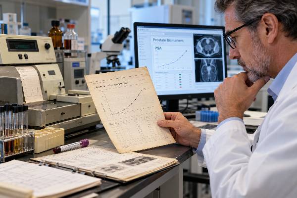
Prostatic acid phosphatase, or PAP, is an enzyme produced in high amounts by prostate tissue. Before prostate-specific antigen became widely used, serum PAP was one of the main blood markers for prostate cancer, especially for advanced or metastatic disease. Today, PAP is not a standard screening test for early prostate cancer because it is less sensitive than PSA and can remain normal in localized disease. An elevated PAP level can occur with prostate cancer—particularly a larger tumor burden or metastatic disease—but it is not specific enough to diagnose cancer on its own. Modern prostate cancer evaluation relies much more heavily on PSA, MRI, biopsy, genomic testing, and PSMA-based imaging. PAP still matters historically and biologically, however. The protein, encoded by the ACP3 gene, remains strongly associated with prostate tissue and has re-emerged as a research target for imaging, immune therapy, and radioligand treatment. That makes PAP unusual: its role as a routine serum marker has faded, while interest in the molecule itself is growing again.
- PAP is an older prostate cancer blood marker that has largely been replaced by PSA for screening, diagnosis, and routine monitoring.
- High PAP can occur in advanced prostate cancer, especially when disease burden is substantial, but a normal PAP does not rule out localized or metastatic cancer.
- PAP is not prostate-cancer-specific enough to diagnose cancer by itself and must be interpreted with PSA, imaging, pathology, and clinical findings.
- There is no widely used modern PAP screening cutoff because laboratories use different methods and the test is no longer central to early-detection pathways.
- PAP is gaining new research interest as ACP3, a prostate-associated target for molecular imaging and potential radioligand or antibody-based therapy.
Table of Contents
- What Prostatic Acid Phosphatase Is
- Why PAP Was Used for Prostate Cancer
- What High PAP Levels Can Mean
- PAP Versus PSA
- How PAP Testing Is Performed
- PAP in Modern Prostate Cancer Care
- New Research on ACP3 Targeting
What Prostatic Acid Phosphatase Is
Acid phosphatases are enzymes that remove phosphate groups from molecules in an acidic environment. Several tissues produce acid phosphatase, but the prostate produces a distinctive prostatic form historically called prostatic acid phosphatase (PAP). The corresponding gene is ACP3, and the protein exists in secreted and membrane-associated forms.
PAP is abundant in prostate epithelial cells and is released into seminal fluid. It can also enter the bloodstream. Prostate cancer cells often continue to express PAP, including many advanced and castration-resistant tumors, which is why the molecule became useful as a tumor-associated marker long before modern molecular imaging existed.
A laboratory PAP test usually refers to measurement of the prostatic component of acid phosphatase in serum. Older methods tried to distinguish prostatic acid phosphatase from total acid phosphatase using chemical inhibition or enzyme activity. Later immunoassays improved specificity by measuring the prostate-associated protein more directly.
The name can be confusing because PAP is not the same as PSA. PSA is a kallikrein-family protease, while PAP is a phosphatase enzyme. They are biologically different molecules and behave differently as blood markers.
PAP also should not be confused with alkaline phosphatase, another enzyme that can be elevated when prostate cancer spreads to bone. Alkaline phosphatase mainly reflects bone and liver activity and remains useful in selected patients with metastatic disease. PAP, by contrast, originates largely from prostate tissue and historically served as a marker of prostate cancer burden.
Because modern laboratories rarely use PAP for screening, a PAP result may appear only in selected oncology settings, research protocols, older records, or specialized diagnostic workups. If a current report includes PAP, the interpretation should use that laboratory’s reference interval and assay method rather than a historical number copied from older literature.
Why PAP Was Used for Prostate Cancer
PAP has an important place in cancer history. In the early and mid-20th century, researchers recognized that serum acid phosphatase activity could become markedly elevated in men with metastatic prostate cancer, particularly when cancer involved bone. For decades, PAP was one of the few biochemical clues available for staging and monitoring prostate malignancy.
The test was especially useful when prostate cancer was diagnosed later than it often is today. Patients frequently presented with locally advanced disease, bone pain, or widespread metastases, situations in which PAP was more likely to be elevated. Falling PAP after treatment could indicate response, while rising levels could suggest progression.
Its main weakness was limited sensitivity for early-stage disease. Many localized prostate cancers do not raise PAP enough to be detected. That made PAP a poor population screening marker. It was more closely associated with tumor burden and advanced disease than with small organ-confined tumors.
PSA changed this landscape. PSA is more sensitive to relatively small changes in prostate tissue and can become elevated much earlier in the disease course. Once PSA testing became widely available in the late 1980s and 1990s, PAP rapidly lost its role as the dominant prostate cancer blood marker.
Modern screening and early-detection strategies therefore center on the prostate-specific antigen test, with MRI and secondary biomarkers used to refine risk. PAP is generally absent from contemporary early-detection algorithms because adding it to PSA has not shown enough incremental value to justify routine use.
Historical importance does not mean PAP is obsolete as a biological target. The same feature that once made it useful as a serum marker—strong expression in prostate tissue and many prostate cancers—now makes ACP3 interesting for targeted imaging and therapy.
What High PAP Levels Can Mean
A high PAP result can be associated with prostate cancer, especially advanced, metastatic, or high-volume disease, but the level is not diagnostic by itself. The clinical meaning depends on why the test was ordered, the assay method, the reference interval, and other findings.
Potential explanations for an elevated prostatic acid phosphatase include:
- prostate cancer with a larger burden of disease;
- metastatic prostate cancer, historically including bone metastases;
- local prostate tissue injury or manipulation;
- benign prostatic disease in some cases;
- analytical or specimen-handling issues, particularly with older enzyme-activity assays.
The relationship between PAP and stage is not exact. Some patients with advanced prostate cancer can have normal PAP, while some elevations occur without metastatic cancer. A single abnormal value therefore cannot establish stage or replace imaging.
There is also no single modern PAP threshold analogous to commonly discussed PSA reference points. Older laboratories reported values using different units and methods, such as enzyme activity units, while newer immunoassays may use mass concentration. A result of “3” in one assay cannot safely be compared with “3” from another historical method.
If PAP is unexpectedly high today, the next question should be why it was measured. In a person without known prostate cancer, PSA, clinical examination, MRI, and standard urologic evaluation are more informative for early detection. In someone with known advanced prostate cancer, the clinician may compare PAP with PSA, imaging, symptoms, alkaline phosphatase, and other markers of disease burden.
Very high or rapidly changing PAP should not be interpreted outside the full clinical picture. Bone pain, weakness, weight loss, urinary obstruction, neurologic symptoms, or other signs of advanced disease require medical evaluation regardless of the biomarker result.
PAP Versus PSA
PAP and PSA are both associated with prostate tissue, but PSA is the superior routine blood marker for most modern clinical purposes.
| Feature | PAP | PSA |
|---|---|---|
| Main historical role | Advanced disease staging and monitoring | Screening, diagnosis support, monitoring, recurrence detection |
| Sensitivity for localized cancer | Relatively low | Higher, though still imperfect |
| Cancer specificity | Limited | Limited; benign prostate conditions also raise it |
| Routine screening use today | No | Yes, after shared decision-making |
| Role after treatment | Occasional or research use | Central marker in most patients |
| Current research interest | ACP3-targeted imaging and therapy | Improved PSA isoforms, density, kinetics, and multivariable models |
PSA’s higher sensitivity made it possible to detect many cancers while still localized. That advantage came with a new problem: PSA can also lead to false-positive testing and overdiagnosis of indolent cancer. Modern care tries to improve specificity with tools such as percent-free PSA, the Prostate Health Index, 4Kscore, urine biomarkers, PSA density, and MRI.
PAP did not disappear because it was meaningless. It disappeared from routine screening because PSA and later tools provided better early-detection performance. A PAP result from an older medical record should therefore be interpreted in the context of the era in which it was measured. Before widespread PSA screening, a markedly elevated PAP often signaled advanced disease; today, patients are often diagnosed before PAP would ever rise.
Another important difference appears after radical prostatectomy. PSA is expected to become extremely low because most PSA-producing tissue has been removed, making tiny recurrences detectable. PAP is not used routinely for that purpose. The sensitivity of modern PSA assays is much better suited to biochemical recurrence monitoring.
How PAP Testing Is Performed
A PAP test is performed on a blood sample. Fasting is usually not required, but preparation instructions depend on the laboratory and clinical context.
Historically, PAP enzyme activity was considered relatively unstable, and specimen handling could affect the result. Prompt processing and laboratory-specific methods were important. Modern immunoassays are more robust, but the test is uncommon enough that clinicians should verify the method if a result will influence care.
Before testing, report recent prostate procedures or manipulation, urinary retention, prostatitis symptoms, and known prostate disease. Older literature describes transient changes after prostate massage or instrumentation, and such factors can complicate interpretation.
No one should delay standard prostate cancer evaluation simply to obtain PAP. If the concern is an elevated PSA or abnormal prostate examination, current diagnostic pathways usually involve repeat PSA when appropriate, MRI, risk calculators, secondary biomarkers, and biopsy rather than PAP.
For advanced disease, PAP may occasionally appear as part of a broader laboratory assessment, but the more routinely followed markers include PSA, complete blood count, metabolic testing, liver enzymes, and alkaline phosphatase. Imaging is used to define the location and extent of metastases.
A normal PAP cannot rule out prostate cancer. This limitation is especially important in localized disease and in modern patients diagnosed earlier through PSA-based evaluation. If a clinician has a substantial cancer concern, a normal PAP should not stop further workup.
PAP in Modern Prostate Cancer Care
The most established modern use of PAP is not as a serum marker but as a tumor-associated antigen. Sipuleucel-T, an autologous cellular immunotherapy used in selected men with metastatic castration-resistant prostate cancer, is manufactured by exposing a patient’s immune cells to a fusion protein that contains prostatic acid phosphatase linked to an immune-stimulating factor. The treatment uses PAP as a target for immune recognition rather than measuring PAP levels to decide whether treatment is working.
This distinction is important. A molecule can be clinically valuable as a therapeutic target even when its blood concentration is not a good screening test. The same pattern is now seen with PSMA, where surface expression on cancer cells is exploited for imaging and radioligand treatment.
For disease localization, the PSMA imaging test has transformed modern prostate cancer staging and recurrence assessment. PSMA PET is much more clinically established than PAP-targeted imaging at present.
PAP can also appear in pathology. Immunohistochemical staining for PAP may help support prostatic origin in selected tumors, although modern pathology often uses a panel of markers such as PSA, NKX3.1, and PSMA depending on the differential diagnosis. Again, tissue staining and serum PAP are different applications.
For patients, the practical message is simple: PAP is usually not a test you need to add to a modern PSA screening panel. If it appears on a current oncology report, ask what specific decision the result is intended to inform.
New Research on ACP3 Targeting
PAP has recently regained attention under its gene/protein name ACP3 because it is highly expressed in most prostate cancers and can remain present in castration-resistant disease. Researchers are developing molecules that bind ACP3 on or around tumor cells for imaging and potentially for targeted radioligand therapy.
A first-in-human PET study reported imaging with [68Ga]Ga-OncoACP3-DOTA, an ACP3-targeting radioligand, and compared it with PSMA-targeted imaging in a small group of patients. The ACP3 tracer showed promising tumor uptake with relatively low activity in salivary glands and kidneys—organs that commonly show substantial uptake with PSMA tracers. These early findings are interesting because off-target radiation to salivary glands and kidneys is one limitation of some PSMA-directed treatments.
However, early imaging research should not be confused with established clinical care. Small first-in-human studies are designed to show feasibility, biodistribution, and preliminary lesion detection. They do not yet prove that ACP3 PET improves survival, replaces PSMA PET, or should be used routinely.
Researchers are also exploring membrane-associated PAP as a target for antibodies and other therapeutic platforms. A 2026 review described ACP3 as a potentially complementary theranostic target for tumors with heterogeneous or low PSMA expression. The idea is attractive because a second target could broaden the number of patients eligible for molecularly directed imaging or therapy.
For now, these applications remain investigational. The historical serum PAP test and emerging ACP3 theranostics are related by the same molecule but belong to different eras of prostate cancer care. One is an older blood marker largely displaced by PSA; the other is a modern attempt to exploit prostate-specific biology for precision imaging and treatment.
That evolution is why PAP remains worth understanding. It shows how a biomarker can move from blood measurement, to immunologic target, to potential theranostic platform as technology changes.
For someone who sees PAP on an old chart, the date and unit are essential. Historical enzyme-activity assays may be reported in units that do not correspond to modern immunoassays, and laboratory reference ranges changed as methods improved. A historical “high PAP” should therefore be interpreted from the original report rather than converted using a current internet reference range. The same caution applies when comparing serial values from different decades or laboratories.
PAP also illustrates a broader tumor-marker principle: a marker can be useful for one disease stage and poor for another. PAP was more informative when prostate cancer was bulky or metastatic, but it was insufficiently sensitive for small localized tumors. PSA had the opposite practical advantage—it often rose earlier—so it became much more useful for detection and post-treatment monitoring. Modern biomarkers continue this progression by trying to identify clinically significant disease rather than simply increasing the number of cancers found.
If PAP is ordered today as part of a specialized evaluation, ask whether the clinician is using it as a serum marker, a pathology marker, or within a research protocol. Those uses are biologically related but clinically distinct, and the result should be interpreted only within the purpose for which it was measured.
References
- The prostatic acid phosphatase in prostate cancer: A novel theranostic target 2026 (Review)
- Prostatic acid phosphatase: a rediscovered target in prostate cancer 2026 (Review)
- Translational and First-in-Human Positron Emission Tomography Targeting Prostatic Acid Phosphatase in Prostate Cancer Using the Ligand [68Ga]Ga-OncoACP3-DOTA 2026
- Transmembrane prostatic acid phosphatase: a therapeutic target in advanced prostate cancer 2024 (Review)
- From prostate specific antigen to genomic signatures: Advances in biomarkers for prostate cancer diagnosis and prognosis 2026 (Review)
- Early Detection of Prostate Cancer: AUA/SUO Guideline Part I: Prostate Cancer Screening 2023 (Guideline)
Disclaimer
This article is for general education and does not replace medical advice or individualized cancer evaluation. PAP is not a standard modern prostate cancer screening test, and reference intervals vary by assay. New ACP3-targeted imaging and treatment approaches are investigational and should not be confused with established PSMA imaging or approved therapies.